

The relationships between lens diameter and ocular biometric parameters: an ultrasound biomicroscopy-based study
- PMID: 38288306
- PMCID: PMC10822951
- DOI: 10.3389/fmed.2023.1306276
The relationships between lens diameter and ocular biometric parameters: an ultrasound biomicroscopy-based study
Abstract
Purpose: This study aims to explore the relationships between lens diameter (LD) measured with ultrasound biomicroscopy (UBM) and ocular biometric parameters.
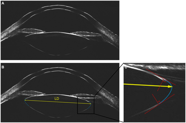
Methods: Ocular biometric parameters including axial length (AL), white-to-white distance (WTW), anterior chamber depth (ACD), lens thickness (LT) and anterior segment length (ASL) were measured with IOL-Master 700, and the direct measurement of LD was conducted through UBM (ArcScan Insight 100). Relationships between LD and ocular biometric parameters were then investigated. Eyes with AL ≥ 28 mm were defined as eyes with extreme myopia, and eyes with AL < 28 mm were defined as eyes without extreme myopia.
Results: A total of 194 eyes from 194 subjects were included. The mean LD was 9.58 ± 0.49 mm, ranging from 8.60 to 10.96 mm. According to univariate analysis, larger LD was associated with elder age, male gender, larger WTW, ACD and ASL (all p < 0.05). Meanwhile, the LD was positively correlated with AL in eyes without extreme myopia (p < 0.05), but not in eyes with extreme myopia (p > 0.05). Backward stepwise regressions revealed that a larger LD was associated with larger WTW, ASL and AL in eyes without extreme myopia (all p < 0.05), while ASL was the only significant variable in eyes with extreme myopia (p < 0.05).
Conclusion: Larger WTW, ASL and AL in eyes without extreme myopia, as well as longer ASL in eyes with extreme myopia indicated a larger LD, which provides guidance in personalized surgical choice and promises ideal visual outcomes.
Keywords: anterior segment length; axial length; lens diameter; ultrasound biomicroscopy; white-to-white distance.
Copyright © 2024 Huang, Qi, Cheng, Liu, Zhang, Du, Lu and Zhu.
Conflict of interest statement
The authors declare that the research was conducted in the absence of any commercial or financial relationships that could be construed as a potential conflict of interest. The author(s) declared that they were an editorial board member of Frontiers, at the time of submission. This had no impact on the peer review process and the final decision.
Figures


Similar articles
-
The use of 35 MHz ultrasonic biomicroscopy (UBM) to assess the biometric parameters of the anterior segment in pediatric cataractous eyes.Indian J Ophthalmol. 2025 May 1;73(5):713-719. doi: 10.4103/IJO.IJO_2429_24. Epub 2025 Apr 24. Indian J Ophthalmol. 2025. PMID: 40272301 Free PMC article.
-
Lens thickness and associated ocular biometric factors among cataract patients in Shanghai.Eye Vis (Lond). 2021 May 31;8(1):22. doi: 10.1186/s40662-021-00245-3. Eye Vis (Lond). 2021. PMID: 34053465 Free PMC article.
-
Ciliary sulcus characteristics in patients with axial myopia using ultrasound biomicroscope.Asia Pac J Ophthalmol (Phila). 2025 Mar-Apr;14(2):100162. doi: 10.1016/j.apjo.2025.100162. Epub 2025 Feb 15. Asia Pac J Ophthalmol (Phila). 2025. PMID: 39961582
-
Distribution of Ocular Anterior and Posterior Segment Lengths Among a Cataract Surgical Population in Shanghai.Front Med (Lausanne). 2021 Sep 23;8:688805. doi: 10.3389/fmed.2021.688805. eCollection 2021. Front Med (Lausanne). 2021. PMID: 34631728 Free PMC article.
-
Associations between anterior segment parameters and rotational stability of a plate-haptic toric intraocular lens.J Cataract Refract Surg. 2021 Nov 1;47(11):1436-1440. doi: 10.1097/j.jcrs.0000000000000653. J Cataract Refract Surg. 2021. PMID: 34675151
References
LinkOut - more resources
Full Text Sources
Research Materials