Review
. 2024 Jan 15:10:1296941.
doi: 10.3389/fmolb.2023.1296941. eCollection 2023.

Recent advances in infectious disease research using cryo-electron tomography

Affiliations
Review

Recent advances in infectious disease research using cryo-electron tomography

Daniel Asarnow et al. Front Mol Biosci. .

Abstract

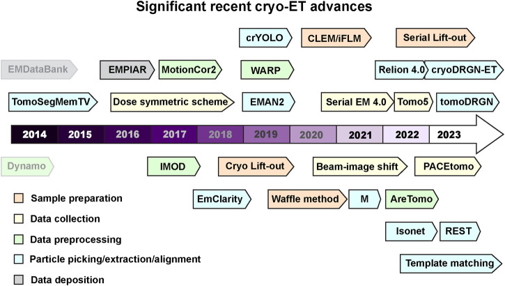
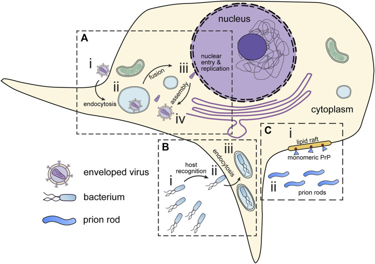
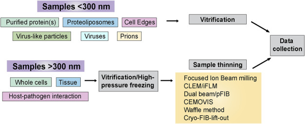
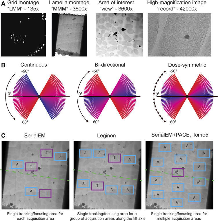
With the increasing spread of infectious diseases worldwide, there is an urgent need for novel strategies to combat them. Cryogenic sample electron microscopy (cryo-EM) techniques, particularly electron tomography (cryo-ET), have revolutionized the field of infectious disease research by enabling multiscale observation of biological structures in a near-native state. This review highlights the recent advances in infectious disease research using cryo-ET and discusses the potential of this structural biology technique to help discover mechanisms of infection in native environments and guiding in the right direction for future drug discovery.

Keywords: bacteria; cryo-EM; cryo-ET; host-pathogen interaction; infectious diseases; pathogen; viruses.


Conflict of interest statement

The authors declare that the research was conducted in the absence of any commercial or financial relationships that could be construed as a potential conflict of interest.

Figures

FIGURE 1
FIGURE 1
Recent milestones in cryo-ET. Significant advances in tools used for cryo-ET are listed according to the recent timeline. The tools are color coded into various categories- sample preparation, data collection, data processing, and deposition. Dynamo and EMDataBank are greyed out to differentiate that these were developed before the timeline mentioned here. Software packages and tools are outlined to distinguish it from methods and other advances. Several of these tools can perform multiple tasks in addition to their color code provided in the figure. All these cryo-ET developments are discussed and referenced in the main text.
FIGURE 2
FIGURE 2
Infectious disease targets for in situ tomography. (A) Viruses attach to the periphery of cells (i). Interaction with host organelles, such as the endosomes are sometimes required for entry (ii). DNA viruses and some RNA viruses must transport genetic material to the nucleus (iii) before assembly in the cytoplasm (iv). (B) Pathogenic bacteria express motility and virulence machinery which enable them to seek out and infect host cells (i). Upon encountering host cells, bacteria deliver virulence factors which promote host cell entry (ii). Following virulence factor delivery, bacteria enter host cells via endocytosis (iii). (C) Prions can interact and seed membranes at lipid rafts on the host cell surface. (i) Monomeric prion proteins (blue) are attached to lipid rafts by glycosylphosphatidylinositol (GPI) anchors (red). (ii) Prion rods occupy the extracellular space. All these have presented interesting targets for in situ tomography.
FIGURE 3
FIGURE 3
Sample preparation flowchart. Sample thickness is an important determinant in deciding the strategy for cryo-ET sample preparation before data collection. This flowchart outlines the freezing methods and post-freezing sample prep requirements before data acquisition.
FIGURE 4
FIGURE 4
Cryo-ET data acquisition. (A) Typical multi-level imaging approach to tomography data acquisition. First images are acquired at the lowest magnification to obtain a grid overview or “low magnification montage (LMM); areas of interest are selected from LMM and imaged at intermediate magnification, often stitched together to produce a “medium magnification montage (MMM)”; targets are selected from MMMs and will be imaged at high magnification while tilting the stage. (B) Comparison of dose distribution among commonly used tilt schemes. Blue slices are earlier tilts in the sequence and have lowest total accumulated dose, red slices are latest tilts and have highest total accumulated dose. (C) Common strategies for object tracking during tilt series acquisition. In SerialEM each acquisition target has an associated tracking/focusing area shifted along the tilt axis. In Leginon multiple acquisition targets along a tilt axis can share a common tracking/focusing area. In PACEtomo within SerialEM a single tracking area can be shared for multiple acquisition areas without being constrained to a tilt axis; this approach is also implemented in TFS Tomo5 software.
FIGURE 5
FIGURE 5
The data pre-processing workflow of cryo-ET with STA. Data pre-processing workflow using the publicly available dataset EMPIAR-10164 as an example. Representative inputs and outputs of each step are shown as an image in the top panel. Input and output file formats are listed below, followed by the software used in the step from motion correction of frames to tomogram generation.
FIGURE 6
FIGURE 6
The data post-processing workflow of cryo-ET with STA. A continuation of Figure 5 for the post-processing workflow of cryo-ET with STA, with the publicly available dataset EMPIAR-10164. Representative inputs and outputs of each step are shown as an image in the top panel. Input and output file formats are listed below, followed by the software used in the step from particle picking to sub-tilt refinement.
FIGURE 7
FIGURE 7
Archiving of volumeEM, cryo-ET and STA data. Tilt series and volumeEM data are archived in EMPIAR, EMPIAR-11456 (unpublished) and EMPIAR-11449 (Suga et al., 2021) shown in this section. Tomograms and subtomogram averages are archived in the EMDB, EMDB-15182 (Calder et al., 2022) and EMD-14590 (Ni et al., 2022) shown in this section. Finally, models are deposited to the PDB 7ZBT shown in this section (model associated with EMD-14590).

References

    1. Al-Amoudi A., Chang J.-J., Leforestier A., McDowall A., Salamin L. M., Norlén L. P. O., et al. (2004). Cryo-electron microscopy of vitreous sections. EMBO J. 23, 3583–3588. 10.1038/sj.emboj.7600366 - DOI - PMC - PubMed
    1. Anderson H. A., Chen Y., Norkin L. C. (1996). Bound simian virus 40 translocates to caveolin-enriched membrane domains, and its entry is inhibited by drugs that selectively disrupt caveolae. Mol. Biol. Cell 7, 1825–1834. 10.1091/mbc.7.11.1825 - DOI - PMC - PubMed
    1. Artikis E., Kraus A., Caughey B. (2022). Structural biology of ex vivo mammalian prions. J. Biol. Chem. 298, 102181. 10.1016/j.jbc.2022.102181 - DOI - PMC - PubMed
    1. Balyschew N., Yushkevich A., Mikirtumov V., Sanchez R. M., Sprink T., Kudryashev M. (2023). Streamlined structure determination by cryo-electron tomography and subtomogram averaging using TomoBEAR. Nat. Commun. 14 6543. 10.1038/s41467-023-42085-w - DOI - PMC - PubMed
    1. Berger C., Dumoux M., Glen T., Yee N. B.-y., Mitchels J. M., Patáková Z., et al. (2023). Plasma FIB milling for the determination of structures in situ . Nat. Commun. 14, 629. 10.1038/s41467-023-36372-9 - DOI - PMC - PubMed

LinkOut - more resources