Review
. 2024 Feb;17(1):e004355.
doi: 10.1161/CIRCGEN.123.004355. Epub 2024 Jan 30.

Mechanisms of RBM20 Cardiomyopathy: Insights From Model Systems

Affiliations
Review

Mechanisms of RBM20 Cardiomyopathy: Insights From Model Systems

Zachery R Gregorich et al. Circ Genom Precis Med. 2024 Feb.

Abstract

RBM20 (RNA-binding motif protein 20) is a vertebrate- and muscle-specific RNA-binding protein that belongs to the serine-arginine-rich family of splicing factors. The RBM20 gene was first identified as a dilated cardiomyopathy-linked gene over a decade ago. Early studies in Rbm20 knockout rodents implicated disrupted splicing of RBM20 target genes as a causative mechanism. Clinical studies show that pathogenic variants in RBM20 are linked to aggressive dilated cardiomyopathy with early onset heart failure and high mortality. Subsequent studies employing pathogenic variant knock-in animal models revealed that variants in a specific portion of the arginine-serine-rich domain in RBM20 not only disrupt splicing but also hinder nucleocytoplasmic transport and lead to the formation of RBM20 biomolecular condensates in the sarcoplasm. Conversely, mice harboring a disease-associated variant in the RRM (RNA recognition motif) do not show evidence of adverse remodeling or exhibit sudden death despite disrupted splicing of RBM20 target genes. Thus, whether disrupted splicing, biomolecular condensates, or both contribute to dilated cardiomyopathy is under debate. Beyond this, additional questions remain, such as whether there is sexual dimorphism in the presentation of RBM20 cardiomyopathy. What are the clinical features of RBM20 cardiomyopathy and why do some individuals develop more severe disease than others? In this review, we summarize the reported observations and discuss potential mechanisms of RBM20 cardiomyopathy derived from studies employing in vivo animal models and in vitro human-induced pluripotent stem cell-derived cardiomyocytes. Potential therapeutic strategies to treat RBM20 cardiomyopathy are also discussed.

Keywords: alternative splicing; biomolecular condensates; cardiomyopathy, dilated; heart failure; induced pluripotent stem cell.


Conflict of interest statement

Disclosures None.

Figures

Figure 1.
Figure 1.
Domain structure and pathogenic sequence variants in human RBM20. A. Schematic showing structure of human RBM20 with the amino acid positions of exon boundaries and domains/amino acid-rich regions indicated. B. Sequence alignment of the RS domain with pathogenic variants reported in the literature listed. *signifies residue that is not conserved throughout vertebrate evolution. The positions of the NLS, identified in our recent work, and the DCM-associated variant hotspot in exon 9 (c.1881–1920) are indicated. C. Sequence alignment showing portion of the glutamate-rich region in RBM20 with pathogenic variants reported in the literature listed. The DCM-associated variant hotspot in exon 11 (c.2721–2760) is shown. Residues conserved throughout vertebrate evolution are highlighted in red. Residues highlighted in blue and green denote conserved positive and negative charge, respectively. For panels B and C, sequences for species at different points throughout vertebrate evolution from zebra fish (D. rerio) to humans (H. sapiens) were downloaded from the NCBI database. Sequence alignments were constructed with the aid of Multalin. References reporting pathological variants listed in panels B and C are provided in Table 1.
Figure 2.
Figure 2.
Forest plot of RBM20 variant frequency in familial DCM/LVNC cases. A. An estimated 4.2% of familial DCM/LVNC cases are associated with RBM20 variants. “Events” refers to the number of individuals in each cohort with pathogenic variants in RBM20 (only variants listed in Table 1 were considered pathological variants) and the “total” refers to the total cohort size (i.e., individuals with DCM/LVNC screened in the study). The red boxes denote a point estimate of the study result (i.e., the proportion of individuals in the given cohort carrying pathogenic variants in RBM20) and the lines represent the 95% confidence intervals of the study result, with each end of the line denoting the boundaries of the confidence interval. CI, confidence interval. B. Additional study details including reported variants that were considered in this analysis and whether the variants co-segregated with disease in the affected families.
Figure 3.
Figure 3.
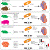
Graphical summary of current RBM20 models, contributing mechanism(s), and phenotypes. Rbm20 KO rodents develop a mild DCM caused by loss of nuclear RBM20 and mis-splicing of RBM20 target genes, such as Ttn and Ca2+-handling genes (e.g. Camk2d, Ryr2, etc.). In contrast to RBM20 KO animals, NLS variant KI and RS domain deletion animals develop a more severe DCM phenotype mimicking that in human patients. In addition to RBM20 target gene mis-splicing, these animals also exhibit abnormal accumulation of RBM20 in the sarcoplasm in RBM20 biomolecular condensates (shown as blue circles in the cell diagram). Surprisingly, despite mis-splicing of major RBM20 target genes (i.e., Ttn, Camk2d, Ldb3), RRM deletion and variant KI models show only systolic dysfunction without evidence of adverse remodeling. Notably, there is no evidence for RBM20 mis-localization in the hearts of these animals. NLS variant KI hiPSC-CMs show evidence of a DCM-like phenotype with impaired contractility and Ca2+-handling abnormalities along with sarcoplasmic RBM20 condensates and mis-splicing consistent with that in NLS variant KI animals.
Figure 4.
Figure 4.
RBM20-dependent regulation of TTN alternative splicing. The TTN gene consists of 363 exons. Alternative splicing occurring in exons encoding the Z-disk (1–28), A-band (252–357), and M-band (358–363) does not significantly alter the size of the titin protein. Exons encoding the I-band region, which consists of the proximal Ig, N2B, middle Ig, N2A, PEVK, and distal Ig segments, are subject to significant alternative splicing under the control of RBM20 that alters titin size to such an extent that it is detectable by gel electrophoresis. At normal levels of RBM20, the N2B pathway is favored such that the ratio of N2B-to-N2BA isoforms in the adult heart is approximately 3-to-7. When RBM20 levels are reduced, N2BA splicing pathways are increased thereby decreasing the ratio of N2B-to-N2BA isoforms in the adult heart. When RBM20 is completely absent as in Rbm20 KO rodents, exon skipping in the middle Ig and PEVK regions does not occur leading to inclusion of these exons and expression of a giant titin isoform (N2BA-G). Adapted from Guo et al. Arrows indicate exons spliced together and solid line connections denote consecutive exons. Arrows with solid and dashed lines indicate validated splicing patterns and putative splicing pathways, respectively.
Figure 5.
Figure 5.
RBM20-dependent regulation of CAMK2D alternative splicing. The CAMK2D gene consists of 22 exons. Exons encoding the kinase domain (1–10), regulatory segment (11–12), and hub domain (20–22) are constitutively expressed. Three exons encoding the variable linker (13–19) are subject to alternative splicing (exons 14–16) under the control of RBM20. At normal levels of RBM20, inclusion of exon 14 is favored resulting in CaMKIIδB being the dominant isoform expressed in the heart with less of the CaMKIIδA, CaMKIIδC, and CaMKIIδ9 isoforms. Conversely, when RBM20 levels are decreased or the protein is absent, exclusion of exon 14 is favored resulting in a shift to CaMKIIδA, which contains the NLS, as the dominant isoform expressed in the heart. Expression of the CaMKIIδ9 isoform is also increased in the hearts of Rbm20 KO animals with less of the CaMKIIδB and CaMKIIδC isoforms.

References

    1. Richardson P, McKenna W, Bristow M, Maisch B, Mautner B, O’Connell J, Olsen E, Thiene G, Goodwin J, Gyarfas I, et al. Report of the 1995 World Health Organization/International Society and Federation of Cardiology Task Force on the Definition and Classification of cardiomyopathies. Circulation. 1996;93:841–842. doi: 10.1161/01.cir.93.5.841 - DOI - PubMed
    1. Hershberger RE, Hedges DJ, Morales A. Dilated cardiomyopathy: the complexity of a diverse genetic architecture. Nat Rev Cardiol. 2013;10:531–547. doi: 10.1038/nrcardio.2013.105 - DOI - PubMed
    1. Schultheiss HP, Fairweather D, Caforio ALP, Escher F, Hershberger RE, Lipshultz SE, Liu PP, Matsumori A, Mazzanti A, McMurray J, et al. Dilated cardiomyopathy. Nat Rev Dis Primers. 2019;5:32. doi: 10.1038/s41572-019-0084-1 - DOI - PMC - PubMed
    1. Maron BJ, Maron MS. Hypertrophic cardiomyopathy. Lancet. 2013;381:242–255. doi: 10.1016/S0140-6736(12)60397-3 - DOI - PubMed
    1. McNally EM, Mestroni L. Dilated Cardiomyopathy: Genetic Determinants and Mechanisms. Circ Res. 2017;121:731–748. doi: 10.1161/CIRCRESAHA.116.309396 - DOI - PMC - PubMed