

Exploring the Safety and Efficacy of Organic Light-Emitting Diode in Skin Rejuvenation and Wound Healing
- PMID: 38288650
- PMCID: PMC10827635
- DOI: 10.3349/ymj.2023.0125
Exploring the Safety and Efficacy of Organic Light-Emitting Diode in Skin Rejuvenation and Wound Healing
Abstract
Purpose: Photobiomodulation (PBM), encompassing low-energy laser treatment and light-emitting diode (LED) phototherapy, has demonstrated positive impacts on skin rejuvenation and wound healing. Organic light-emitting diodes (OLEDs) present a promising advancement as wearable light sources for PBM. However, the biological and biochemical substantiation of their skin rejuvenation and wound healing effects remains limited. This study aimed to ascertain the safety and efficacy of OLEDs as a next-generation PBM modality through comprehensive in vitro and in vivo investigations.
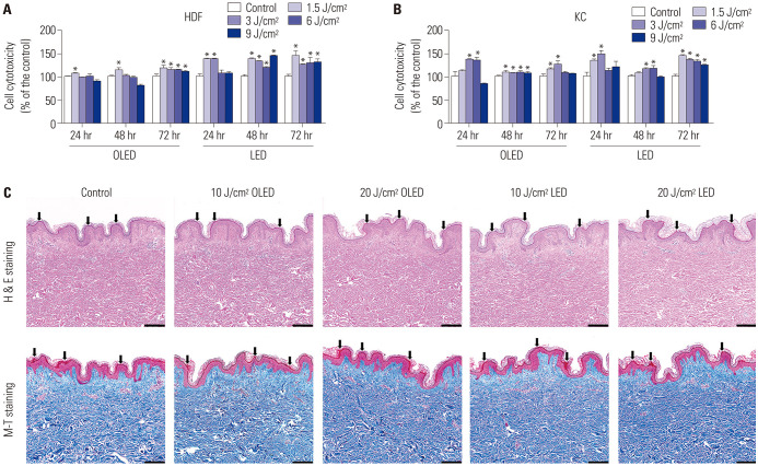
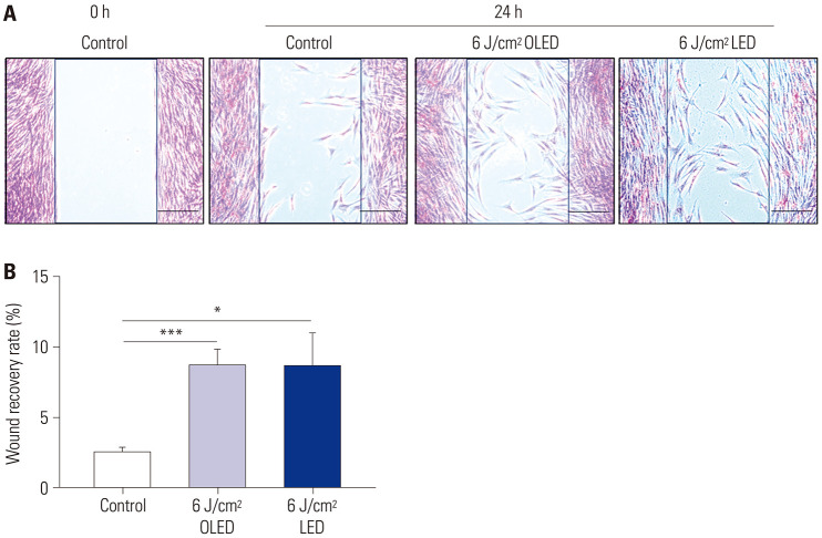
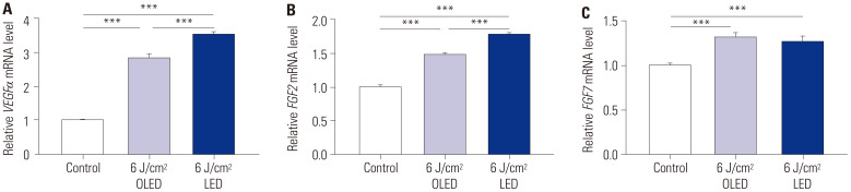
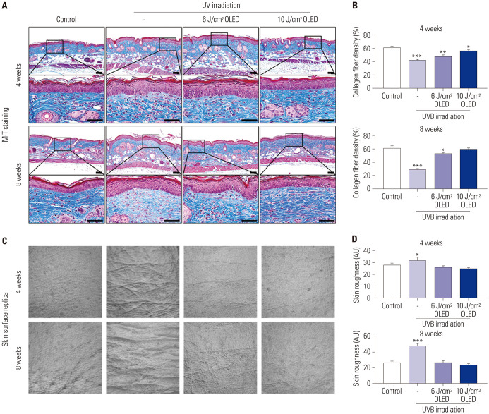
Materials and methods: Cell viability assays and human ex vivo skin analyses were performed after exposure to OLED and LED irradiation to examine their safety. Subsequent evaluations examined expression levels and wound healing effects in human dermal fibroblasts (HDFs) using quantitative reverse transcription-polymerase chain reaction, enzyme-linked immunosorbent assay, and wound healing assays post-irradiation. Additionally, an in vivo study was conducted using a ultra violet (UV)-irradiated animal skin model to explore the impact of OLED exposure on dermal collagen density and wrinkles, employing skin replica and tissue staining techniques.
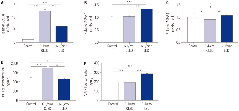
Results: OLED irradiation had no significant morphological effects on human skin tissue, but caused a considerably higher expression of collagen than the control and LED-treated groups. Moreover, OLED irradiation reduced the expression levels of matrix metalloproteinases (MMPs) more effectively than did LED on HDFs. OLED irradiation group in HDFs had significantly higher expression levels of growth factors compared to the control group, but similar to those in the LED irradiation group. In addition, OLED irradiation on photo-aged animal skin model resulted in increased collagen fiber density in the dermis while reducing ultra violet radiation-mediated skin wrinkles and roughness, as shown in the skin replica.
Conclusion: This study established comparable effectiveness between OLED and LED irradiation in upregulating collagen and growth factor expression levels while downregulating MMP levels in vitro. In the UV-irradiated animal skin model, OLED exposure post UV radiation correlated with reduced skin wrinkles and augmented dermal collagen density. Accelerated wound recovery and demonstrated safety further underscore OLEDs' potential as a future PBM modality alongside LEDs, offering promise in the realms of skin rejuvenation and wound healing.
Keywords: Organic light-emitting diode; light-emitting diode; photobiomodulation; skin rejuvenation; wound healing.
© Copyright: Yonsei University College of Medicine 2024.
Conflict of interest statement
The authors have no potential conflicts of interest to disclose.
Figures
References
-
- Houreld NN. The use of lasers and light sources in skin rejuvenation. Clin Dermatol. 2019;37:358–364. - PubMed
-
- Arany PR. Craniofacial wound healing with photobiomodulation therapy: new insights and current challenges. J Dent Res. 2016;95:977–984. - PubMed
-
- Huang A, Nguyen JK, Ho D, Jagdeo J. Light emitting diode phototherapy for skin aging. J Drugs Dermatol. 2020;19:359–364. - PubMed
MeSH terms
Substances
Grants and funding
LinkOut - more resources
Full Text Sources
