. 2024 Mar 5;12(3):e0286523.
doi: 10.1128/spectrum.02865-23. Epub 2024 Jan 30.

Comparative analysis of Porphyromonas gingivalis A7436 and ATCC 33277 strains reveals differences in the expression of heme acquisition systems

Affiliations

Comparative analysis of Porphyromonas gingivalis A7436 and ATCC 33277 strains reveals differences in the expression of heme acquisition systems

Michał Śmiga et al. Microbiol Spectr. .

Abstract

Porphyromonas gingivalis strains exhibit different phenotypes in vitro, different virulence potential in animal models, and different associations with human diseases, with strains classified as virulent/more virulent (e.g., A7436 and W83) or as less virulent/avirulent (e.g., ATCC 33277). In this study, we comparatively analyzed the A7436 and ATCC 33277 strains to better understand their variability. Global gene expression analysis in response to heme and iron limitation revealed more pronounced differences in the A7436 than in the ATCC 33277 strain; however, in both strains, the largest changes were observed in genes encoding hypothetical proteins, genes whose products participate in energy metabolism, and in genes encoding proteins engaged in transport and binding proteins. Our results confirmed that variability between P. gingivalis strains is due to differences in the arrangement of their genomes. Analysis of gene expression of heme acquisition systems demonstrated that not only the availability of iron and heme in the external environment but also the ability to store iron intracellularly can influence the P. gingivalis phenotype. Therefore, we assume that differences in virulence potential may also be due to differences in the production of systems involved in iron and heme acquisition, mainly the Hmu system. In addition, our study showed that hemoglobin, in a concentration-dependent manner, differentially influences the virulence potential of P. gingivalis strains. We conclude that iron and heme homeostasis may add to the variability observed between P. gingivalis strains.

Importance: Periodontitis belongs to a group of multifactorial diseases, characterized by inflammation and destruction of tooth-supporting tissues. P. gingivalis is one of the most important microbial factors involved in the initiation and progression of periodontitis. To survive in the host, the bacterium must acquire heme as a source of iron and protoporphyrin IX. P. gingivalis strains respond differently to changing iron and heme concentrations, which may be due to differences in the expression of systems involved in iron and heme acquisition. The ability to accumulate iron intracellularly, being different in more and less virulent P. gingivalis strains, may influence their phenotypes, production of virulence factors (including proteins engaged in heme acquisition), and virulence potential of this bacterium.

Keywords: HmuY; IhtB; Porphyromonas gingivalis; gingipain; heme; heme acquisition; hemoglobin; iron; strain variation; virulence.


Conflict of interest statement

The authors declare no conflict of interest.

Figures
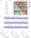
Fig 1
Fig 1
Comparative analysis of selected genomes of P. gingivalis strains. (A) A phylogenetic tree was constructed based on nucleotide sequences of the hmu operon present in strains listed in Table 1. Sequence for P. gulae (DSM15663 strain) was used as an out-group. (B) A heat map was constructed based on nucleotide sequences of the hmuY gene and nucleotide sequences of the entire hmu operon. The color gradient from green to red shows the percentage of identity from lowest (green) to highest (red). (C) Linearized genome maps of P. gingivalis A7436, ATCC 33277, and W83 strains demonstrating the location of selected genes. The genomes were visualized with Proksee (https://proksee.ca/). (D) Graphical presentation of the arrangement of genes in pairs of selected genomes. The hmu operon location is marked on the graphs as a reference. The changes in the slope of the curve represent the inversion of chromosome fragments of the strain indicated at the Y-axis in comparison to the reference strain at the X-axis.
Fig 2
Fig 2
General summary of microarray data of gene expression in P. gingivalis A7436 and ATCC 33277 strains grown in iron- and heme-depleted conditions (DIP medium) in comparison to iron- and heme-replete conditions (Hm medium). Data are shown from three independent biological replicates. Up- and down-regulated genes are grouped according to their function. The inset shows the Venn diagram showing either the number of genes whose expression changed in both tested strains or the number of genes whose expression is strain-specific.
Fig 3
Fig 3
Analysis of production of selected proteins engaged in heme acquisition in P. gingivalis. RT-qPCR was used to determine differences in the expression of hmuY and ihtB genes during late exponential (10 hours; 10 h) and early stationary (24 hours; 24 h) growth phases. Results are shown as relative hmuY or ihtB mRNA levels in comparison to the 16S rRNA gene (hmuY/16S rRNA or ihtB/16S rRNA) (A) or as expression fold change in the ATCC 33277 strain in comparison to the A7436 strain (33277 vs A7436) (B). Comparative analysis of cell-associated HmuY, IhtB, and RgpB proteins in P. gingivalis cultured in liquid media (C) and on anaerobic blood agar (ABA) plates (D) using Western blotting and densitometric analysis. Results were normalized to the A7436 strain grown in a liquid medium supplemented with iron and heme (C) or results are shown as relative protein production fold change in the ATCC 33277 strain in comparison to the A7436 strain (D). The results are shown as mean ± standard error (mean ± SE). *P < 0.05, **P < 0.01, ****P < 0.0001.
Fig 4
Fig 4
Comparison of RgpB produced by P. gingivalis A7436, 33277, and W83 strains. (A) Amino acid sequence alignment. (B) Gene length analysis. The whole DNA sequence of the rgpB gene was amplified using genomic DNA of A7436, 33277, or W83 strains and primers listed in Table S4. (C) Differences in RgpB molecular weight were examined in whole cell lysates by sodium dodecyl sulfate-polyacrylamide gel electrophoresis and Western blotting using anti-RgpB antibody. (D) Differences in the production of glycans were examined using reactivity with JACALIN (the lectin preferentially recognizes galactose linked with β−1,3 linkage to N-acetylgalactosamine and mono- or di-sialylated form of this structure).
Fig 5
Fig 5
Phenotypic analysis of P. gingivalis A7436 and ATCC 33277 (33277) strains. (A) Growth of bacteria in liquid culture media containing iron and heme (iron- and heme-replete conditions, Hm medium) or without heme and supplemented with the iron chelator 2,2-dipyridyl (iron- and heme-depleted conditions, DIP medium). (B) Recovery of bacteria after prior heme and iron starvation. Bacteria were cultured for two passages in a DIP medium and then transferred to a fresh DIP medium supplemented with heme (DIP + Hm) or hemoglobin (DIP + Hb). Growth without an iron source was used as a control (DIP medium). Bacterial growth was monitored over time by measuring the optical density at 600 nm (statistical analysis is presented in Table S3). (C) The intracellular amount of iron was measured using a ferrozine-based assay. (D) Hemolytic activity was examined on anaerobic blood agar (ABA) plates after 5 days by visual inspection, semi-quantitatively evaluated by densitometric analysis, and shown as the relative hemolytic activity in the 33277 strain in relation to the A7436 strain (the latter set as 1.0). To better visualize and analyze hemolysis, bacterial colonies were removed. BM medium was used as a negative hemolysis control. (E) Gingipain activities of whole P. gingivalis cultures were measured using arginine-specific (Rgp) and lysine-specific (Kgp) substrates. Enzymatic activity was determined as an increase in absorbance at 405 nm per 1 minute per 1 µL of bacterial culture of optical density at 600 nm equal to 1. (F) Heme-binding ability by P. gingivalis cells. (G) Biofilm formation on the abiotic surface was examined using crystal violet staining and absorbance measurement at 570 nm. Insets in the graphs show a relative increase in heme binding (F) or biofilm formation (G) in iron- and heme-depleted conditions (DIP medium) in comparison to iron and heme-replete conditions (Hm medium). *P < 0.05, **P < 0.01, ***P < 0.001, ****P < 0.0001.
Fig 6
Fig 6
Influence of hemoglobin on interaction with host cells of P. gingivalis A7436 (A), W83 (B), and ATCC 33277 (C) strains. The ability to interact with, adhere to, and invade host cells was analyzed using a P. gingivalis-gingival keratinocytes co-culture model. The number of viable bacteria was shown as the number of colony-forming units (CFU) per mL of the culture medium. Adhesion—live bacteria attached to keratinocytes; invasion—live bacteria that invaded keratinocytes; interaction—the total number of live bacteria that adhered to and invaded keratinocytes. (D) Summary of the correlation between hemoglobin concentration and P. gingivalis interaction with human keratinocytes. The average interaction of P. gingivalis with keratinocytes in samples without hemoglobin was set as 100% for each tested strain. As a protein control, purified apo-SgGAPDH protein instead of hemoglobin was used. Results are shown as mean ± SD (A through C) or as mean ± SE (D). Hb—hemoglobin; SgGAPDH—glyceraldehyde-3-phosphate dehydrogenase from Streptococcus gordonii. *P < 0.05, **P < 0.01, ****P < 0.0001.

Similar articles

Cited by

References

    1. Hajishengallis G, Diaz PI. 2020. Porphyromonas gingivalis: immune subversion activities and role in periodontal dysbiosis. Curr Oral Health Rep 7:12–21. doi:10.1007/s40496-020-00249-3 - DOI - PMC - PubMed
    1. Hajishengallis G, Lamont RJ. 2012. Beyond the red complex and into more complexity: the polymicrobial synergy and dysbiosis (PSD) model of periodontal disease etiology. Mol Oral Microbiol 27:409–419. doi:10.1111/j.2041-1014.2012.00663.x - DOI - PMC - PubMed
    1. Holt SC, Ebersole JL. 2005. Porphyromonas gingivalis, Treponema denticola, and Tannerella forsythia: the “red complex”, a prototype polybacterial pathogenic consortium in periodontitis”. Periodontol 2000 38:72–122. doi:10.1111/j.1600-0757.2005.00113.x - DOI - PubMed
    1. Deng ZL, Szafrański SP, Jarek M, Bhuju S, Wagner-Döbler I. 2017. Dysbiosis in chronic periodontitis: key microbial players and interactions with the human host. Sci Rep 7:3703. doi:10.1038/s41598-017-03804-8 - DOI - PMC - PubMed
    1. Deng ZL, Sztajer H, Jarek M, Bhuju S, Wagner-Döbler I. 2018. Worlds apart – transcriptome profiles of key oral microbes in the periodontal pocket compared to single laboratory culture reflect synergistic interactions. Front Microbiol 9:124. doi:10.3389/fmicb.2018.00124 - DOI - PMC - PubMed

LinkOut - more resources