Review
. 2024 Jan 30;81(1):67.
doi: 10.1007/s00018-023-05096-w.

MuSCs and IPCs: roles in skeletal muscle homeostasis, aging and injury

Affiliations
Review

MuSCs and IPCs: roles in skeletal muscle homeostasis, aging and injury

Haiyan Jiang et al. Cell Mol Life Sci. .

Abstract

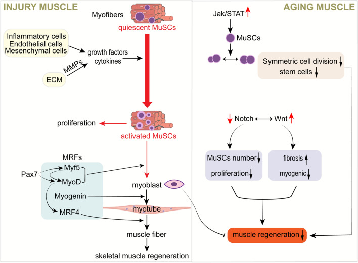
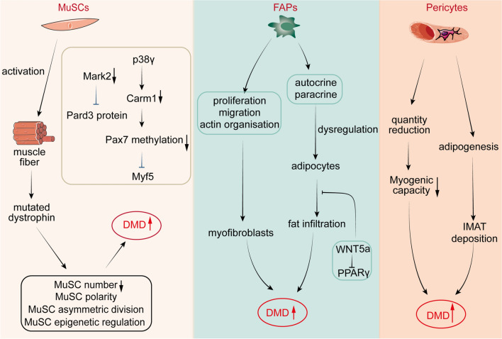
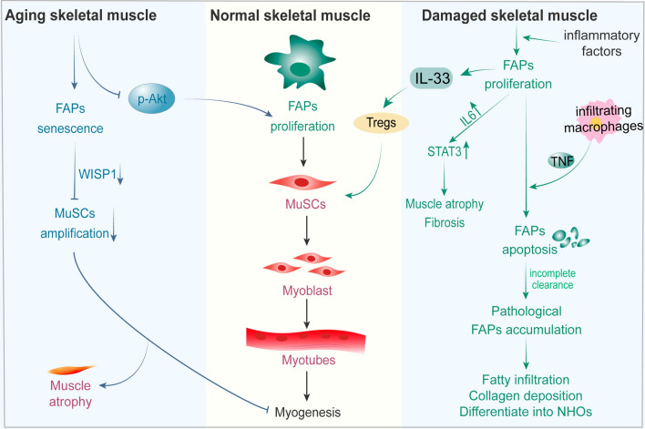
Skeletal muscle is a highly specialized tissue composed of myofibres that performs crucial functions in movement and metabolism. In response to external stimuli and injuries, a range of stem/progenitor cells, with muscle stem cells or satellite cells (MuSCs) being the predominant cell type, are rapidly activated to repair and regenerate skeletal muscle within weeks. Under normal conditions, MuSCs remain in a quiescent state, but become proliferative and differentiate into new myofibres in response to injury. In addition to MuSCs, some interstitial progenitor cells (IPCs) such as fibro-adipogenic progenitors (FAPs), pericytes, interstitial stem cells expressing PW1 and negative for Pax7 (PICs), muscle side population cells (SPCs), CD133-positive cells and Twist2-positive cells have been identified as playing direct or indirect roles in regenerating muscle tissue. Here, we highlight the heterogeneity, molecular markers, and functional properties of these interstitial progenitor cells, and explore the role of muscle stem/progenitor cells in skeletal muscle homeostasis, aging, and muscle-related diseases. This review provides critical insights for future stem cell therapies aimed at treating muscle-related diseases.

Keywords: Fibro-adipogenic progenitors; Interstitial progenitor cells; Muscle stem cells; Pericytes; Skeletal muscle.


Conflict of interest statement

The authors declare that they have no competing interests.

Figures

Fig. 1
Fig. 1
Role of MuSCs in skeletal muscle injury and aging. Growth factors and cytokines released by inflammatory cells, endothelial cells and mesenchymal cells lead to the activation of MuSCs. MRFs then regulate the differentiation of MuSCs, leading to the formation of new myotubes by myoblasts, which eventually mature into myofibrils and cause muscle regeneration. During the ageing process, activation of the JAK-STAT and WNT signalling pathways and inhibition of Notch activity lead to a gradual decrease in the number of MuSCs and differentiation of MuSCs from myogenic to fibrogenic
Fig. 2
Fig. 2
Regulation of MuSCs, FAPs, and pericytes in DMD. MuSCs express mutated dystrophin, creating anomalies in MuSC polarity, asymmetric division, and epigenetic regulation. FAPs and pericytes drive muscle fibrosis and fat infiltration
Fig. 3
Fig. 3
Role of FAPs in skeletal muscle injury and aging. Upon muscle injury, FAPs become activated and have a dual role. On one hand, FAPs can regulate the proliferation and differentiation of MuSCs through modulation of cytokine secretion, thus directly promoting muscle regeneration. On the other hand, FAPs can also drive muscle fibrosis and adipose tissue accumulation if improperly regulated. Ageing pushes FAPs into a fibrotic state, while a decrease in levels of phosphorylated Akt leads to a decrease in their proliferative capacity; Furthermore, a reduction in WISP1 production in FAP cells leads to an impaired muscle-forming capacity in MuSCs, resulting in muscle atrophy

Similar articles

Cited by

References

    1. Huang L, Li M, Deng C, Qiu J, Wang K, Chang M, Zhou S, Gu Y, Shen Y, Wang W, et al. Potential therapeutic strategies for skeletal muscle atrophy. Antioxidants (Basel, Switzerland) 2022;12(1):44. - PMC - PubMed
    1. Chen X, Ji Y, Liu R, Zhu X, Wang K, Yang X, Liu B, Gao Z, Huang Y, Shen Y, et al. Mitochondrial dysfunction: roles in skeletal muscle atrophy. J Transl Med. 2023;21(1):503. doi: 10.1186/s12967-023-04369-z. - DOI - PMC - PubMed
    1. Zhang H, Qi G, Wang K, Yang J, Shen Y, Yang X, Chen X, Yao X, Gu X, Qi L, et al. Oxidative stress: roles in skeletal muscle atrophy. Biochem Pharmacol. 2023;214:115664. doi: 10.1016/j.bcp.2023.115664. - DOI - PubMed
    1. Sun J, Zhou H, Chen Z, Zhang H, Cao Y, Yao X, Chen X, Liu B, Gao Z, Shen Y, et al. Altered m6A RNA methylation governs denervation-induced muscle atrophy by regulating ubiquitin proteasome pathway. J Transl Med. 2023;21(1):845. doi: 10.1186/s12967-023-04694-3. - DOI - PMC - PubMed
    1. Wong A, Pomerantz JH. The role of muscle stem cells in regeneration and recovery after denervation: a review. Plast Reconstr Surg. 2019;143(3):779–788. doi: 10.1097/PRS.0000000000005370. - DOI - PubMed