Review
. 2024 Jun;67(4):887-900.
doi: 10.1007/s10840-024-01744-8. Epub 2024 Jan 30.

Ebstein's anomaly: an electrophysiological perspective

Affiliations
Review

Ebstein's anomaly: an electrophysiological perspective

Anunay Gupta et al. J Interv Card Electrophysiol. 2024 Jun.

Abstract

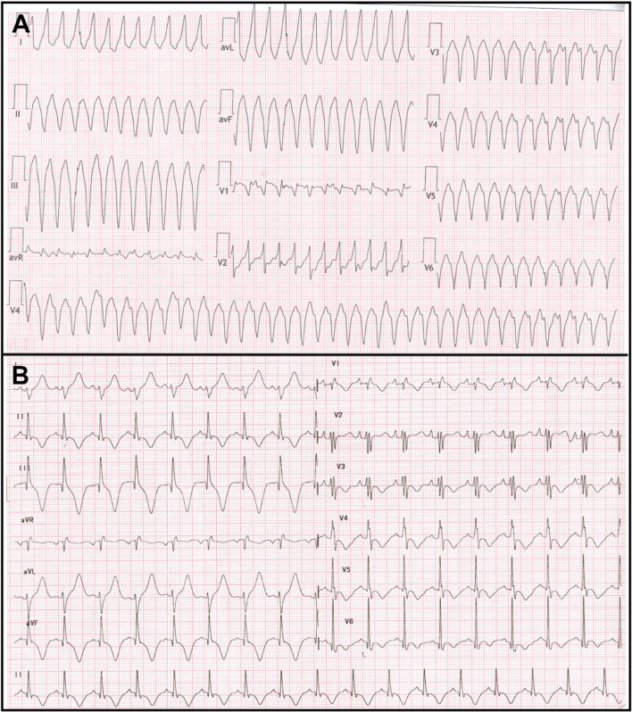
Ebstein's anomaly of the tricuspid valve (EA) is an uncommon congenital cardiac malformation. It can present with atrioventricular tachycardia (AVRT), atrioventricular nodal re-entrant tachycardia (AVNRT), atrial arrhythmias, and rarely with ventricular tachycardia. The 12-lead electrocardiogram (ECG) is critically important and often diagnostic even prior to an electrophysiology study (EPS). Due to its complex anatomy, it poses particular challenges for mapping and ablation, even for an experienced electrophysiologist. In this review, we aim to provide insight into the electrophysiological perspective of EA and an in-depth analysis of the various arrhythmias encountered in diverse clinical scenarios.

Keywords: Ablation; Arrhythmia; Ebstein anomaly.


Conflict of interest statement

Dr Saurabh Kumar is supported by the NSW Health Early Mid-Career Fellowship. Timothy Campbell has received speakers’ honoraria for Biosense Webster. All other authors declare no competing interests.

Figures

Fig. 1
Fig. 1
Illustration of a heart with Ebstein’s anomaly highlighting the anatomical abnormalities relevant to the electrophysiologist. The right atrium (RA) is dilated and so is the tricuspid annulus. The septal tricuspid leaflet is apically displaced, and there is a fibromuscular ridge instead (arrow) which forms the boundary of the triangle of Koch which is smaller compared to normal hearts. The coronary sinus (CS) ostium is dilated, and the AV node (AVN) is irregular, and its body can reach the upper border of the CS ostium unlike in normal hearts where only its inferior extension reaches here. ASD, atrial septal defect; aRV, atrialised RV; LA, left atrium; LV, left ventricle; RV, right ventricle
Fig. 2
Fig. 2
The standard 12-lead electrocardiogram in an 11-year-old boy with severe Ebstein’s anomaly demonstrating peaked P waves, prolonged PR interval, and a splintered QRS complex. The leads V1 and aVR have similar morphologies of the QRS complex (see text for details)
Fig. 3
Fig. 3
12-lead ECG in a patient with Ebstein anomaly showing normal PR interval and no delta waves with the absence of RBBB in lead V1 (A). This is suggestive of right-sided pathway. Patient underwent successful ablation of the right posterior bypass tract, and the postablation ECG showed typical RBBB pattern in lead V1 (B)
Fig. 4
Fig. 4
Still frames from a right ventriculogram in the right anterior oblique 20 degree view in a patient with Ebstein’s anomaly. The black arrows (A) mark the atrioventricular groove whereas the white asterisk (B) marks the apically displaced tricuspid valve leaflet
Fig. 5
Fig. 5
Images using CARTO system (Biosense Webster) with intracardiac echocardiography. A Vision Wire was placed in the right coronary artery to mark the plane of the true annulus. Panels A and B represent the AP and the LAO views, respectively. Panel C shows the plane of the ultrasound beam used to derive the image in the panel D. The red line denotes the plane of the tricuspid annulus, while the yellow tags and the connecting white line marks the plane of the displaced tricuspid leaflet (arrow in D). Also marked are the right atrium (RA), the atrialised right ventricle (aRV), and the right ventricle (RV)
Fig. 6
Fig. 6
A The activation map with a PentaRay high-density mapping catheter during an orthodromic AVRT showing the earliest activation in the aRV. A 0.014-inch Vision Wire guidewire (Biotronik SE and Co. KG, Berlin, Germany) is seen floating adjacent and posterior to the annulus. Also marked is the aortic root (Ao) and the RA. Panel B shows the voltage map of the aRV with some low voltage areas (arrow). Panel C shows the successful site of ablation in the posterior annulus, and the corresponding signals are shown in the panel D. Note that the atrial signal (arrow), though the earliest, is not fused with the aRV signal perhaps due to a slow conducting pathway
Fig. 7
Fig. 7
Electrograms during an orthodromic AVRT in a patient with Ebstein’s anomaly. Panel A shows the signals in the mapping catheter as it maps the tricuspid annulus. The EGMs representing ventricle, the atrialised RV, and the atria are marked V, aRV, and A, respectively. The arrow marks the His potential preceding the ventricular EGM in the His catheter. In panel B, the mapping catheter is positioned at the site of successful ablation at the posteroseptal tricuspid annulus. The EGMs from the aRV and the A are fused (double-headed arrow), and the pathway potential is seen (arrows). In both the panels, RFd and RFp represent the distal and proximal bipoles of the mapping catheter. HBd, HBm, and HBp represents the distal, mid, and proximal bipoles of the hexapolar His bundle catheter. CSp to CSd represent the proximal and distal bipoles of the coronary sinus decapolar catheter with the other bipoles sequentially numbered. The RFd and CS78 have been displayed adjacent to the surface electrocardiogram leads to make it easier for timing the ventricular and the atrial EGMs
Fig. 8
Fig. 8
A Twelve lead ECG of a young adult with ventricular tachycardia which was successfully ablated from atrialised right ventricle. B The baseline ECG after tachycardia termination showed presence of RBBB in lead V1 and memory T waves in inferolateral leads. No SVT was induced during EP study

References

    1. Watson H. Natural history of Ebstein’s anomaly of tricuspid valve in childhood and adolescence. An international co-operative study of 505 cases. Br Heart J. 1974;36(5):417–27. doi: 10.1136/hrt.36.5.417. - DOI - PMC - PubMed
    1. Van Lingen B, Bauersfeld SR. The electrocardiogram in Ebstein’s anomaly of the tricuspid valve. Am Heart J. 1955;50(1):13–23. doi: 10.1016/0002-8703(55)90250-7. - DOI - PubMed
    1. Attenhofer Jost CH, Connolly HM, Dearani JA, Edwards WD, Danielson GK. Ebstein’s anomaly. Circulation. 2007;115(2):277–285. doi: 10.1161/CIRCULATIONAHA.106.619338. - DOI - PubMed
    1. Becker AE, Becker MJ, Edwards JE. Pathologic spectrum of dysplasia of the tricuspid valve. Features in common with Ebstein’s malformation. Arch Pathol. 1971;91(2):167–78. - PubMed
    1. Lev M, Liberthson RR, Joseph RH, Seten CE, Eckner FA, Kunske RD, et al. The pathologic anatomy of Ebstein’s disease. Arch Pathol. 1970;90(4):334–343. - PubMed

MeSH terms

LinkOut - more resources