. 2024 Jan 3:102:skae021.
doi: 10.1093/jas/skae021.

The effect and mechanism of selenium supplementation on the proliferation capacity of bovine endometrial epithelial cells exposed to lipopolysaccharide in vitro under high cortisol background

Affiliations

The effect and mechanism of selenium supplementation on the proliferation capacity of bovine endometrial epithelial cells exposed to lipopolysaccharide in vitro under high cortisol background

Hanqing Li et al. J Anim Sci. .

Abstract

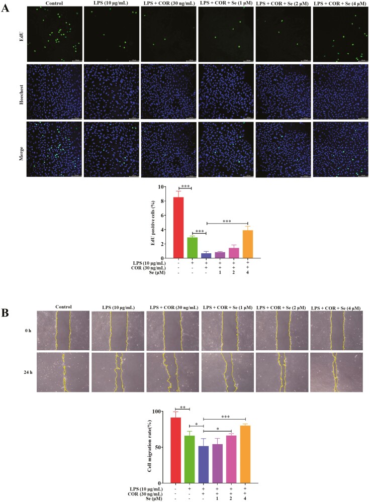
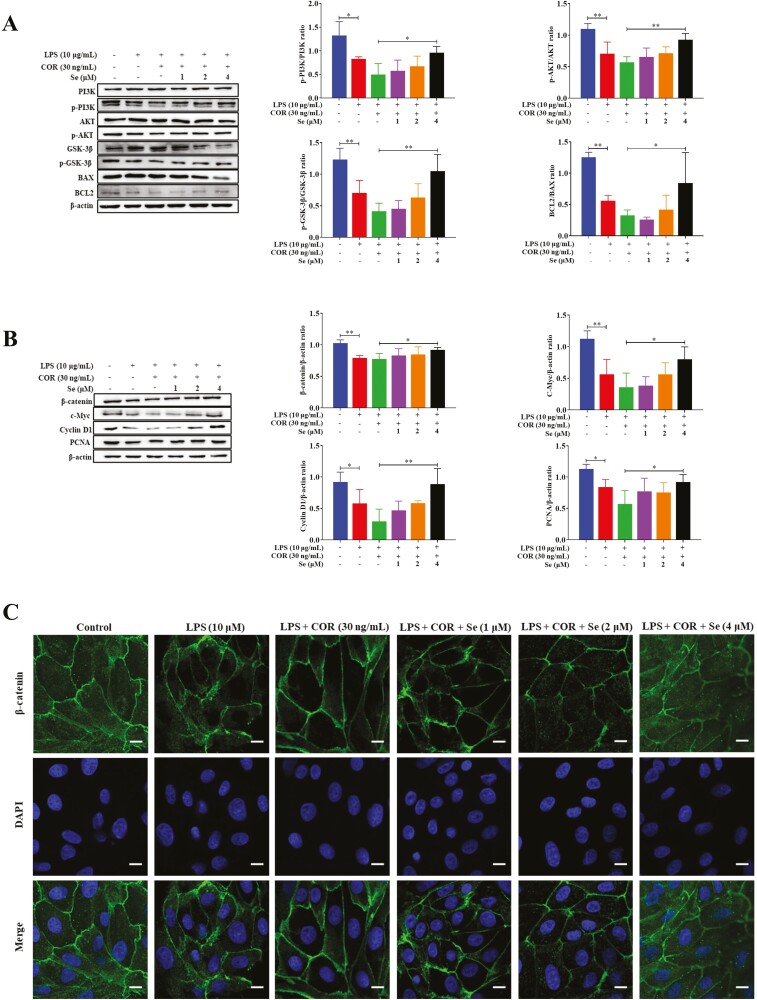
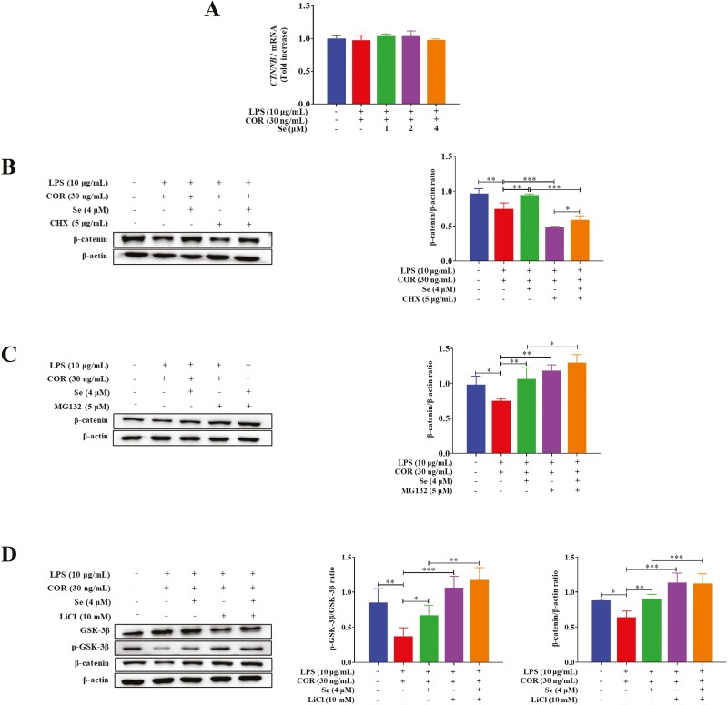
Bovine endometritis severely inhibits uterine repair and causes considerable economic loss. Besides, parturition-induced high cortisol levels inhibit immune function, reduce cell proliferation, and further inhibit tissue repair. Selenium (Se) is an essential trace element for animals to maintain normal physiological function and has powerful antioxidant functions. This study investigated whether Se supplementation reduces endometrial damage and promotes tissue repair in cows with endometritis under stress and explored the underlying mechanism. Primary bovine endometrial epithelial cells were isolated and purified from healthy cows. The cells were treated with different combinations of lipopolysaccharide (LPS), cortisol, and various concentrations of Se. Data showed that LPS stimulation inhibited cell proliferation and increased cell apoptosis. High levels of cortisol further exacerbated these effects. Flow cytometry, scratch wound healing tests, and 5-ethynyl-2'-deoxyuridine (EdU) proliferation assays showed that Se supplementation promoted cell cycle progression, cell migration, and cell proliferation in the presence of LPS and cortisol. The quantitative PCR results showed that the expression of related growth factors was increased after Se supplementation. After administering various inhibitors, we further demonstrated that Se supplementation decreased the activity of glycogen synthetase kinase 3β (GSK-3β) through the phosphatidylinositol 3-kinase (PI3K)/protein kinase B (AKT) signaling pathway to reduce the degradation of β-catenin except the Wnt signal to promote cell proliferation. In conclusion, Se supplementation attenuated the cell damage induced by LPS at high cortisol levels and increased cell proliferation to promote uterine repair by elevating the mRNA expression of TGFB3 and VEGFA and activating the PI3K/AKT/GSK-3β/β-catenin signaling pathway.

Keywords: bovine endometrial epithelial cells; cortisol; endometritis; lipopolysaccharide; proliferation; selenium.

Plain language summary

After parturition, endometritis is a common bovine disease, which hinders endometrial repair and reduces bovine economic value. Besides, parturition-induced high cortisol levels cause immunosuppression, aggravate infection, and further inhibit cell proliferation and tissue repair. As an essential trace element, adding selenium to feed helps to maintain the normal physiological function of animals. This study developed a cellular model using lipopolysaccharide (LPS) and cortisol to simulate cows with endometritis in stress conditions. The results showed that Se supplementation attenuated bovine endometrial epithelial cell damage and promoted their proliferation in the presence of LPS and high cortisol levels, which are positively correlated with the concentration of Se. Besides, this study proved another molecular mechanism for Se to regulate β-catenin except for the Wnt signal by affecting the β-catenin degradation pathway.


Conflict of interest statement

The authors declare no real or perceived conflicts of interest.

Figures

Graphical Abstract
Graphical Abstract
Figure 1.
Figure 1.
The effect of Se on the LPS-inhibited cell cycle distribution of the BEECs at high COR levels. The cell cycle distribution was detected by flow cytometry after treated with LPS or co-treated with LPS and COR or co-treated with LPS, COR, and Se (1, 2, and 4 μM). *, P < 0.05, **, P < 0.01, and ***, P < 0.001. All data were presented as the means ± SEM (n = 3).
Figure 2.
Figure 2.
Se enhanced LPS-inhibited migration and proliferation of BEECs at high COR levels. (A) The effect of Se on the cell migration rate of the BEECs by using the scratch wound healing assay. The cell migration rate = (scratch width at 0 h − scratch width at 24 h)/scratch width at 0 h × 100%. The cells were observed under light microscopy at 100× magnification. (B) EdU assay of the cell proliferation ability in BEECs. (A and B) The cells were treated with LPS or co-treated with LPS and COR or co-treated with LPS, COR, and Se (1, 2, and 4 μM). *, P < 0.05, **, P < 0.01, and ***, P < 0.001. The scale bar = 100 μm. All data were presented as the means ± SEM (n = 3).
Figure 3.
Figure 3.
Effects of Se on the gene expressions of CCN2 (A), TGFB1 (B), TGFB3 (C), and VEGFA (D) in BEECs. The cells were treated with LPS or co-treated with LPS and COR or co-treated with LPS, COR, and Se (1, 2, and 4 μM). *, P < 0.05, **, P < 0.01, and ***, P < 0.001. All data were presented as the means ± SEM (n = 3).
Figure 4.
Figure 4.
Effects of Se on the PI3K/AKT (A) and Wnt/β-catenin (B) signaling pathways in BEECs. The phosphorylation levels the protein levels were detected by Western blotting. (C) The effect of Se on the nuclear-transport of β-catenin in BEECs. The β-catenin levels were evaluated by confocal microscopy. The scale bar = 10 μm. (A, B, and C) The cells were treated with LPS or co-treated with LPS and COR or co-treated with LPS, COR and Se (1, 2 and 4 μM). *, P < 0.05, **, P < 0.01, and ***, P < 0.001. All data were presented as the means ± SEM (n = 3).
Figure 5.
Figure 5.
Se inhibited the LPS-induced activity of GSK-3β and accelerated the degradation of β-catenin protein through ubiquitin–proteasome pathway at high COR levels. (A) Effects of Se on the gene expressions of CTNNB1 in BEECs. The cells were co-treated with LPS and COR or co-treated with LPS, COR, and Se (1, 2, and 4 μM). (B and C) Effects of Se on the β-catenin protein levels of BEECs. (D) Effects of Se on the phosphorylation levels of GSK-3β and the protein levels of β-catenin in BEECs. (B, C, and D) The cells were co-treated with LPS and COR or co-treated with LPS, COR, and Se (4 μM) in/not in the presence of CHX (B), MG-132 (C), or LiCl (D). The phosphorylation levels of GSK-3β and the protein levels of β-catenin were detected by Western blotting. *, P < 0.05, **, P < 0.01 and ***, P < 0.001. All data were presented as the means ± SEM (n = 3).
Figure 6.
Figure 6.
Effects of Se on the PI3K/AKT/GSK-3β/β-catenin signaling pathway. The cells were treated with co-treated with LPS and COR or co-treated with LPS, COR, and Se (4 μM) in/not in the presence of Ly294002. The phosphorylation levels the protein levels were detected by Western blotting. *, P < 0.05, **, P < 0.01, and ***, P < 0.001. All data were presented as the means ± SEM (n = 3).
Figure 7.
Figure 7.
(A) The effect of Se on the cell migration rate of the BEECs by using the scratch wound healing assay. The cell migration rate = (scratch width at 0 h − scratch width at 24 h)/scratch width at 0 h × 100%. The cells were observed under light microscopy at 100× magnification. (B) EdU assay of the cell proliferation ability in BEECs. (A and B) The cells were treated with co-treated with LPS and COR or co-treated with LPS, COR, and Se (4 μM) in/not in the presence of Ly294002. *, P < 0.05, **, P < 0.01, and ***, P < 0.001. All data were presented as the means ± SEM (n = 3).

References

    1. Alabdullah, H. A., L. K. Fox, J. M. Gay, and G. M. Barrington. 2018. Interactive effects of dexamethasone and opsonized Mycoplasma bovis on bovine neutrophil function in vitro. Vet. Immunol. Immunopathol. 196:18–21. doi:10.1016/j.vetimm.2017.12.008 - DOI - PubMed
    1. Allison, R. D., and R. A. Laven. 2000. Effect of vitamin E supplementation on the health and fertility of dairy cows: a review. Vet. Rec. 147:703–708 - PubMed
    1. Amit, S., A. Hatzubai, Y. Birman, J. S. Andersen, E. Ben-Shushan, M. Mann, Y. Ben-Neriah, and I. Alkalay. 2002. Axin-mediated CKI phosphorylation of beta-catenin at Ser 45: a molecular switch for the Wnt pathway. Genes Dev. 16:1066–1076. doi:10.1101/gad.230302 - DOI - PMC - PubMed
    1. Behrens, J., B. A. Jerchow, M. Würtele, J. Grimm, C. Asbrand, R. Wirtz, M. Kühl, D. Wedlich, and W. Birchmeier. 1998. Functional interaction of an axin homolog, conductin, with beta-catenin, APC, and GSK3beta. Science 280:596–599. doi:10.1126/science.280.5363.596 - DOI - PubMed
    1. Bradham, D. M., A. Igarashi, R. L. Potter, and G. R. Grotendorst. 1991. Connective tissue growth factor: a cysteine-rich mitogen secreted by human vascular endothelial cells is related to the SRC-induced immediate early gene product CEF-10. J. Cell Biol. 114:1285–1294. doi:10.1083/jcb.114.6.1285 - DOI - PMC - PubMed

MeSH terms