. 2024 Jan 30;22(1):e3002462.
doi: 10.1371/journal.pbio.3002462. eCollection 2024 Jan.

Evaluating protein cross-linking as a therapeutic strategy to stabilize SOD1 variants in a mouse model of familial ALS

Affiliations

Evaluating protein cross-linking as a therapeutic strategy to stabilize SOD1 variants in a mouse model of familial ALS

Md Amin Hossain et al. PLoS Biol. .

Abstract

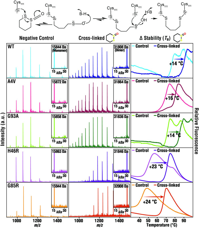
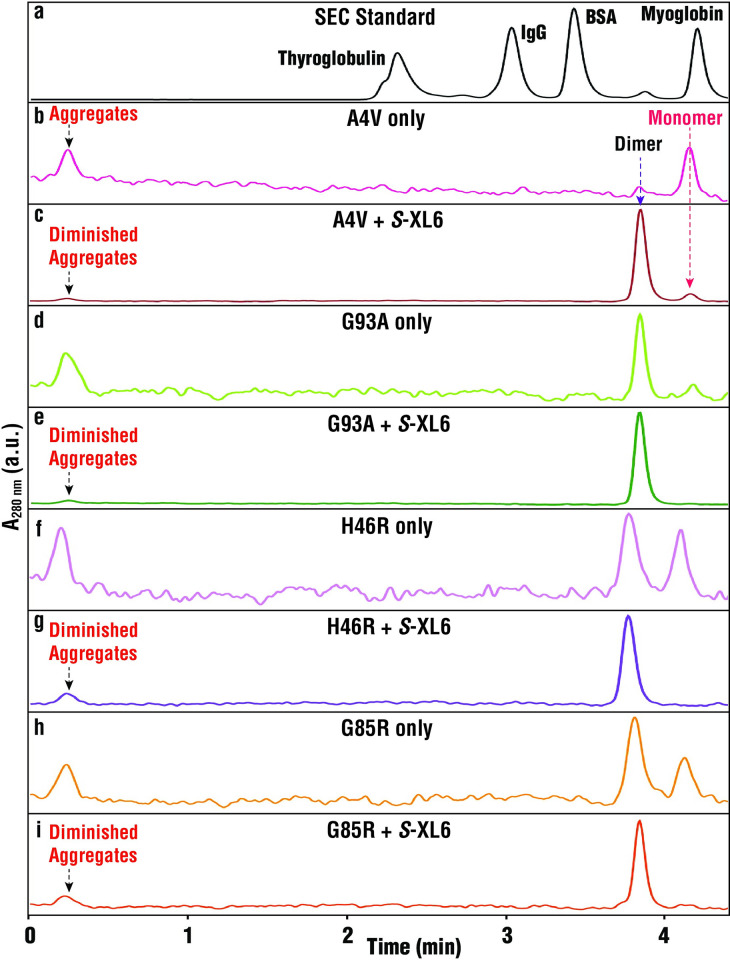
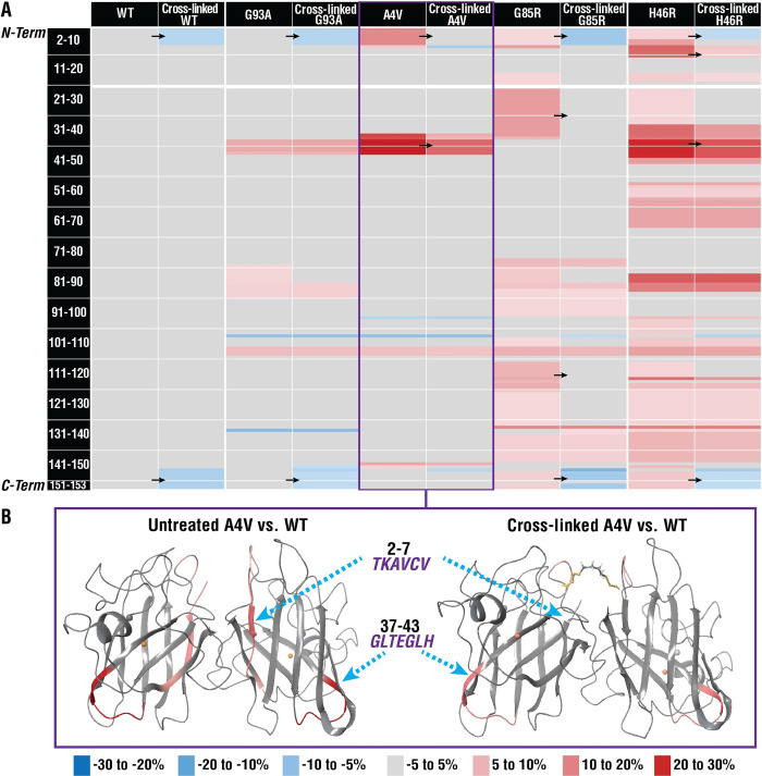
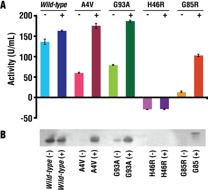
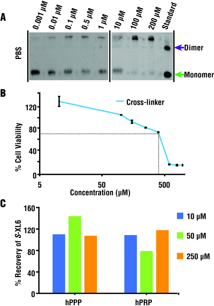
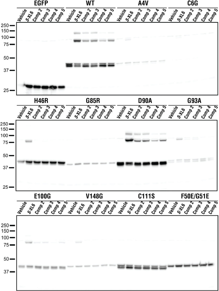
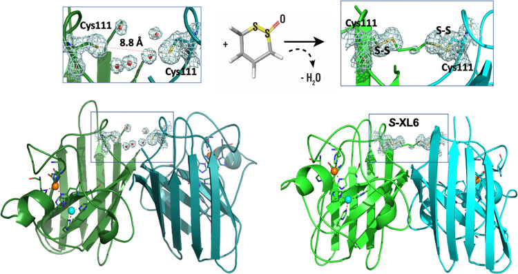
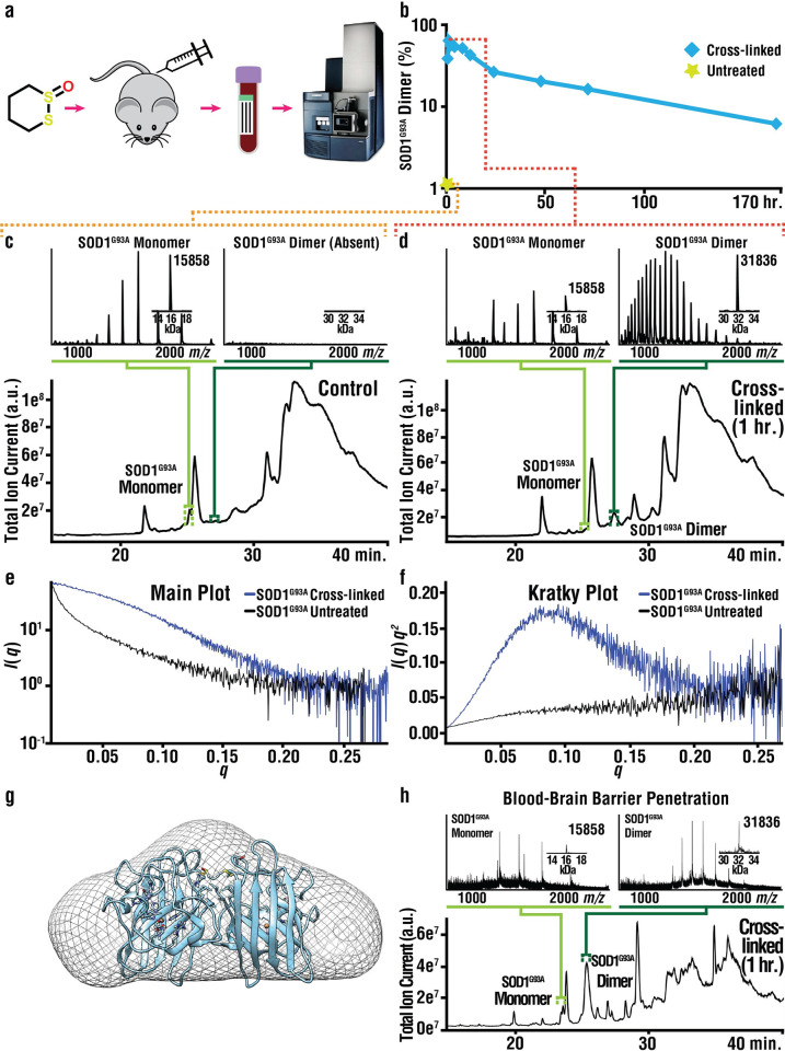
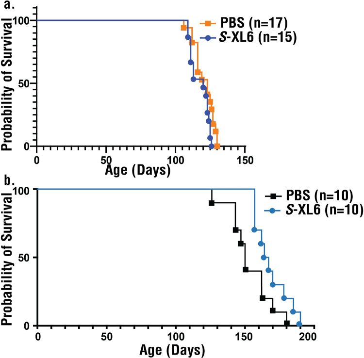
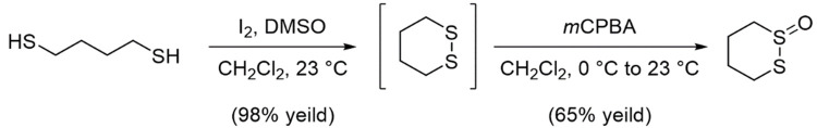
Mutations in the gene encoding Cu-Zn superoxide dismutase 1 (SOD1) cause a subset of familial amyotrophic lateral sclerosis (fALS) cases. A shared effect of these mutations is that SOD1, which is normally a stable dimer, dissociates into toxic monomers that seed toxic aggregates. Considerable research effort has been devoted to developing compounds that stabilize the dimer of fALS SOD1 variants, but unfortunately, this has not yet resulted in a treatment. We hypothesized that cyclic thiosulfinate cross-linkers, which selectively target a rare, 2 cysteine-containing motif, can stabilize fALS-causing SOD1 variants in vivo. We created a library of chemically diverse cyclic thiosulfinates and determined structure-cross-linking-activity relationships. A pre-lead compound, "S-XL6," was selected based upon its cross-linking rate and drug-like properties. Co-crystallographic structure clearly establishes the binding of S-XL6 at Cys 111 bridging the monomers and stabilizing the SOD1 dimer. Biophysical studies reveal that the degree of stabilization afforded by S-XL6 (up to 24°C) is unprecedented for fALS, and to our knowledge, for any protein target of any kinetic stabilizer. Gene silencing and protein degrading therapeutic approaches require careful dose titration to balance the benefit of diminished fALS SOD1 expression with the toxic loss-of-enzymatic function. We show that S-XL6 does not share this liability because it rescues the activity of fALS SOD1 variants. No pharmacological agent has been proven to bind to SOD1 in vivo. Here, using a fALS mouse model, we demonstrate oral bioavailability; rapid engagement of SOD1G93A by S-XL6 that increases SOD1G93A's in vivo half-life; and that S-XL6 crosses the blood-brain barrier. S-XL6 demonstrated a degree of selectivity by avoiding off-target binding to plasma proteins. Taken together, our results indicate that cyclic thiosulfinate-mediated SOD1 stabilization should receive further attention as a potential therapeutic approach for fALS.


Conflict of interest statement

The authors have declared that no competing interests exist.

Figures

Fig 1
Fig 1. Cyclic thiosulfinate S-XL6 cross-links SOD1 variants via Cys111 residues on adjacent monomers.
(Top) Crystal structure of wild-type SOD1 (PDB ID: 1SPD [5], cartoon representation generated with Maestro 11.8) highlighting opposing Cys111 residues on both monomer A (blue) and monomer B (green) with a representation of the S-XL6 cross-linked SOD1 dimer. Cu and Zn molecules are represented by orange and gray spheres, respectively. (A) Trypsin digest and MALDI-ToF-MS analysis of SOD1A4V gave a combined sequence coverage of 94%, including peaks corresponding to the Cys57-Cys146 disulfide linked peptides (blue) and the Cys111- containing peptide (purple) linked by S-XL6. N-terminal acetylation is shown in blue text, cysteines are shown in red text, and the SOD1A4V mutation is underlined. (B) Examining the higher mass range revealed multiply charged forms of SOD1A4V including the S-XL6 linked dimer, as well as 2 different peaks corresponding to Cys111-linked peptides. (C) MALDI-FTICR-MS analysis of pepsin digested SOD1H46R. The top panel shows SOD1H46R control sample and middle panel exhibits peaks corresponding to Cys111 linked peptides via S-XL6. The cross-linked peptides are shown in black text with cysteines highlighted in red. The bottom panel shows confirmation of cross-linked peptides using deuterated S-XL6 (mass shift of 8 Da). MALDI, matrix-assisted laser desorption ionization; MS, mass spectrometry; SOD1, superoxide dismutase 1.
Fig 2
Fig 2. S-XL6-mediated cross-linking and stabilization of wild-type and fALS SOD1 variants.
Top: chemical mechanism of action of S-XL6. The cross-linking reaction proceeds through an initial thiolate-disulfide interchange between a cysteine thiolate and the cyclic thiosulfinate, generating a sulfenic acid intermediate upon opening of the ring structure. This sulfenic acid intermediate forms a cross-link by rapid condensation with a second cysteine thiolate. Mass spectra of untreated (left column, average mass [± 1 Da]) and S-XL6 cross-linked (middle column, average mass [± 2 Da]) proteins are consistent with cross-linking. DSF results (right column) indicate that cross-linking increased the thermal stability of SOD1 and its variants. We quantify ΔTu as the difference in unfolding temperature measured between the major inflections of the untreated and cross-linked samples. The unfolding temperature of the untreated and S-XL6 cross-linked SOD1 proteins are as follows: wild type (WT) from 75.9°C to 90.2°C (ΔTu ~14°C); SOD1A4V from 62.9°C to 79.3°C (ΔTu ~16°C); SOD1G93A from 70.6°C to 85.0°C (ΔTu ~14°C); SOD1H46R from 46.3°C to 69.3°C (ΔTu ~23°C); SOD1G85R from 40.8°C to 65.1°C (ΔTu ~24°C). Note: two inflections can be observed when there is a mixture of partially (lower ΔTu) and fully metalated (higher ΔTu) SOD1 proteoforms, which applies to the following proteins: untreated wild-type SOD1 unfolds at 75.9°C and 87.6°C; untreated SOD1G93A unfolds at 70.6°C and 84.4°C. All samples were analyzed in technical triplicate (standard errors <0.3°C). fALS, familial amyotrophic lateral sclerosis; SOD1, superoxide dismutase 1.
Fig 3
Fig 3. SEC demonstrates that the monomeric population of fALS variants is decreased by S-XL6 treatment and that this is associated with reduced aggregation.
Top: SEC results for the molecular weight calibration standards, Thyroglobulin (660 kDa), IgG (150 kDa), BSA (66 kDa), and myoglobin (17 kDa). Bottom: SEC results for the same variants as Figs 2 and 4, with and without S-XL6 treatment. The peak labeled “aggregates” elute near the void volume of the SEC column and are therefore too large (>1,000 kDa) for MW determination. fALS, familial amyotrophic lateral sclerosis; SEC, size exclusion chromatography.
Fig 4
Fig 4. Cross-linking enhances the structure of fALS SOD1 variants.
Differences in deuterium uptake (ΔU, legend shown below part b) of untreated and cross-linked variants for the 4-h time point compared to the wild-type SOD1 (WT) protein are reported here. (A) Black arrows indicate areas where prominent shifts in ΔU were observed (e.g., residues 2–7 for SOD1A4V untreated 13.8% to 1.2% cross-linked, for full results see S1 Fig). (B) ΔU for untreated and cross-linked SOD1A4V mapped onto the cartoon representation of wild-type SOD1 structure (PDB ID: 1SPD) [5], generated with Maestro 11.8. ΔU for all time points (15 s, 50 s, 500 s, 1 h, 4 h) are reported in S1 Fig. fALS, familial amyotrophic lateral sclerosis; SOD1, superoxide dismutase 1.
Fig 5
Fig 5. Enzymatic activity of SOD1 variants after S-XL6 treatment.
Plate-based biochemical (A) and gel-based (B) assays were used to assess the impact of cross-linking on the enzymatic activity of SOD1 variants. Upon treatment with cross-linker the activity of SOD1A4V, SOD1G93A, and SOD1G85R increased to that of wild-type SOD1. Treatment with crosslinker did not affect the enzymatically inactive SOD1H46R. “+” and “–” denote samples treated with S-XL6 and untreated samples, respectively. The data underlying this figure can be found in S1 Data. SOD1, superoxide dismutase 1.
Fig 6
Fig 6. Cross-linking at sub-toxic concentrations within cells.
(A) Western blot analysis using a SOD1-selective antibody indicates that cross-linking proceeds in HEP G2 cells with an EC50 of approximately 5 μm (in PBS buffer). (B) Low cytotoxicity of S-XL6 (LC50 approximately 446 μm) was observed using the MTT (3-(4,5-Dimethylthiazol 2-yl)-2,5-diphenyltetrazolium bromide) cytotoxicity assay (triplicate sample analysis). Results are shown as percentage of viable cells compared to a vehicle control. Note: cell viability <70% (dotted line) is generally considered as the threshold for cytotoxicity, and viabilities >100% indicate a trophic effect. Controls included EMEM with 0.1% DMSO (dimethyl sulfoxide, negative control) and 500 μm of chlorpromazine (positive control). (C) Plasma protein binding was assayed in human platelet rich (hPRP) and platelet poor plasma (hPPP) using an LC-MS/MS assay at 3 concentrations (10, 50, and 250 μm) and no binding of S-XL6 was observed. Likewise, S-XL6 was not bound to purified alpha 1-acid glycoprotein and human serum albumin. The data underlying this figure can be found in S1 Data. LC-MS/MS, liquid chromatography-tandem mass spectrometry; MS, mass spectrometry; SOD1, superoxide dismutase 1.
Fig 7
Fig 7. S-XL6 outperforms leading cyclic thiosulfinates, binds a variety of fALS variants, and requires the Cys111 residue in cellulo.
C-terminally EGFP-labeled SOD1 (wild-type SOD1, 8 fALS variants, a Cys111Ser variant, and a constitutively monomeric F50E/G51E variant) were expressed in NSC-34 cells. S-XL6 was the most effective compound of the top-five 6-membered cyclic thiosulfinates identified through mass spectrometry screening of NCI compounds and our own medicinal chemistry efforts (labeled “Comp 2–5”). Compounds 2 and 3 are epimers of the 4,5-hydroxyl derivates of S-XL6; the identity of compounds 4 and 5 are unpatented and cannot be disclosed here, and 20 μm S-XL6 engaged all fALS variants other than G85R and wild-type SOD1, but not Cys111Ser or F50E/G51E. EGFP-wild-type and EGFP-D90A SOD1 formed an off-product (unintended) high-molecular weight species consistent with trimeric SOD1. S-XL6 treatment did not affect the cellular viability or aggregation of G93A SOD1. S-XL6 treatment did not affect the viability but did accelerate the aggregation of wild-type SOD1 (S3 Fig). fALS, familial amyotrophic lateral sclerosis; SOD1, superoxide dismutase 1.
Fig 8
Fig 8. Cyclic thiosulfinate S-XL6 cross-links wild-type SOD1 via Cys111 residues on adjacent monomers.
On the left crystal structure of as-isolated wild-type SOD1 at 1.77 Å resolution (cartoon representation generated with Pymol) highlighting opposing Cys111 residues on both monomer A (dark green) and monomer B (dark blue) with multiple water molecule present at dimer interface (PDB ID 8Q6M). Right hand panels show the crystal structure of S-XL6 cross-linked wild-type SOD1 at 1.67Å resolution (PDB ID 8CCX) clearly displaying additional density for S atoms from S-XL6 on both monomers A (light green) and B (cyan). 2FoFc electron density maps for the structures are shown as gray mesh, contoured at 1 σ level around Cys111, water molecules at the SOD1 dimer interface and cross-linker. Waters are indicated as small red spheres, Cu and Zn molecules are represented by cyan and orange spheres, respectively. These water molecules are largely excluded upon the incorporation of S-XL6. The data underlying this figure can be found at rcsb.org (PDB IDs 8Q6M and 8CCX). SOD1, superoxide dismutase 1.
Fig 9
Fig 9. S-XL6 cross-links SOD1 in an ALS mouse model and reduces aggregation in vitro.
(A) Schematic of the pharmacodynamic workflow. (B) Pharmacodynamic profiling of RBC proteins by LC-MS analysis indicates the formation of an S-XL6 cross-linked dimer in treated SOD1G93A mice. Hemizygous fALS SOD1G93A mice were dosed once at 10 mg/kg with S-XL6 via tail vein injection, blood was collected periodically over a 7-day period, and LC-MS analysis (C and D) was performed to assess percentage of cross-linked SOD1 (details in S1 Table). Spectra represent a 6-s average at peak apex. (E and F) In vitro SAXS experiments for SOD1G93A: Semi-log plot of the scattering intensity (I, log scale) as a function of momentum transfer, q; and Kratky plot [q2I(q) versus q]. The plot shape for untreated SOD1G93A is consistent with a heterogenous mixture of unfolded proteins including amorphous aggregates, whereas plot shape for cross-linked SOD1G93A is consistent with a folded, globular structure. The estimated radius of gyration is approximately 20 Å. (G) A 3-D reconstruction of cross-linked SOD1G93A is superimposed upon the SOD1G93A crystal structure (PDB: 3GZO). (H) Target engagement via LC-MS analysis in transgenic (Tg) SOD1G93A mouse brain dosed once at 30 mg/kg with S-XL6 via subcutaneous injection. The data underlying this figure can be found in S1 Data. ALS, amyotrophic lateral sclerosis; fALS, familial amyotrophic lateral sclerosis; LC-MS, liquid chromatography-mass spectrometry; RBC, red blood cell; SOD1, superoxide dismutase 1.
Fig 10
Fig 10. Survival studies of fALS mice treated with subcutaneously administered S-XL6.
(a) Treatment with S-XL6 in B6SJL G93A (hybrid line) mice began at age 60 days in cohorts of 40 mice (18 untreated and 18 treated until end-stage; 2 mice from each group dosed for ca. 50 days before being killed for target engagement assessment in brain). After 1 week at a 20 mg/kg dose target engagement in blood was monitored as shown in Fig 9. Based upon these results, to improve target engagement to >50% peak effect, the dose was increased to 50 mg/kg. No survival improvement was observed in B6SJL mice. Body weight, disease score, and all paws’ grip score analysis for B6SJL mice are given in S8 Fig. (b) B6 G93A/YFP (congenic line) mice were dosed on weekdays starting from day 108 to endpoint and demonstrated a modest survival improvement. Immediately after dosing mice exhibited lethargy and shaking that subsided after 20 min. The data underlying this figure can be found in S1 Data. fALS, familial amyotrophic lateral sclerosis.
Fig 11
Fig 11. Synthesis of cyclic thiosulfinate 1,2-dithiane-1-oxide (S-XL6).

Similar articles

Cited by

References

    1. Abalkhail H, Mitchell J, Habgood J, Orrell R, de Belleroche J. A new familial amyotrophic lateral sclerosis locus on chromosome 16q12.1-16q12.2. Am J Hum Genet. 2003;73:383–389. doi: 10.1086/377156 - DOI - PMC - PubMed
    1. Chiò A, Logroscino G, Hardiman O, Swingler R, Mitchell D, Beghi E, et al.. Prognostic factors in ALS: A critical review. Amyotroph Lateral Scler. 2009;10:310–323. doi: 10.3109/17482960802566824 - DOI - PMC - PubMed
    1. Henden L, Twine NA, Szul P, McCann EP, Nicholson GA, Rowe DB, et al.. Identity by descent analysis identifies founder events and links SOD1 familial and sporadic ALS cases. NPJ Genom Med. 2020;5:32. doi: 10.1038/s41525-020-00139-8 - DOI - PMC - PubMed
    1. Renton AE, Chiò A, Traynor BJ. State of play in amyotrophic lateral sclerosis genetics. Nat Neurosci. 2014;17:17–23. doi: 10.1038/nn.3584 - DOI - PMC - PubMed
    1. Deng HX, Hentati A, Tainer JA, Iqbal Z, Cayabyab A, Hung WY, et al.. Amyotrophic lateral sclerosis and structural defects in Cu,Zn superoxide dismutase. Science. 1993;261:1047. doi: 10.1126/science.8351519 - DOI - PubMed

MeSH terms

Supplementary concepts