. 2024 Jan 30;17(821):eadi7861.
doi: 10.1126/scisignal.adi7861. Epub 2024 Jan 30.

A plasma membrane-associated form of the androgen receptor enhances nuclear androgen signaling in osteoblasts and prostate cancer cells

Affiliations

A plasma membrane-associated form of the androgen receptor enhances nuclear androgen signaling in osteoblasts and prostate cancer cells

Hema Kalyanaraman et al. Sci Signal. .

Abstract

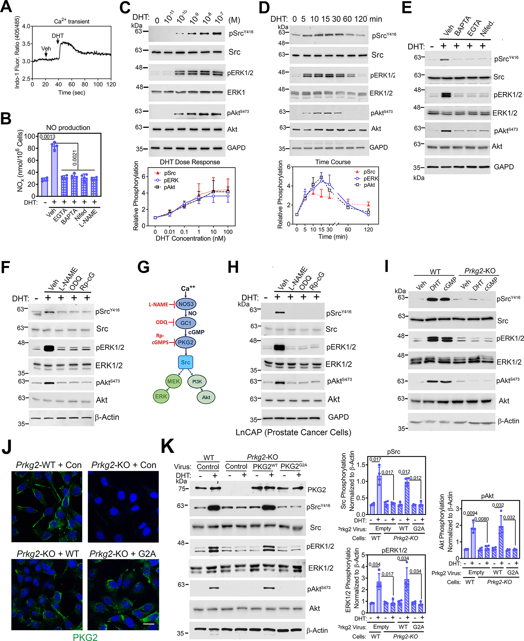
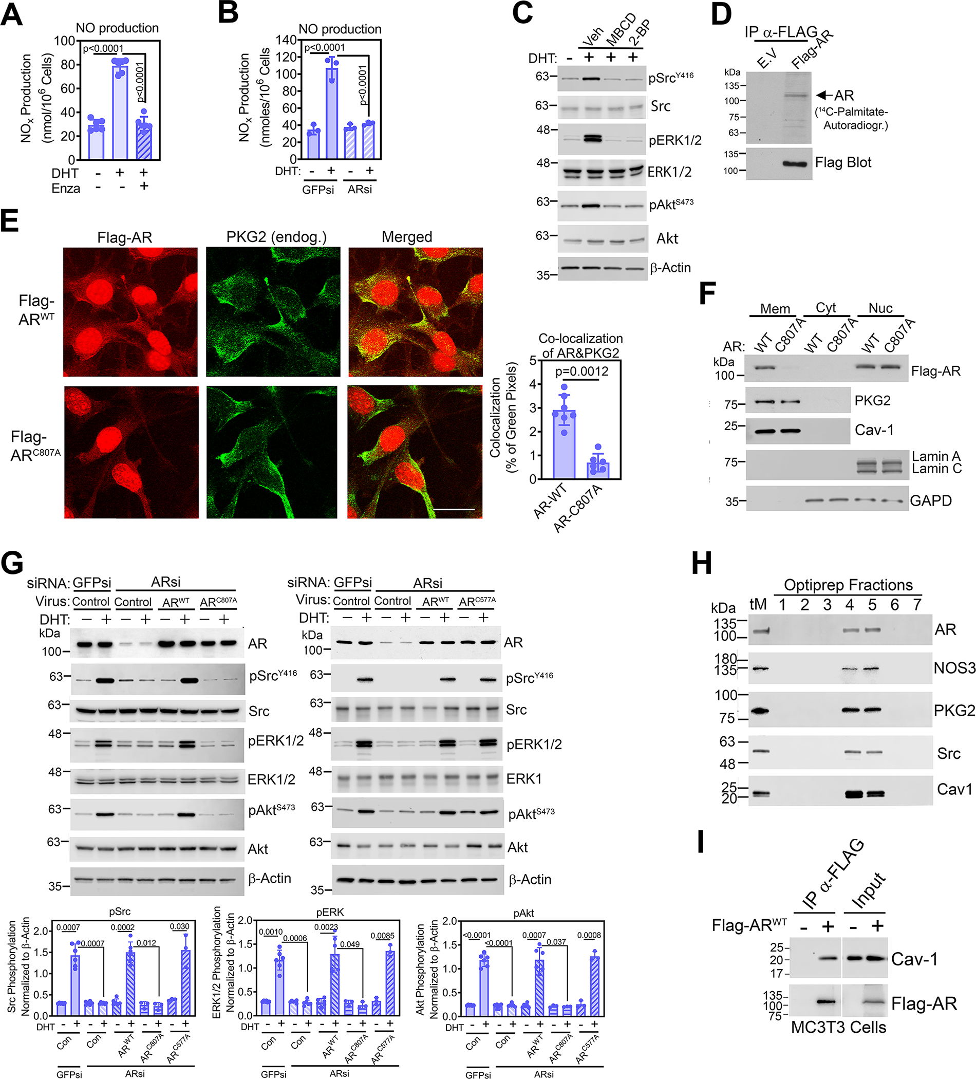
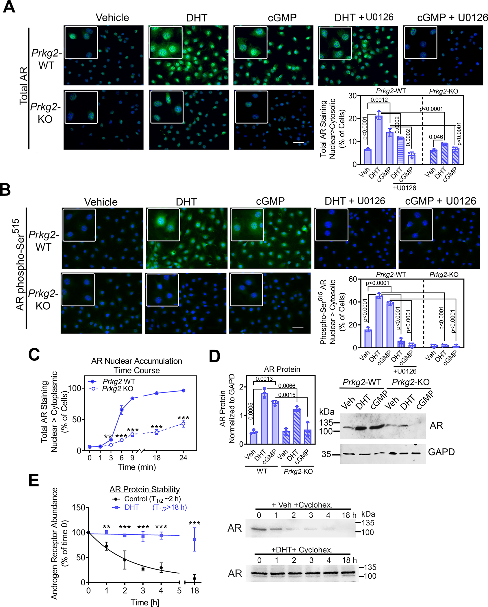
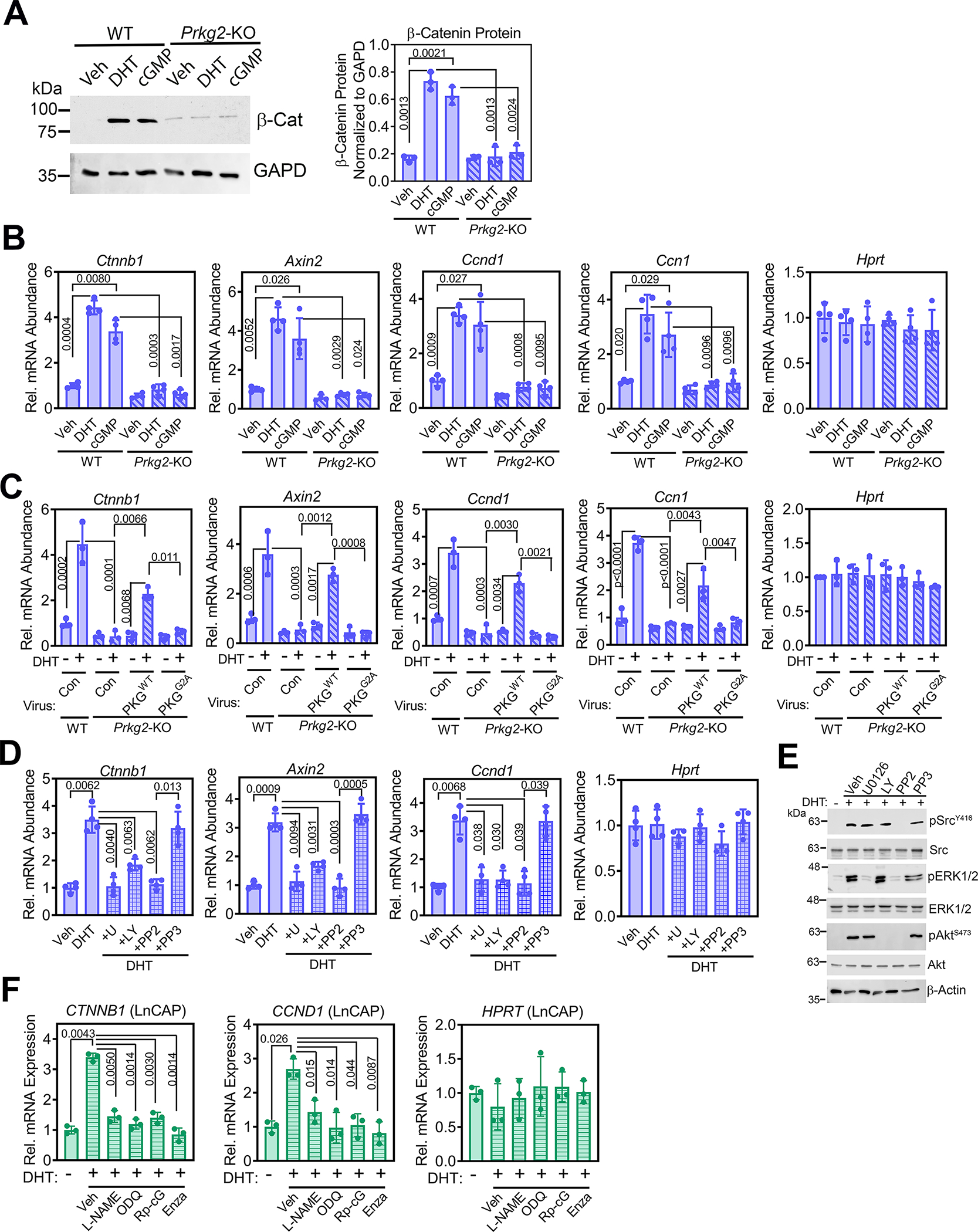
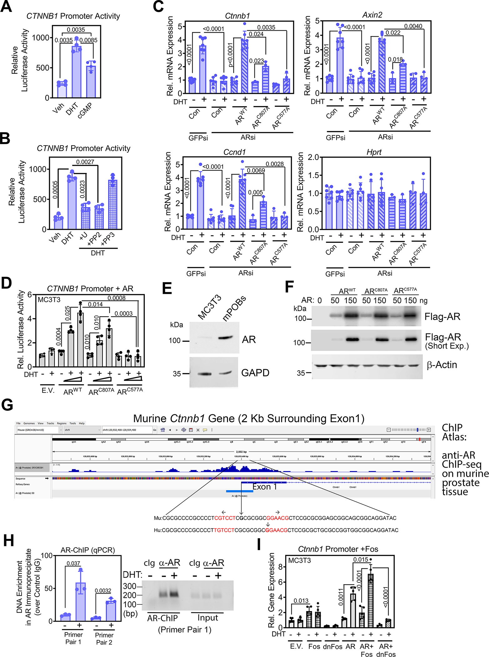
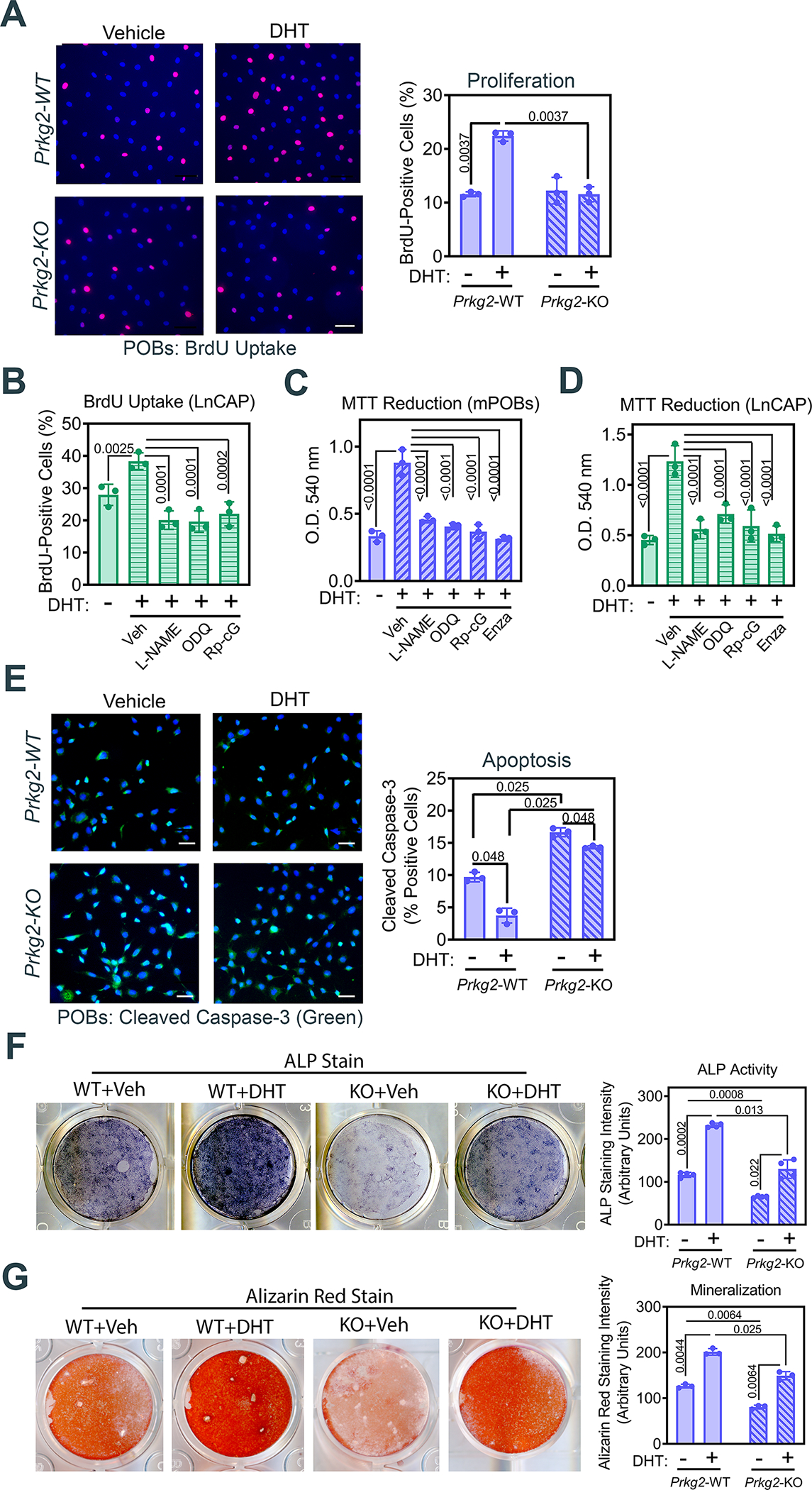
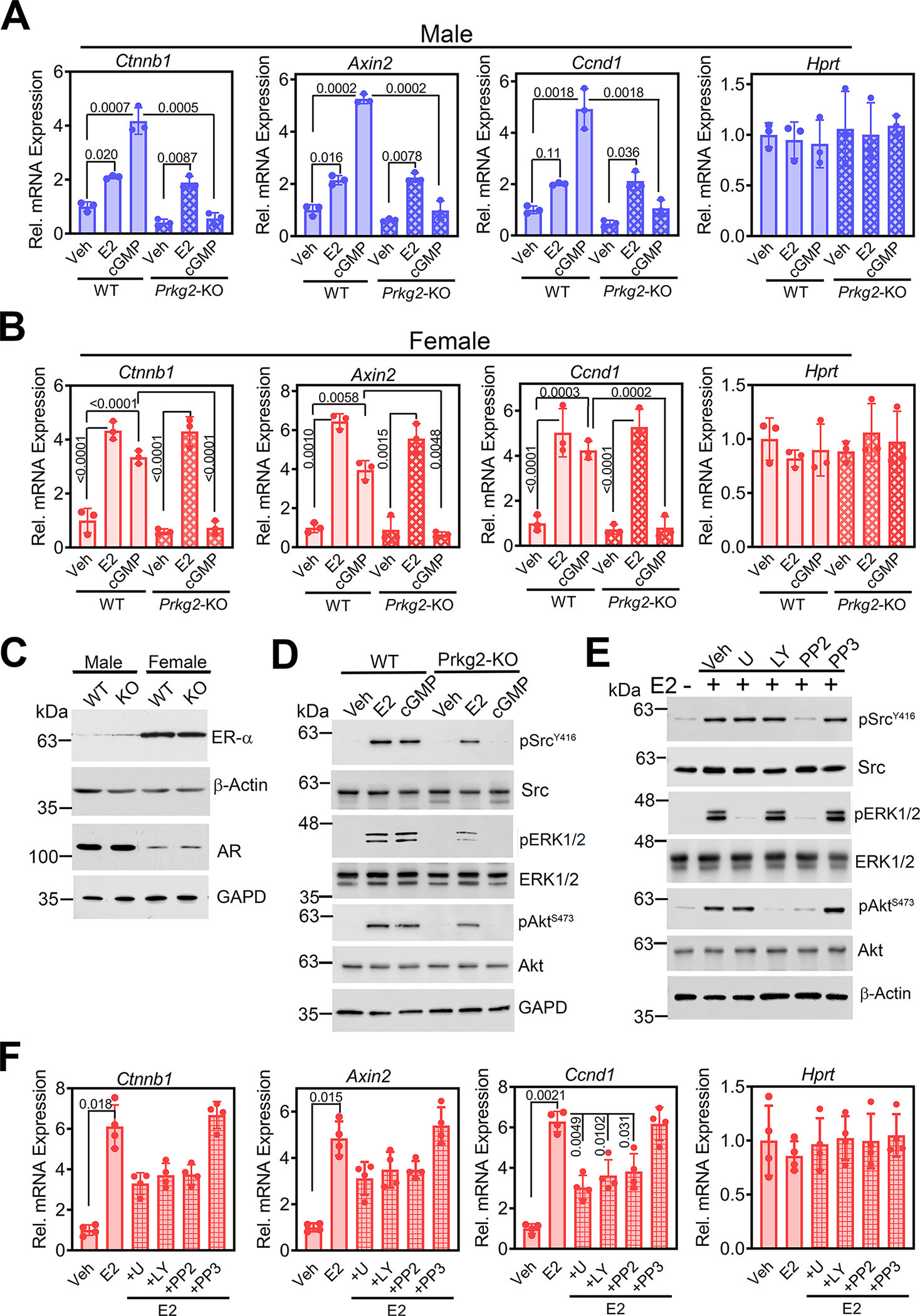
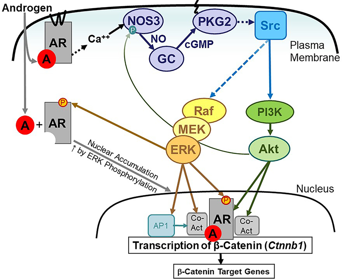
Androgen binding to the androgen receptor (AR) in the cytoplasm induces the AR to translocate to the nucleus, where it regulates the expression of target genes. Here, we found that androgens rapidly activated a plasma membrane-associated signaling node that enhanced nuclear AR functions. In murine primary osteoblasts, dihydrotestosterone (DHT) binding to a membrane-associated form of AR stimulated plasma membrane-associated protein kinase G type 2 (PKG2), leading to the activation of multiple kinases, including ERK. Phosphorylation of AR at Ser515 by ERK increased the nuclear accumulation and binding of AR to the promoter of Ctnnb1, which encodes the transcription factor β-catenin. In male mouse osteoblasts and human prostate cancer cells, DHT induced the expression of Ctnnb1 and CTNN1B, respectively, as well as β-catenin target genes, stimulating the proliferation, survival, and differentiation of osteoblasts and the proliferation of prostate cancer cells in a PKG2-dependent fashion. Because β-catenin is a master regulator of skeletal homeostasis, these results explain the reported male-specific osteoporotic phenotype of mice lacking PKG2 in osteoblasts and imply that PKG2-dependent AR signaling is essential for maintaining bone mass in vivo. Our results suggest that widely used pharmacological PKG activators, such as sildenafil, could be beneficial for male and estrogen-deficient female patients with osteoporosis but detrimental in patients with prostate cancer.


Conflict of interest statement

Competing interests: The authors declare that they have no competing interests.

Figures

Fig. 1.
Fig. 1.. Androgens rapidly induce NO production and activate Src, ERK, and Akt through membrane-associated PKG2.
(A) Quantification of the 405 nm/485 nm fluorescence ratio in male WT POBs loaded with the intracellular Ca2+ indicator Indo1-AM and treated with vehicle (Veh) followed by 1 nM DHT. (B) Quantification of NO derivatives in the media of male WT POBs pretreated with vehicle, EGTA, BAPTA-AM, nifedipine (Nifed), or L-NAME before treatment with DHT for 30 min. (C and D) Immunoblotting for total and phosphorylated (p) forms of Src, ERK1/2, and Akt in male WT POBs treated with the indicated DHT concentrations for 10 min (C), or with 1nM DHT for the indicated amounts of time (D). GAPD is a loading control. Amounts of pSrcY416, pERKY204, and pAktS473 were quantified by densitometry. (E and F) Immunoblotting for the indicated proteins in male WT POBs pretreated with Ca2+ modulators (E) or pretreated with L-NAME, ODQ, or Rp-8-CPT-PET-cGMPS (Rp-cG) (F) and treated with 1 nM DHT for 10 min. GAPD and β-actin are loading controls. (G) Schematic showing the Ca2+-induced activation of NOS3 and the NO-cGMP-PKG2 pathway resulting in Src, ERK, and Akt activation, with pathway inhibitors shown in red. (H) Immunoblotting of LnCAP cells pretreated with L-NAME, ODQ, or Rp-8-CPT-PET-cGMPS and treated with 1 nM DHT for 10 min. (I) Immunoblotting of POBs from male Prkg2-WT and OB Prkg2-KO mice after treatment with DHT or 8-CPT-cGMP (cGMP) for 10 min. (J) Immunofluorescence showing PKG2 localization in POBs from male Prkg2-WT and OB Prkg2-KO mice that were infected with virus expressing LacZ (control) or WT, membrane-associated PKG2 (PKG2WT) or mutant, cytosolic PKG2 (PKG2G2A) before DHT stimulation. Scale bar, 25 μm. (K) Immunoblotting for and quantification of the indicated proteins in cells treated as in (J). All data are representative of at least three independent experiments; n=4 for (B) and (J); graphs show means ± S.D. P values for the indicated comparisons by ANOVA. For (E) to (I), quantification of three independent experiments is shown in fig. S1B, D, and E and S2A. Separate, identical gels loaded with equal amounts of cell lysates were run for analysis of total and phospohorylated forms of Src, ERK, and Akt.
Fig. 2.
Fig. 2.. Rapid androgen signaling occurs through a membrane-associated AR.
(A and B) Quantification of NO derivatives in the media of male WT POBs pre-treated with enzalutamide (A), or transfected with siRNAs targeting AR (ARsi) or green fluorescent protein (GFPsi, control) (B); some cells received DHT for 30 min. (C) Immunoblotting for total and phosphorylated (p) forms of Src, ERK1/2, and Akt in male WT POBs pretreated with methyl-β-cyclodextrin (MBCD) or 2-bromopalmitate (2-BP) and treated with DHT for 10 min. Actin is a loading control. (D) Autoradiograph of and Western blotting for Flag in Flag immunoprecipitates from MC3T3 cells transfected with empty vector (E.V.) or Flag-tagged AR labelled with 14C-palmitate for 4 h. (E) Immunofluorescence staining for Flag (red) and endogenous PKG2 (green) in POBs transfected with Flag-tagged WT AR (ARWT) or AR with a mutation in a conserved membrane localization motif (ARC807A) and treated with DHT for 18 h. Scale bar, 25 μm. Quantification of AR and PKG2 colocalization, indicated by yellow pixels in the merged image, is shown in the graph; 7 images from 3 independent experiments were analyzed, representing ~30 cells per condition. (F) Immunoblotting for the indicated proteins in membrane (Mem), cytosol (Cyt), and nuclear (Nuc) fractions of POBs infected with virus encoding Flag-tagged ARWT or ARC807A and treated with DHT for 18 h. Caveolin-1 (Cav-1) and lamin A/C are markers for plasma membranes and nuclei, respectively. GAPD is a loading control for cytosol. (G) Immunoblotting for the indicated proteins in POBs transfected with siRNAs targeting GFP or AR and then infected with LacZ virus (control) or virus encoding Flag-tagged WT AR (ARWT), ARC807A with a mutation in a conserved membrane localization motif (left), or ARC577A with a mutation in the first DNA-binding zinc finger motif (right). Cells received DHT for 10 min; three independent experiments with each AR mutant are summarized in the bar graphs below. (H) Immunoblotting for the indicated proteins in POB membranes from a Percoll gradient (total membranes, tM) that were further fractionated over a density gradient of Optiprep. Caveolin-1 is a marker of caveolar fractions. (I) Flag immunoprecipitates from MC3T3 cells transfected with empty vector or Flag-tagged ARWT were blotted with antibodies specific for caveolin-1 or Flag. 10% input lysate was analyzed in parallel. All data are representative of three independent experiments, except n=6 for (A), n=7 for (E), and n=2 for (D). Graphs show means ± S.D.; p values for the indicated comparisons by one-way ANAOVA (A), two-tailed Mann-Whitney test (B), or two-way ANOVA (B and G). For (C), quantification of three independent experiments is shown in fig. S3D. Separate, identical gels loaded with equal amounts of cell lysates were run for analysis of total and phospohorylated forms of Src, ERK, and Akt.
Fig. 3.
Fig. 3.. Rapid androgen signaling through PKG2-dependent activation of ERK enhances AR nuclear accumulation.
(A and B) Immunofluorescence staining for total AR (A) or AR phosphorylated on Ser515 (B) in POBs from male Prkg2-WT mice or OB Prkg2-KO mice. Cells were treated for 4 h with vehicle, DHT, or the cGMP analog 8-CPT-cGMP, and some cells were pre-treated with the MEK inhibitor U1026. Graphs show the percentage of cells in which nuclear fluorescence was greater than cytosolic fluorescence; at least 200 cells from 3 independent experiments were analyzed per condition, and the threshold for cytosolic fluorescence intensity was defined in untreated WT cells. Effects of U1026 in WT cells were analyzed separately by one-way ANOVA; comparison between WT and KO cells treated with DHT or cGMP was by two-way ANOVA. (C) WT and KO POBs were treated with DHT for the indicated times, with nuclear and cytosolic fluorescence staining for total AR assessed as in (A). (D) Immunoblotting for and quantification of total AR protein in whole cell lysates of WT and KO POBs treated for 24 h with vehicle, DHT or 8-CPT-cGMP. GAPD is a loading control. (E) AR protein abundance assessed by immunoblotting at the indicated times after addition of cycloheximide at time 0 to male WT POBs; cells were treated with vehicle or DHT for 4 h prior to and during incubation with cycloheximide. The graphs in (C) to (E) show means ± S.D of three independent experiments; p values for the indicated comparisons by two-way ANOVA, with **p<0.01 and ***p<0.001 for the comparison between WT and KO cells (C) or between vehicle and DHT (E) at each timepoint.
Fig. 4.
Fig. 4.. Androgen stimulation of β-catenin and its target genes requires signaling by PKG2, Src, Erk, and Akt.
(A) Immunoblotting for and quantification of total β-catenin protein in POBs from male Prkg2-WT and OB Prkg2-KO mice treated with DHT or 8-CPT-cGMP for 24 h. GAPD is a loading control. (B) Relative mRNA abundance of Ctnnb1 and β-catenin target genes (Axin2, Ccnd1, and Ccn1) normalized to 18S RNA in POBs from male Prkg2-WT and OB Prkg2-KO mice treated with DHT or 8-CPT-cGMP for 24 h. Hprt served as a control. Mean ΔCt values obtained in vehicle-treated Prkg2-WT cells were assigned a value of one. (C) Quantification of the indicated mRNAs in Prkg2-KO osteoblasts reconstituted with virus encoding LacZ (con), membrane-associated WT PKG2 (PKGWT), or cytosolic mutant PKG2 (PKGG2A); cells were treated as in (B). (D) Quantification of the indicated mRNAs in WT POBs pre-treated with U0126, LY29004, PP2, or PP3 (inactive PP2 analog) prior to treatment with DHT for 24 h. (E) Immunoblotting for total and phosphorylated (p) forms of Src, ERK1/2, and Akt in male WT POBs pretreated with U0126, LY29004, PP2, or PP3 and treated with DHT for 10 min; quantification of 3 independent experiments is in fig. S5E. Actin is a loading control. (F) Quantification of Ctnnb1 and Ccnd1 mRNAs in LnCAP prostate cancer cells pre-treated with L-NAME, ODQ, Rp-8-CPT-PET-cGMPS (Rp-cG), or enzalutamide (enza), prior to receiving DHT for 24 h. All graphs show means ± S.D. of three (A, C, E, and F) or four (B and D) independent experiments; p values for the indicated comparisons by two-way (A to C) or one-way (D and F) ANOVA. Separate, identical gels loaded with equal amounts of cell lysates were run for analysis of total and phospohorylated forms of Src, ERK, and Akt.
Fig. 5.
Fig. 5.. DHT increases Ctnnb1 transcription by inducing AR binding near the transcription start site.
(A) Luciferase activity from a human CTNNB1 promoter–driven luciferase reporter in male WT POBs after 36 h treatment with vehicle, DHT, or 8-CPT-cGMP. (B) Luciferase activity from the CTNNB1 reporter in POBs pre-treated with U0126, PP2, or PP3 prior to treatment with DHT. (C) Relative abundance of Ctnnb1 mRNA and β-catenin target genes (Axin2, Ccnd1), normalized to 18S RNA, in male WT POBs. Cells were transfected with siRNAs targeting GFP or AR and received LacZ virus (Con) or virus encoding Flag-tagged ARWT, membrane binding-deficient ARC807A, or DNA binding-deficient ARC577A prior to treatment with vehicle or DHT for 24 h. Hprt served as a control. Mean ΔCt values obtained in vehicle-treated cells were assigned a value of one. (D) Luciferase activity in MC3T3 cells co-transfected with the CTNNB1 reporter and either empty vector (E.V.) or increasing amounts of ARWT, ARC807A, or ARC577A; cells were treated with vehicle or DHT for 36 h. (E) Immunoblotting for endogenous AR in untransfected MC3T3 cells and male WT POBs. GAPD is a loading control. (F) Immunoblotting for the Flag-tagged AR constructs in MC3T3 cells transfected as in (D). Quantification in shown fig. S6D. Actin is a loading control. (G) Published ChIP-seq results for AR binding to sequences surrounding exon 1 of the Ctnnb1 gene in murine prostate tissue (50). The enlargement shows conserved murine and human sequences with the transcription start sites marked by vertical arrows, and an inverted repeat of an ARE-like motif highlighted in red. (H) Quantification of Ctnnb1 enrichment in chromatin immunoprecipitation using an AR-specific antibody from POBs treated with vehicle or DHT for 6 h. Sequences near the Ctnnb1 transcription start site were amplified by two independent primer pairs. QPCR results are expressed as a fold increase over the signal obtained with control IgG; PCR products were also analyzed by agarose gel electrophoresis (right). (I) Luciferase activity in MC3T3 cells co-transfected with the CTNNB1 reporter and empty vector (E.V.) or vectors encoding c-Fos, a dominant-negative Fos (dnFos), and/or ARWT as indicated and treated with vehicle or DHT for 36 h. Bar graphs show means ± S.D. of 3 (H), 4 (A, B, C, D) or 5 (I) independent experiments; in (C), 3 independent experiments comparing ARWT to ARC807 and 4 experiments comparing ARWT to ARC577A were combined. All blots are representative of 3 independent experiments. P values for the indicated comparisons by one-way ANOVA (A and B) or two-way ANOVA (C, D, and I); for H, comparison by two-tailed Welch’s t test.
Fig. 6.
Fig. 6.. Androgen stimulation of cell proliferation, mitochondrial activity, and survival requires NO-cGMP-PKG2 signaling.
(A) Immunofluorescence staining for and quantification of bromo-deoxyuridine (BrdU) incorporation into S-phase nuclei in POBs from male Prkg2-WT or OB Prkg2-KO mice treated with DHT for 24 h. Scale bar, 50 μm. (B) Quantification of immunofluorescence staining for BrdU uptake in LnCAP prostate cancer cells pretreated with L-NAME, ODQ, or Rp-8-CPT-PET-cGMPS (Rp-cG), prior to receiving BrdU and DHT or vehicle for 24 h. (C and D) Mitochondrial MTT reduction to formazan measured spectrophotometrically in WT POBs (C) and LnCAP cells (D) pretreated with L-NAME, ODQ, Rp-8-CPT-PET-cGMPS, or enzalutamide, and treated with DHT for 24 h. (E) Immunofluorescence staining for and quantification of cleaved caspase-3 in serum-starved POBs from male Prkg2-WT or OB Prkg2-KO mice treated with DHT for 24 h. Scale bar, 50 μm. (F and G) Quantification of alkaline phosphatase (ALP) activity (F) and alizarin red staining (G) in Prkg2-WT or OB Prkg2-KO POBs cultured in differentiation medium and treated with vehicle or DHT for 14 d (F) or 21 d (G), respectively. All graphs show means ± S.D. of 3 (A to E, G) or 4 (F) independent experiments. P values for the indicated comparisons by one-way ANOVA (B to D) or two-way ANOVA (A, E, F and G).
Fig. 7.
Fig. 7.. Estrogen induction of β-catenin and its target genes is PKG2-independent.
(A and B) Relative abundance of Ctnnb1 mRNA and the β-catenin target genes Axin2 and Ccnd1 normalized to 18S RNA in POBs from male (A) or female (B) Prkg2-WT or OB Prkg2-KO mice treated with 17β-estradiol (E2) or 8-CPT-cGMP (cGMP) for 24 h. Hprt served as a control. Mean ΔCt values in vehicle-treated WT cells were assigned a value of one. (C) Immunoblotting for ER-α and AR in whole-cell extracts of male and female WT and KO POBs. β-actin and GAPD are loading controls. Quantification of three independent experiments is shown in fig. S8B. (D) Immunoblotting for total and phosphorylated (p) forms of Src, ERK1/2, and Akt in female Prkg2-WT and Prkg2-KO POBs treated with E2 or 8-CPT-cGMP for 10 min. Quantification of 5 independent experiments is shown in fig. S8D. (E) Immunoblots of the indicated proteins in female WT POBs pre-treated with U1026, LY294002, PP2, or PP3 and treated with E2 for 10 min. Quantification of 2 independent experiments is shown in fig. S8E. (F) Relative abundance of Ctnnb1, Axin2, and Ccnd1 transcripts in female WT POBs pretreated with the indicated drugs and treated with E2 for 24 h. Graphs show means ± S.D. of 3 (A and B) or 4 (F) independent experiments; P values for the indicated comparisons by one-way (F) or two-way ANOVA (A and B). Separate, identical gels loaded with equal amounts of cell lysates were run for analysis of total and phospohorylated forms of Src, ERK, and Akt.
Fig. 8.
Fig. 8.. Transcriptional regulation of β-catenin through PKG2-mediated cooperation between membrane-associated and nuclear androgen receptors.
Androgen (A) binding to the membrane-associated AR in osteoblasts increases the intracellular Ca2+ concentration, which activates NOS3 to produce NO. NO activates guanylyl cyclase-1 (GC) to produce cGMP, which activates membrane-bound PKG2. As we have previously shown, PKG2 activates Src in a Shp1/2–dependent manner to increase ERK1/2 activity through Raf and MEK (15) and Akt activity through phosphoinositol-3-kinase (PI3K) (30). ERK-mediated phosphorylation of AR on Ser515 enhances AR nuclear accumulation. Akt can phosphorylate AR, and ERK and Akt can phosphorylate transcriptional co-activators to increase AR transcriptional activity (40). A positive feedback mechanisms enhances NOS activity through Akt-mediated phosphorylation of NOS3. AR binds near the transcription start site of the Ctnnb1 gene to increase transcription, thereby promoting the expression of β-catenin target genes In addition, PKG2-dependent ERK activation increases c-Fos, which can bind to the Ctnnb1 promoter as part of the AP-1 complex (52) and cooperate with AR to enhance transcription.

Similar articles

Cited by

References

    1. Vanderschueren D, Laurent MR, Claessens F, Gielen E, Lagerquist MK, Vandenput L, Borjesson AE, Ohlsson C, Sex steroid actions in male bone. Endocr Rev 35, 906–960 (2014). - PMC - PubMed
    1. Levin ER, Hammes SR, Nuclear receptors outside the nucleus: extranuclear signalling by steroid receptors. Nat Rev Mol Cell Biol 17, 783–797 (2016). - PMC - PubMed
    1. Mauvais-Jarvis F, Lange CA, Levin ER, Membrane-Initiated Estrogen, Androgen, and Progesterone Receptor Signaling in Health and Disease. Endocr Rev 43, 720–742 (2022). - PMC - PubMed
    1. Ahluwalia A, Hoa N, Ge L, Blumberg B, Levin ER, Mechanisms by Which Membrane and Nuclear ER Alpha Inhibit Adipogenesis in Cells Isolated From Female Mice. Endocrinology 161, (2020). - PubMed
    1. Thomas P, Membrane Androgen Receptors Unrelated to Nuclear Steroid Receptors. Endocrinology 160, 772–781 (2019). - PubMed