. 2024 Apr 3;226(4):iyae015.
doi: 10.1093/genetics/iyae015.

A missense SNP in the tumor suppressor SETD2 reduces H3K36me3 and mitotic spindle integrity in Drosophila

Affiliations

A missense SNP in the tumor suppressor SETD2 reduces H3K36me3 and mitotic spindle integrity in Drosophila

Jovan S Brockett et al. Genetics. .

Abstract

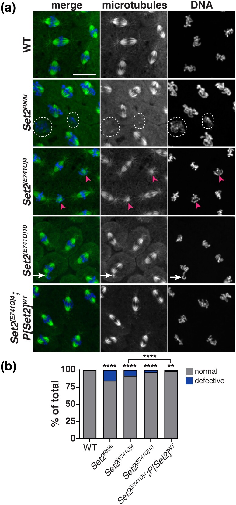
Mutations in SETD2 are among the most prevalent drivers of renal cell carcinoma (RCC). We identified a novel single nucleotide polymorphism (SNP) in SETD2, E902Q, within a subset of RCC patients, which manifests as both an inherited or tumor-associated somatic mutation. To determine if the SNP is biologically functional, we used CRISPR-based genome editing to generate the orthologous mutation within the Drosophila melanogaster Set2 gene. In Drosophila, the homologous amino acid substitution, E741Q, reduces H3K36me3 levels comparable to Set2 knockdown, and this loss is rescued by reintroduction of a wild-type Set2 transgene. We similarly uncovered significant defects in spindle morphogenesis, consistent with the established role of SETD2 in methylating α-Tubulin during mitosis to regulate microtubule dynamics and maintain genome stability. These data indicate the Set2 E741Q SNP affects both histone methylation and spindle integrity. Moreover, this work further suggests the SETD2 E902Q SNP may hold clinical relevance.

Keywords: Drosophila; H3K36me3; SETD2; SNP; models of human disease; renal cell carcinoma.


Conflict of interest statement

Conflicts of interest The author(s) declare no conflict of interest.

Figures

Fig. 1.
Fig. 1.
Functional domains and location of E902Q in the human SETD2 protein and the homologous E741Q in Drosophila melanogaster Set2-RA protein. a) The missense SNP described in this report (rs58906143, E902Q) location (arrow) is in the amino half of the protein, while the 5 known functional domains (AWS, SET, post-SET, WW, and SRI domains) are in the carboxy half. Note the domain architecture is similar in comparison to Drosophila melanogaster Set2. The orthologous E741Q mutation is shown (arrow). (Not drawn to scale.) b) A partial alignment of hSETD2 and Drosophila Set2-RA (dSet2) proteins with the conserved glutamic acid (E) highlighted.
Fig. 2.
Fig. 2.
Drosophila Set2 E741Q does not impair fitness. a) Quantification of the fecundity rate, measured as the number of eggs laid per female over 2-h, from the indicated age-matched genotypes. Each dot represents a measurement over 3 consecutive days. Mean ± SD for WT = 5.5 ± 0.8, Set2RNAi = 5.7 ± 0.4, Set2CTRL31= 5.5 ± 0.4, and Set2[E741Q]4= 4.9 ± 0.2 eggs laid per female. b) Quantification of hatch rate. Each dot represents the percent of unhatched embryos from N∼200 embryos from 3 independent replicate experiments from the indicated genotypes. Mean ± SD for WT = 13 ± 3.3, Set2RNAi = 17 ± 5, Set2CTRL31= 8.2 ± 1.3, and Set2[E741]4= 17.7 ± 5.3% unhatched embryos. n.s., not significant by a Kruskal–Wallis test followed by a Dunn's multiple comparison post-test.
Fig. 3.
Fig. 3.
H3K36me3 levels in Drosophila Set2 E741Q mutants. Immunoblots of relative H3K36me3 levels within 0–1 day total ovarian extracts relative to the βTub loading control, as quantified and normalized to the WT control (below). n.s., not significant or *P < 0.05 by one-way ANOVA followed by Dunnett's multiple comparison post-test.
Fig. 4.
Fig. 4.
Drosophila Set2 E741Q reduces H3K36me3 in the female germline. a) Images show maximum intensity projections of germaria from the indicated genotypes stained with DAPI (blue), Vasa (red), and H3K36me3 (green) antibodies. Boxed regions contain GSCs and are enlarged in the insets, where ovals mark representative GSCs used for H3K36me3 intensity measurements. b) Quantification of the H3K36me3 intensity in GSCs of the indicated genotypes. Each dot represents a measurement from a single GSC. Data were normalized to the mean intensity of Set2RNAi; n.s., not significant and ****P < 0.0001 by a Kruskal–Wallis test followed by a Dunn's multiple comparison post-test relative to WT. c) Quantification of the H3K36me3 intensity in GSCs of the indicated genotypes. Each dot represents a measurement from a single GSC. Data were normalized to the mean intensity of Set2[E741Q]4; **P < 0.01 by an unpaired, 2-tailed t-test. Scale bars: 10 μm and (insets) 5 μm.
Fig. 5.
Fig. 5.
Drosophila Set2 supports spindle morphogenesis. a) Maximum intensity projected images of fixed syncytial blastoderm embryos stained to visualize mitotic spindles using αTub (microtubules; green) antibodies and DAPI to label DNA (blue). Examples of spindle inactivation (dashed circles), monopolar spindles (arrowheads), and loss of bipolarity (arrow) are shown. b) Quantification of spindle defects (i.e. bent, inactivated, or multipolar) tabulated from N = 8/1977 spindles from n = 10 WT embryos; 136/880 spindles from n = 8 Set2RNAi embryos; N = 98/1183 spindles from n = 9 Set2[E741Q]4 embryos N = 31/998 spindles from n = 10 Set2[E741Q]10 embryos; and N = 20/1643 spindles from n = 20 Set2[E741Q]4; P[Set2]WT NC 11–13 embryos. Significance determined by chi-square test relative to WT. Scale bar: 10 μm.
Fig. 6.
Fig. 6.
Drosophila Set2 E741Q affects mitotic fidelity in cycling embryos. a) Maximum projected still images from videos of cycling Drosophila syncytial embryos of the indicated genotypes expressing UASp-GFP-αTubulin (GFP-αTub) to visualize microtubules. Time is displayed as minutes:s and is relative to NEB onset. Dashed lines highlight errors in microtubule organization throughout the timelapse. For Set2[E741Q]4, multiple clusters of mitotic errors are labeled. b) Defective spindles (i.e. bent, inactivated, or multipolar) were quantified from N = 8/364 spindles from n = 5 control embryos; N = 36/324 spindles from n = 6 Set2RNAi embryos; N = 32/256 spindles from n = 4 Set2[E741Q]4 embryos; and N = 109/1608 spindles from n = 5 Set2[E741Q]10 NC 10–13 embryos. Statistical significance was determined by chi-square test relative to the control; ***, P < 0.001; ****, P < 0.0001. Scale bars: 10 μm.

References

    1. Apweiler R, Attwood TK, Bairoch A, Bateman A, Birney E, Biswas M, Bucher P, Cerutti L, Corpet F, Croning MD, et al. 2000. InterPro–an integrated documentation resource for protein families, domains and functional sites. Bioinformatics. 16(12):1145–1150. doi:10.1093/bioinformatics/16.12.1145. - DOI - PubMed
    1. 1000 Genomes Project Consortium; Auton A, Brooks LD, Durbin RM, Garrison EP, Kang HM, Korbel JO, Marchini JL, McCarthy S, McVean GA, et al. 2015. A global reference for human genetic variation. Nature. 526(7571):68–74. doi:10.1038/nature15393. - DOI - PMC - PubMed
    1. Bannister AJ, Schneider R, Myers FA, Thorne AW, Crane-Robinson C, Kouzarides T. 2005. Spatial distribution of di- and tri-methyl lysine 36 of histone H3 at active genes. J Biol Chem. 280(18):17732–17736. doi:10.1074/jbc.M500796200. - DOI - PubMed
    1. Benner L, Castro EA, Whitworth C, Venken KJT, Yang H, Fang J, Oliver B, Cook KR, Lerit DA. 2019. Drosophila heterochromatin stabilization requires the zinc-finger protein small ovary. Genetics. 213(3):877–895. doi:10.1534/genetics.119.302590. - DOI - PMC - PubMed
    1. Buszczak M, Paterno S, Spradling AC. 2009. Drosophila stem cells share a common requirement for the histone H2B ubiquitin protease scrawny. Science. 323(5911):248–251. doi:10.1126/science.1165678. - DOI - PMC - PubMed

Publication types

MeSH terms

LinkOut - more resources