. 2024 Mar 1;326(3):L280-L291.
doi: 10.1152/ajplung.00300.2023. Epub 2024 Jan 30.

Early-life pulmonary viral infection leads to long-term functional and lower airway structural changes in the lungs

Affiliations

Early-life pulmonary viral infection leads to long-term functional and lower airway structural changes in the lungs

Carrie-Anne Malinczak et al. Am J Physiol Lung Cell Mol Physiol. .

Abstract

Early-life respiratory virus infections have been correlated with enhanced development of childhood asthma. In particular, significant numbers of respiratory syncytial virus (RSV)-hospitalized infants go on to develop lung disease. It has been suggested that early-life viral infections may lead to altered lung development or repair that negatively impacts lung function later in life. Our data demonstrate that early-life RSV infection modifies lung structure, leading to decreased lung function. At 5 wk postneonatal RSV infection, significant defects are observed in baseline pulmonary function test (PFT) parameters consistent with decreased lung function as well as enlarged alveolar spaces. Lung function changes in the early-life RSV-infected group continue at 3 mo of age. The altered PFT and structural changes induced by early-life RSV were mitigated in TSLPR-/- mice that have previously been shown to have reduced immune cell accumulation associated with a persistent Th2 environment. Importantly, long-term effects were demonstrated using a secondary RSV infection 3 mo following the initial early-life RSV infection and led to significant additional defects in lung function, with severe mucus deposition within the airways, and consolidation of the alveolar spaces. These studies suggest that early-life respiratory viral infection leads to alterations in lung structure/repair that predispose to diminished lung function later in life.NEW & NOTEWORTHY These studies outline a novel finding that early-life respiratory virus infection can alter lung structure and function long-term. Importantly, the data also indicate that there are critical links between inflammatory responses and subsequent events that produce a more severe pathogenic response later in life. The findings provide additional data to support that early-life infections during lung development can alter the trajectory of airway function.

Keywords: RSV; lung function.


Conflict of interest statement

No conflicts of interest, financial or otherwise, are declared by the authors.

Figures

None
Graphical abstract
Figure 1.
Figure 1.
Neonatal respiratory syncytial virus (RSV) infection leads to severe pulmonary function deficiencies. Neonatal male mice were infected at 7 days of age (EL-RSV) and pulmonary function tests (PFT) were performed using plethysmography of tracheotomized mice at 5 wk postinfection compared with sex- and age-matched controls. AC: measurements of lung compliance and resistance. DF: flow-volume relationships measuring dynamic lung properties. G and H: static inhalation and exhalation capacities of the lung. n = 6 mice/group; *P < 0.05, **P < 0.01, ***P < 0.001, ****P < 0.0001. dPpl, compliant pressure; dPmax, maximum pressure; FVC, forced vital capacity; Cchord, Chord compliance; Cfvc50, compliance at 50% vital capacity.
Figure 2.
Figure 2.
Alveolarization and extracellular matrix (ECM) components are altered following neonatal respiratory syncytial virus (RSV) infection. Neonatal male mice were infected at 7 days of age (EL-RSV), and lungs were examined at 5 wk postinfection compared with sex- and age-matched controls. A and B: whole lungs were gravity inflated from a height of ∼25 cm using 10% formalin for 5 min and then removed intact from the sternum. Hematoxylin & eosin (H&E) staining was performed and images captured at ×200 magnification. Representative images shown. C and D: H&E stained lung sections were blind-coded and four images captured from the upper left, lower left, upper right, and lower right sections of the lung and alveolar size, and number were quantified using Fiji software. E and F: immunohistochemistry staining was performed on five-micrometer lung sections to detect alveolar type-1 cells (green) and alveolar type-2 cells (red). G: whole lungs were collected and processed into single cell suspension and alveolar type-2 cells were quantified using flow cytometry. *P < 0.05, **P < 0.01; n = 5 mice/group.
Figure 3.
Figure 3.
Neonatal respiratory syncytial virus (RSV) infection leads to long-term severe pulmonary function deficiencies. Neonatal male mice were infected at 7 days of age (EL-RSV) and pulmonary function tests (PFT) were performed using plethysmography of tracheotomized mice at 3 mo postinfection compared with sex- and age-matched controls. AC: measurements of lung compliance and resistance. DF: flow-volume relationships measuring dynamic lung properties. G and H: static inhalation and exhalation capacities of the lung. ***P < 0.001, ****P < 0.0001; n = 4 mice/group. dPpl, compliant pressure; dPmax, maximum pressure; FEV, forced expiratory volume; FVC, forced vital capacity; Cchord, Chord compliance; Cfvc50, compliance at 50% vital capacity.
Figure 4.
Figure 4.
Early-life respiratory syncytial virus (RSV) infection induced lung changes are mitigated in TSLPR−/− mice. Neonatal Balb/c wild-type (WT) and TSLPR−/− male mice were infected at 7 days of age (EL-RSV), and pulmonary function tests (PFT) were performed using plethysmography of tracheotomized mice at 5 wk postinfection compared with sex- and age-matched controls. AC: measurements of lung compliance and resistance. D and E: flow-volume relationships measuring dynamic lung properties. F and G: static capacities of the lung. HK: whole lungs were gravity inflated from a height of ∼25 cm using 10% formalin for 5 min and then removed intact from the sternum. Hematoxylin & eosin (H&E) staining was performed and images captured at ×200 magnification. Representative images shown. L: H&E stained lung sections were blind-coded and four images captured from the upper left, lower left, upper right, and lower right sections of the lung, and alveolar size was quantified using Fiji software. *P < 0.05, **P < 0.01, ***P < 0.001, ****P < 0.0001; n = 5 mice/group. dPpl, compliant pressure; dPmax, maximum pressure; FEV, forced expiratory volume; FVC, forced vital capacity; Cchord, Chord compliance; Cfvc50, compliance at 50% vital capacity.
Figure 5.
Figure 5.
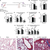
Neonatal respiratory syncytial virus (RSV) infection primes for severe lung functional defects and structural destruction upon secondary RSV infection. Neonatal male mice were infected at 7 days of age and a second RSV infection performed at 3 mo post-early-life infection (EL-RSV + Adult RSV) and compared with age-matched adult mice given a single RSV infection at 3 mo of age (Adult only RSV) and naïve mice. Analysis was then performed at 7 days post adult RSV infection. A: experimental design. BI: pulmonary function tests (PFT) were performed using plethysmography of tracheotomized mice. JL: the two middle lobes of the right lung were removed and fixed in formalin and embedded in paraffin. Five-micrometer sections were stained with trichrome to visualize collagen deposition and alveolar consolidation. Representative images shown at ×200 magnification. *P < 0.05, **P < 0.01, ***P < 0.001, ****P < 0.0001; n= 5 mice/group. dPpl, compliant pressure; dPmax, maximum pressure; FEV, forced expiratory volume; FVC, forced vital capacity; Cchord, Chord compliance; Cfvc50, compliance at 50% vital capacity. [Image A created with a licensed version of BioRender.com.]
Figure 6.
Figure 6.
Previous neonatal respiratory syncytial virus (RSV) infection leads to enhanced recruitment of inflammatory cells and mucus deposition upon reinfection with RSV 3 mo later. Neonatal male mice were infected at 7 days of age and a second RSV infection performed at 3 mo post-early-life infection (EL-RSV + Adult RSV) and compared with age-matched adult mice given a single RSV infection at 3 mo of age (Adult only RSV) and naïve mice. Analysis was then performed at 7 days post adult RSV infection. AC: the two middle lobes of the right lung were removed and fixed in formalin and embedded in paraffin. Five-micrometer sections were stained with periodic-acid Schiff (PAS) to visualize mucus deposition. Representative images shown at ×200 magnification. D: slides containing PAS-stained lung sections were blind coded and scored by an individual observer to quantify mucus on a scale of 1–4. 1 = Minimal/No Mucus; 2 = Slight: Multiple airways with goblet cell hyperplasia and mucus; 3 = Moderate: Multiple airways with significant mucus and some plugging; 4 = Severe: significant Mucus plugging. EG: the two middle lobes of the right lung were removed and fixed in formalin and embedded in paraffin. Five-micrometer sections were stained with hematoxylin & Eosin (H&E) to visualize inflammatory immune cell infiltration. Representative images shown at ×200 magnification. HK: the left lung was removed and dissociated into a single cell suspension and stained for flow cytometry analysis to quantify immune cell populations within the lung. *P < 0.05, **P < 0.01; n = 5 mice/group. DC, dendritic cells; ILC2, innate lymphoid type-2 cells.

References

    1. Welliver RC. Review of epidemiology and clinical risk factors for severe respiratory syncytial virus (RSV) infection. J Pediatr 143, 5 Suppl: S112–S117, 2003. doi:10.1067/s0022-3476(03)00508-0. - DOI - PubMed
    1. Wright M, Piedimonte G. Respiratory syncytial virus prevention and therapy: past, present, and future. Pediatr Pulmonol 46: 324–347, 2011. doi:10.1002/ppul.21377. - DOI - PubMed
    1. Lambert L, Sagfors AM, Openshaw PJ, Culley FJ. Immunity to RSV in early-life. Front Immunol 5: 466, 2014. doi:10.3389/fimmu.2014.00466. - DOI - PMC - PubMed
    1. Welliver RC. Respiratory syncytial virus and other respiratory viruses. Pediatr Infect Dis J 22, Suppl 2: S6–S10, 2003. doi:10.1097/01.inf.0000053880.92496.db. - DOI - PubMed
    1. Bont L, Kimpen JL. Immunological mechanisms of severe respiratory syncytial virus bronchiolitis. Intensive Care Med 28: 616–621, 2002. doi:10.1007/s00134-002-1256-z. - DOI - PubMed

LinkOut - more resources