

doi: 10.1093/europace/euae034.
Wall shear stress in outflow tract premature ventricular contraction location assessed through 4D-flow MRI
Affiliations
- PMID: 38290435
- PMCID: PMC10849830
- DOI: 10.1093/europace/euae034
Item in Clipboard
Wall shear stress in outflow tract premature ventricular contraction location assessed through 4D-flow MRI
Europace.
.
No abstract available
Keywords: 4D-flow MRI; Cardiac imaging; Premature ventricular contraction; Right ventricular outflow tract; Wall shear stress.
Conflict of interest statement
Conflict of interest: none declared.
Figures
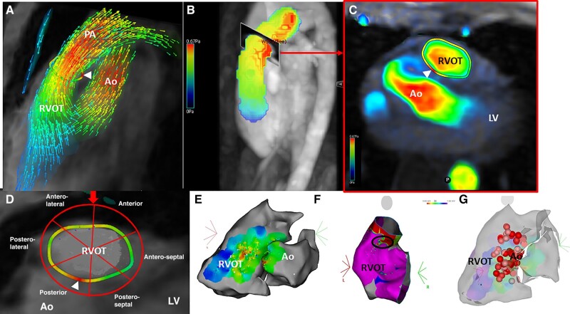
Illustration of the right ventricular ejection pathway segmentation method and correlation with electrophysiological findings. (A) Colorimetric streamline of the systolic flows in the aorta and the right ventricular ejection pathway. White arrowhead: position for the wall shear stress (WSS) analysis under the pulmonary annulus. (B) Colorimetric flow mapping in the RVOT and PA. Arrow: position for the analysis slice transverse to the main axis of the PA, under the pulmonary valve. (C) Transverse slice of the RVOT under the PA (position on B). Circumferential WSS analysis in colorimetric mapping. White arrowhead: area of relative heterogeneity of the WSS. (D) From the section obtained in C, red arrow, segmentation according to the six-segment method with the same angular arc of the subvalvular RVOT walls. Posterior adjacent to the aorta. Anterior proximal to the left internal mammary. Posterior and anterior septal on the LV side. Posterior and anterior lateral on the RV free wall side. White arrowhead: area of focal heterogeneity in WSS of the posterior–septal wall of the RVOT in this patient. (E and F) Colocalization on the same patient of the PVC focus on electrophysiological ablation maps with activation (E) and amplitude (F) map showing the common focus at the posterior–septal segment of the RVOT with the lowest EGM amplitude (black circle). (G) Final ablation map showing a colocalization with the WSS hyperpessure patch. Ao, aorta; LV, left ventricle; PA, pulmonary artery; RVOT, right ventricular outflow tract.
Similar articles
-
Magnetic resonance imaging abnormalities in the basal interventricular septum of patients with left ventricular outflow tract arrhythmias.J Cardiovasc Electrophysiol. 2019 Jul;30(7):1042-1052. doi: 10.1111/jce.13951. Epub 2019 Apr 29. J Cardiovasc Electrophysiol. 2019. PMID: 30983055
-
Altered ascending aortic wall shear stress in patients with corrected atrioventricular septal defect: a comprehensive cardiovascular magnetic resonance and 4D flow MRI evaluation.Cardiol Young. 2019 May;29(5):637-642. doi: 10.1017/S1047951119000374. Epub 2019 May 29. Cardiol Young. 2019. PMID: 31138335
-
Cardiac magnetic resonance imaging findings in patients with right ventricular outflow tract premature contractions.Eur Heart J. 1997 Dec;18(12):2002-10. doi: 10.1093/oxfordjournals.eurheartj.a015212. Eur Heart J. 1997. PMID: 9447331
-
Noninvasive Mapping of Premature Ventricular Contractions by Merging Magnetocardiography and Computed Tomography.JACC Clin Electrophysiol. 2019 Oct;5(10):1144-1157. doi: 10.1016/j.jacep.2019.06.010. Epub 2019 Jul 31. JACC Clin Electrophysiol. 2019. PMID: 31648739
-
Modern mapping and ablation of idiopathic outflow tract ventricular arrhythmias.Rev Cardiovasc Med. 2022 Mar 16;23(3):103. doi: 10.31083/j.rcm2303103. Rev Cardiovasc Med. 2022. PMID: 35345270 Review.
References
-
- von Rotz M, Aeschbacher S, Bossard M, Schoen T, Blum S, Schneider Set al. . Risk factors for premature ventricular contractions in young and healthy adults. Heart 2017;103:702–7. - PubMed
-
- Gorenek B, Fisher JD, Kudaiberdieva G, Baranchuk A, Burri H, Campbell KBet al. . Premature ventricular complexes: diagnostic and therapeutic considerations in clinical practice: a state-of-the-art review by the American College of Cardiology Electrophysiology Council. J Interv Card Electrophysiol 2020;57:5–26. - PubMed
MeSH terms
LinkOut - more resources
Full Text Sources
