. 2024 Apr:116:111069.
doi: 10.1016/j.cellsig.2024.111069. Epub 2024 Jan 28.

Transmembrane protein TMEM97 and epigenetic reader BAHCC1 constitute an axis that supports pro-inflammatory cytokine expression

Affiliations

Transmembrane protein TMEM97 and epigenetic reader BAHCC1 constitute an axis that supports pro-inflammatory cytokine expression

Jing Li et al. Cell Signal. 2024 Apr.

Abstract

Pro-inflammatory cytokine production by the retinal pigment epithelium (RPE) is a key etiology in retinal degenerative diseases, yet the underlying mechanisms are not well understood. TMEM97 is a scarcely studied transmembrane protein recently implicated in retinal degeneration. BAH domain coiled coil 1 (BAHCC1) is a newly discovered histone code reader involved in oncogenesis. A role for TMEM97 and BAHCC1 in RPE inflammation was not known. Here we found that they constitute a novel axis regulating pro-inflammatory cytokine expression in RPE cells. Transcriptomic analysis using a TMEM97-/- ARPE19 human cell line and the validation via TMEM97 loss- and gain-of-function revealed a profound role of TMEM97 in promoting the expression of pro-inflammatory cytokines, notably IL1β and CCL2, and unexpectedly BAHCC1 as well. Moreover, co-immunoprecipitation indicated an association between the TMEM97 and BAHCC1 proteins. While TMEM97 ablation decreased and its overexpression increased NFκB (p50, p52, p65), the master transcription factor for pro-inflammatory cytokines, silencing BAHCC1 down-regulated NFκB and downstream pro-inflammatory cytokines. Furthermore, in an RPE-damage retinal degeneration mouse model, immunofluorescence illustrated down-regulation of IL1β and CCL2 total proteins and suppression of glial activation in the retina of Tmem97-/- mice compared to Tmem97+/+ mice. Thus, TMEM97 is a novel determinant of pro-inflammatory cytokine expression acting via a previously unknown TMEM97- > BAHCC1- > NFκB cascade. SYNOPSIS: Retinal pigment epithelium (RPE) inflammation can lead to blindness. We identify here a previously uncharacterized cascade that underlies RPE cell production of pro-inflammatory cytokines. Specifically, transmembrane protein TMEM97 positively regulates the recently discovered histone code reader BAHCC1, which in turn enhances pro-inflammatory cytokine expression via the transcription factor NFκB.

Keywords: BAHCC1; NFκB; Pro-inflammatory cytokines; Retinal pigment epithelial cell; TMEM97.


Conflict of interest statement

Declaration of competing interest The authors have declared no competing interests and no industry or financial relationship to disclose.

Figures

Figure 1.
Figure 1.. Immunofluorescence of retinal IL1β and CCL2 is reduced in Tmem97−/− mice compared to Tmem97+/+ mice treated with NaIO3.
A and B. Representative images. Tmem97+/+ and Tmem97−/− mice received a single tail-vein injection of NaIO3 (30 mg/kg), and 3 days later the mice were euthanized for retinal cryosection preparation and immunostaining of IL1β or CCL2. RPE: Retinal pigment epithelium; ONL: outer nuclear layer; INL: inner nuclear layer; GCL: ganglion cell layer. Scale bar: 50 μm. Red: IL1β total protein in A or CCL2 total protein in B. Blue: DAPI stained nuclei. Note: The antibody cannot distinguish the pro-protein and mature protein of IL1β or CCL2, thus immunofluorescence signals the total protein level, which refers to the mix of the pro-protein and the cleaved mature form. C. Quantification: Immunostained cell area was measured using Image-J. Fluorescence intensity values from 3-4 sections were averaged for each animal, and the averages from all animals in each group were averaged again to produce a mean ± SEM; n = 3 or 4 mice. Multiple t-test: ***P < 0.001, ****P < 0.0001.
Figure 2.
Figure 2.. TMEM97 positively regulates IL1β and CCL2 expression in ARPE19 cells
TMEM97+/+ and TMEM97−/− ARPE19 cells were cultured until ~80% confluency in the normal growth medium and then incubated without or with 5 mM NaIO3. For qRT-PCR, data are presented as Mean ± SD (n = 3 repeats). For immunoblot assay, data are presented as Mean ± SEM (n = 3 independent repeat experiments). Statistics: One-way ANOVA/Tukey test; *P < 0.05, **P<0.001, ***P < 0.001 (all compared to the first value bar); #P<0.05, ##P<0.01, ###P<0.001 (pairwise comparison). mRNA and protein levels are relative units. WT: TMEM97+/+ cells. KO: TMEM97−/− cells. EV: Empty vector. OE: TMEM97 overexpression in TMEM97+/+ ARPE19 cells. A and B. Time course of the NaIO3 treatment of TMEM97+/+ and TMEM97−/− ARPE19 cells. C and D. Effect of TMEM97 loss (TMEM97−/−) or gain (overexpression) of function on mRNA levels of IL1β, IL18, and CCL2. E and F. Changes in protein levels of pro-IL1β, pro-IL18 and pro-CCL2 due to TMEM97 ablation in ARPE19 cells. The colored bands on the blots are pre-stained molecular weight markers. G. Differential changes of pro-IL1β and pro-IL18 protein levels in response to TMEM97 ablation.
Figure 3.
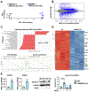
Figure 3.. Transcriptomic analysis of the impact of TMEM97 ablation in ARPE19 cells.
TMEM97+/+ (WT) and TMEM97−/− (KO) ARPE19 cells were cultured in the normal growth medium and then collected for total RNA extraction followed by bulk RNA-seq. Triplicate cell cultures of WT or KO cells were used.
  1. Data clustering. Triplicate WT cultures are labeled as CK1, CK2, and CK3. Triplicate KO cultures are labeled as of S2R1, S2R2, and S2R3.

  2. MA plot showing distribution of differentially expressed genes.

  3. Pathway analysis of the RNA-seq data. Top 20 identified KEGG pathways including the 4 upregulated and 16 downregulated are presented. GSEA: Gene set enrichment analysis. NES, normalized enrichment score.

  4. Heatmap. Presented are the down-regulated genes that are included in the top 100 differentially expressed genes with highest q values (Padj, adjusted p value). Inflammation-associated genes are labeled on the right side of the heatmap.

  5. Western blot validation of cyclin D1 (CCND1) expression. TMEM97 WT and KO ARPE19 cells were incubated without or with 5 mM NaIO3 for 24h prior to cell collection for immunoblotting. Data are presented as mean ± SEM (n = 3 independent repeat experiments). Statistics: One-way ANOVA/Tukey test; ***P<0.001. The RNA-seq data are FPKM values.
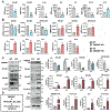
Figure 4.
Figure 4.. TMEM97 positively regulates NFκB levels in ARPE19 cells
TMEM97+/+ and TMEM97−/− ARPE19 cells cultured to ~80% confluency were used for qRT-PCR and immunoblot assays. For the former, data are presented as mean ± SD (n = 3 repeats). For the latter, data are presented as mean ± SEM (n = 3 independent repeat experiments). Statistics: Student t-test; *P < 0.05, **P<0.01, ***P < 0.001. mRNA and protein levels are relative units (r.u.). WT: TMEM97+/+ cells. KO: TMEM97−/− cells. EV: Empty vector. OE: TMEM97 overexpression in TMEM97+/+ ARPE19 cells.
  1. RNA-seq data indicating down-regulation of pro-inflammatory genes in TMEM97 KO cells. Data are presented as FPKM values. Each bar represents mean ± SEM of triplicate samples used for RNA-seq.

  2. Effect of TMEM97 loss and gain of function on the mRNA levels of NFκB genes and pro-inflammatory cytokines.

  3. Effect of TMEM97 loss and gain of function on the protein levels of NFκB family members and pro-inflammatory cytokines.

  4. Schematic depiction of the proposed TMEM97-> NFκB-> cytokines cascade.

Figure 5.
Figure 5.. TMEM97 controls the expression of BAHCC1 which in turn regulates the NFκB/cytokine pathway
TMEM97+/+ and TMEM97−/− ARPE19 cells at ~80% confluency were used for qRT-PCR and immunoblot assays. For BAHCC1 silencing, ARPE19 cells were transduced with lentivirus to express scrambled or BAHCC1-specific human shRNA. mRNA data are presented as mean ± SD (n = 3 repeats). Protein data are presented as mean ± SEM (n = 3 independent repeat experiments). Statistics: Student t-test; *P < 0.05, **P<0.01, ***P < 0.001. mRNA and protein levels are relative units (r.u.). WT: TMEM97+/+ cells. KO: TMEM97−/− cells. EV: Empty vector. OE: TMEM97 overexpression ARPE19 cell line.
  1. TMEM97 KO and OE markedly reduces and elevates BAHCC1 levels, respectively. RNA-seq data are presented as FPKM values (mean ± SEM).

  2. BAHCC1 silencing down-regulates NFκB mRNA and protein levels.

  3. BAHCC1 silencing down-regulates mRNA and protein levels or pro-inflammatory cytokines.

  4. Co-immunoprecipitation (co-IP) of BAHCC1 with TMEM97. Control (empty vector) and TMEM97 OE cell lines were cultured. IP was performed with an antibody against endogenous TMEM97 or non-specific IgG for control. Shown on the blot are 3 separate repeat experiments. GAPDH was detected with input samples.

Figure 6.
Figure 6.. IBA-1 and GFAP immunofluorescence in the mouse retina is reduced due to TMEM97 ablation.
A and B. Representative images. TMEM97+/+ and TMEM97−/− mice received a single tail-vein injection of NaIO3 (30 mg/kg). The mice were euthanized 3 and 7 days later for retinal cryosection preparation and immunofluorescence staining. RPE: retinal pigment epithelium; ONL: outer nuclear layer; INL: inner nuclear layer; GCL: ganglion cell layer. Scale bar: 50 μm. Green: IBA-1 in A or GFAP in B. Blue: DAPI stained nuclei. C. Quantification: Immunostained cell area was measured using Image-J. Fluorescence intensity values from 3-4 sections were averaged for each animal, and the averages from all animals in each group were averaged again to produce a mean ± SEM; n = 3 or 4 mice. Multiple t-test: *P<0.05, **P < 0.01, ***P < 0.001.
Figure 7.
Figure 7.. Schematic depiction of the working model of TMEM97-> BAHCC1 regulating pro-inflammatory cytokines expression.
In vitro: TMEM97 influences transcriptional activities in the nucleus through an yet-to-be-identified intermediate player that shuttles between the cytosol and the nucleus, and/or by interacting with the chromatin or chromatin associated proteins such as BAHCC1. In vivo: TMEM97 ablation blunts the activation of Muller glia and microglia in the retina of oxidant-treated mice.

References

    1. Handa JT, Bowes Rickman C, Dick AD, Gorin MB, Miller JW, Toth CA, Ueffing M, Zarbin M and Farrer LA. A systems biology approach towards understanding and treating non-neovascular age-related macular degeneration. Nature communications. 2019;10:3347. - PMC - PubMed
    1. Holtkamp GM, Kijlstra A, Peek R and de Vos AF. Retinal pigment epithelium-immune system interactions: cytokine production and cytokine-induced changes. Prog Retin Eye Res. 2001;20:29–48. - PubMed
    1. Wright CB and Ambati J. Dry Age-Related Macular Degeneration Pharmacology. Handb Exp Pharmacol. 2017;242:321–336. - PMC - PubMed
    1. Datta S, Cano M, Ebrahimi K, Wang L and Handa JT. The impact of oxidative stress and inflammation on RPE degeneration in non-neovascular AMD. Prog Retin Eye Res. 2017;60:201–218. - PMC - PubMed
    1. Nazari H, Zhang L, Zhu D, Chader GJ, Falabella P, Stefanini F, Rowland T, Clegg DO, Kashani AH, Hinton DR and Humayun MS. Stem cell based therapies for age-related macular degeneration: The promises and the challenges. Prog Retin Eye Res. 2015;48:1–39. - PubMed

Publication types