Mycosynthesis of selenium nanoparticles using Penicillium tardochrysogenum as a therapeutic agent and their combination with infrared irradiation against Ehrlich carcinoma
- PMID: 38291218
- PMCID: PMC10827740
- DOI: 10.1038/s41598-024-52982-9
Mycosynthesis of selenium nanoparticles using Penicillium tardochrysogenum as a therapeutic agent and their combination with infrared irradiation against Ehrlich carcinoma
Abstract
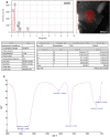
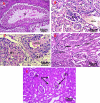
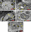
Over the past years, the assessment of myco-fabricated selenium nanoparticles (SeNPs) properties, is still in its infancy. Herein, we have highly stable myco-synthesized SeNPs using molecularly identified soil-isolated fungus; Penicillium tardochrysogenum OR059437; (PeSeNPs) were clarified via TEM, EDX, UV-Vis spectrophotometer, FTIR and zeta potential. The therapeutic efficacy profile will be determined, these crystalline PeSeNPs were examined for antioxidant, antimicrobial, MIC, and anticancer potentials, indicating that, PeSeNPs have antioxidant activity of (IC50, 109.11 μg/mL) using DPPH free radical scavenging assay. Also, PeSeNPs possess antimicrobial potential against Penicillium italicum RCMB 001,018 (1) IMI 193,019, Methicillin-Resistant Staphylococcus aureus (MRSA) ATCC 4330 and Porphyromonas gingivalis RCMB 022,001 (1) EMCC 1699; with I.Z. diameters and MIC; 16 ± 0.5 mm and MIC 500 µg/ml, 11.9 ± 0.6 mm, 500 µg/ml and 15.9±0.6 mm, 1000 µg/ml, respectively. Additionally, TEM micrographs were taken for P. italicum treated with PeSeNPs, demonstrating the destruction of hyphal membrane and internal organelles integrity, pores formation, and cell death. PeSeNP alone in vivo and combined with a near-infrared physiotherapy lamp with an energy intensity of 140 mW/cm2 showed a strong therapeutic effect against cancer cells. Thus, PeSeNPs represent anticancer agents and a suitable photothermal option for treating different kinds of cancer cells with lower toxicity and higher efficiency than normal cells. The combination therapy showed a very large and significant reduction in tumor volume, the tumor cells showed large necrosis, shrank, and disappeared. There was also improvement in liver ultrastructure, liver enzymes, and histology, as well as renal function, urea, and creatinine.
© 2024. The Author(s).
Conflict of interest statement
The authors declare no competing interests.
Figures


Similar articles
-
Selenium Nanoparticles as Candidates for Antibacterial Substitutes and Supplements against Multidrug-Resistant Bacteria.Biomolecules. 2021 Jul 14;11(7):1028. doi: 10.3390/biom11071028. Biomolecules. 2021. PMID: 34356651 Free PMC article.
-
Comparative Study of Antimicrobial and Antioxidant Potential of Olea ferruginea Fruit Extract and Its Mediated Selenium Nanoparticles.Molecules. 2022 Aug 15;27(16):5194. doi: 10.3390/molecules27165194. Molecules. 2022. PMID: 36014433 Free PMC article.
-
Exploring the antimicrobial, antioxidant, anticancer, biocompatibility, and larvicidal activities of selenium nanoparticles fabricated by endophytic fungal strain Penicillium verhagenii.Sci Rep. 2023 Jun 3;13(1):9054. doi: 10.1038/s41598-023-35360-9. Sci Rep. 2023. PMID: 37270596 Free PMC article.
-
Green and ecofriendly biosynthesis of selenium nanoparticles using Urtica dioica (stinging nettle) leaf extract: Antimicrobial and anticancer activity.Biotechnol J. 2022 Feb;17(2):e2100432. doi: 10.1002/biot.202100432. Epub 2021 Nov 21. Biotechnol J. 2022. PMID: 34747563
-
Biosynthesis of Selenium Nanoparticles (via Bacillus subtilis BSN313), and Their Isolation, Characterization, and Bioactivities.Molecules. 2021 Sep 13;26(18):5559. doi: 10.3390/molecules26185559. Molecules. 2021. PMID: 34577029 Free PMC article.
Cited by
-
Colistin-Conjugated Selenium Nanoparticles: A Dual-Action Strategy Against Drug-Resistant Infections and Cancer.Pharmaceutics. 2025 Apr 24;17(5):556. doi: 10.3390/pharmaceutics17050556. Pharmaceutics. 2025. PMID: 40430850 Free PMC article.
-
Potential Applications and Risks of Supranutritional Selenium Supplementation in Metabolic Dysfunction-Associated Steatotic Liver Disease: A Critical Review.Nutrients. 2025 Jul 30;17(15):2484. doi: 10.3390/nu17152484. Nutrients. 2025. PMID: 40806069 Free PMC article. Review.
References
-
- Abo-Neima S, Ahmed AA, El-Sheekh MM, Makhlof EM. Polycladia myrica-based delivery of selenium nanoparticles in combination with radiotherapy induces potent in vitro antiviral and in vivo anticancer activities against Ehrlich ascites tumor. Frontiers in Molecular Biosciences. 2023;10:1120422. doi: 10.3389/fmolb.2023.1120422. - DOI - PMC - PubMed
-
- El-Sheekh M, Elshobary M, Ismail E, Metwally M. Application of a novel biological-nanoparticle pretreatment to Oscillatoria acuminata biomass and coculture dark fermentation for improving hydrogen production. Microbial Cell Factories. 2023;22:34. doi: 10.1186/s12934-023-02036-y. - DOI - PMC - PubMed
-
- Panda M. K., Singh Y. D., Behera R. K., Dhal N. K. (2020). “Biosynthesis of nanoparticles and their potential application in food and agricultural sector,” in Green nanoparticles, nanotechnology in the life sciences. Editors Patra J., Fraceto L., Das G., Campos E. (Cham: Springer), 213–225. Part of the Nanotechnology in the Life Sciences book series (NALIS). http://www.springer.com/series/15921
-
- Elshahawy I, Abouelnasr HM, Lashin SM, Darwesh OM. First report of Pythium aphanidermatum infecting tomato in Egypt and its control using biogenic silver nanoparticles. J. Plant Prot. Res. 2018;15:137–151. doi: 10.24425/122929. - DOI
MeSH terms
Substances
Supplementary concepts
LinkOut - more resources
Full Text Sources
Medical
Miscellaneous

