Review
. 2024 Jan 30;12(1):17.
doi: 10.1186/s40478-024-01721-2.

Potential prognostic determinants for FET::CREB fusion-positive intracranial mesenchymal tumor

Affiliations
Review

Potential prognostic determinants for FET::CREB fusion-positive intracranial mesenchymal tumor

Frank M Mezzacappa et al. Acta Neuropathol Commun. .

Abstract

Intracranial mesenchymal tumor (IMT), FET::CREB fusion-positive is a provisional tumor type in the 2021 WHO classification of central nervous system tumors with limited information available. Herein, we describe five new IMT cases from four females and one male with three harboring an EWSR1::CREM fusion and two featuring an EWSR1::ATF1 fusion. Uniform manifold approximation and projection of DNA methylation array data placed two cases to the methylation class "IMT, subclass B", one to "meningioma-benign" and one to "meningioma-intermediate". A literature review identified 74 cases of IMTs (current five cases included) with a median age of 23 years (range 4-79 years) and a slight female predominance (female/male ratio = 1.55). Among the confirmed fusions, 25 (33.8%) featured an EWSR1::ATF1 fusion, 24 (32.4%) EWSR1::CREB1, 23 (31.1%) EWSR1::CREM, one (1.4%) FUS::CREM, and one (1.4%) EWSR1::CREB3L3. Among 66 patients with follow-up information available (median: 17 months; range: 1-158 months), 26 (39.4%) experienced progression/recurrences (median 10.5 months; range 0-120 months). Ultimately, three patients died of disease, all of whom underwent a subtotal resection for an EWSR1::ATF1 fusion-positive tumor. Outcome analysis revealed subtotal resection as an independent factor associated with a significantly shorter progression free survival (PFS; median: 12 months) compared with gross total resection (median: 60 months; p < 0.001). A younger age (< 14 years) was associated with a shorter PFS (median: 9 months) compared with an older age (median: 49 months; p < 0.05). Infratentorial location was associated with a shorter overall survival compared with supratentorial (p < 0.05). In addition, the EWSR1::ATF1 fusion appeared to be associated with a shorter overall survival compared with the other fusions (p < 0.05). In conclusion, IMT is a locally aggressive tumor with a high recurrence rate. Potential risk factors include subtotal resection, younger age, infratentorial location, and possibly EWSR1::ATF1 fusion. Larger case series are needed to better define prognostic determinants in these tumors.

Keywords: FET::CREB fusion; Fluorescence in situ hybridization; Genome-wide DNA methylation profiling; Intracranial mesenchymal tumor; Next-generation sequencing.


Conflict of interest statement

The authors have no competing interests to declare.

Figures

Fig. 1
Fig. 1
Representative images of MRI T1 post contrast (T1 + C), T2 FLAIR, H&E, and immunohistochemical stains for Cases 1–5. On MRI imaging, all five tumors were supratentorial, well-circumscribed, avidly enhancing lesions with surrounding vasogenic edema; however, on H&E stained sections, they showed heterogenous morphology. Cases 1 and 2 were composed of spindled/stellate tumor cells with a dense lymphocytic infiltrate, while Cases 3 and 4 featured sheets of epithelioid cells and highly-packed small round blue cells, respectively. Case 5 was distinct for its relatively low cellularity and a prominent myxoid stroma. By immunohistochemistry, desmin was positive in all cases except Case 5, and all five cases showed at least focal positivity for EMA
Fig. 2
Fig. 2
Representative images of fluorescence in situ hybridization for Cases 1, 2, 3, and 5. Yellow arrows indicate fusions. EWSR1::CREM fusion was detected in Cases 1 and 3 and EWSR1::ATF1 in Cases 2 and 5
Fig. 3
Fig. 3
UMAP embedding of DNA methylation array data for Cases 1, 2, 3, and 4. Unsupervised clustering was performed on four samples using the NCI reference set (n = 7467) consisting of 198 classes. Cases 1 and 2 were placed to the methylation class “intracranial mesenchymal tumor subclass B (ICMT_B)”, Case 3 to “meningioma subclass benign_3 (MNG_BEN_3)”, and Case 4 to “meningioma subclass intermediate_A (MNG_INT_A)”
Fig. 4
Fig. 4
Age, sex, location, and fusion partners of the 74 cases of IMT reported to date with the five new cases included. The tumors predominantly occurred in children and young adults (median age 23 years old; A with a female predominance (F:M = 1.56; B Fifty-nine (79.7%) tumors were supratentorial, 12 (16.2%) were infratentorial, two (2.7%) were at the tentorium with both supratentorial and infratentorial extension, and the location of the remaining one (1.4%) was unclear (C). Among the 74 tumors with documented fusions, 25 (33.8%) featured an EWSR1::ATF1 fusion, 24 (32.4%) EWSR1::CREB1, 23 (31.1%) EWSR1::CREM, one (1.4%) EWSR1::CREB3L3, and the last one (1.4%) FUS::CREM fusion (D)
Fig. 5
Fig. 5
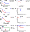
Kaplan–Meier analysis of the 66 IMT cases with available outcome data based on extent of resection, age, tumor location, and fusion partners. A, B Patients who underwent subtotal resection (STR) had a significantly shorter PFS and OS compared with patients who underwent gross total resection (GTR; p = 0.0003 and 0.0346, respectively). C, D Patients younger than 14 years old showed a shorter PFS (p = 0.0218) but not OS (p = 6738) compared with patients of or older than 14 years. E, F Patients with infratentorial tumors did not demonstrate a shorter PFS (p = 0.8507) but did show a shorter OS (p = 0.0345) compared with patients with supratentorial tumors. G, H No significant difference in PFS (p = 0.1766) was observed among tumors with different fusion partners; however, patients with EWSR1::ATF1 fused tumors had a shorter OS (p = 0.0247) compared with patients with tumors harboring other fusions
Fig. 6
Fig. 6
Multivariate Cox regression analysis of the 66 IMT cases with available outcome data. Subtotal resection (STR; HR: 5.6, confidence interval: 2.3–13.6, p < 0.001) and a young age < 14 years (HR: 3.3, confidence interval: 1.4–7.7, p = 0.006) remained as independent risk factors leading to a shorter progression free survival

References

    1. Acosta AM, Bridge JA, Dal Cin PS, Sholl LM, Cornejo KM, Fletcher CDM, Ulbright TM. Inflammatory and nested testicular sex cord tumor: a novel neoplasm with aggressive clinical behavior and frequent EWSR1::ATF1 gene fusions. Am J Surg Pathol. 2023;47:504–517. doi: 10.1097/pas.0000000000002022. - DOI - PubMed
    1. Adams JW, Malicki D, Levy M, Crawford JR. Rare intracranial EWSR1-rearranged myxoid mesenchymal tumour in a teenager. BMJ Case Rep. 2021 doi: 10.1136/bcr-2021-245282. - DOI - PMC - PubMed
    1. Aghajan Y, Malicki DM, Levy ML, Crawford JR. Atypical central neurocytoma with novel EWSR1-ATF1 fusion and MUTYH mutation detected by next-generation sequencing. BMJ Case Rep. 2019 doi: 10.1136/bcr-2018-226455. - DOI - PMC - PubMed
    1. Alshareef MA, Almadidy Z, Baker T, Perry A, Welsh CT, Vandergrift WA., 3rd Intracranial angiomatoid fibrous histiocytoma: case report and literature review. World Neurosurg. 2016;96:403–409. doi: 10.1016/j.wneu.2016.09.059. - DOI - PubMed
    1. Aryee MJ, Jaffe AE, Corrada-Bravo H, Ladd-Acosta C, Feinberg AP, Hansen KD, Irizarry RA. Minfi: a flexible and comprehensive Bioconductor package for the analysis of Infinium DNA methylation microarrays. Bioinformatics. 2014;30:1363–1369. doi: 10.1093/bioinformatics/btu049. - DOI - PMC - PubMed

Publication types

MeSH terms

Substances