A Foot in the Right Direction: Metatarsal Osteoblastoma-A Rare Case and its Management
- PMID: 38292114
- PMCID: PMC10823824
- DOI: 10.13107/jocr.2024.v14.i01.4130
A Foot in the Right Direction: Metatarsal Osteoblastoma-A Rare Case and its Management
Abstract
Introduction: Osteoblastoma is a rare, benign, bone-forming tumor accounting for <1% of all primary bone tumors. It has a predilection for the posterior elements of the spine and metaphysis and diaphysis of long bones. The occurrence of this tumor in the metatarsal region is rare. We report such the case of a metatarsal osteoblastoma which was treated with wide excision and non-vascularized fibular autograft: a reliable method of reconstruction.
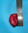
Case report: A 25-year-old woman presented with progressive pain and swelling over the right foot for 4 years. On examination, there was a gross swelling over the fourth metatarsal region over the dorsum of the foot. Radiographs revealed a osteoblastic lesion of the fourth metatarsal bone expanding into the intermetatarsal region. Magnetic resonance imaging (MRI) revealed an expansile altered signal intensity lesion which was hypointense on both T1 and T2 - weighted images with no soft-tissue component. With a working diagnosis of locally aggressive bone-forming tumor, she underwent wide excision of the tumor with reconstruction using a non-vascularized fibular autograft. Intraoperative samples sent for histopathological examination confirmed the diagnosis of osteoblastoma. After 2 years of follow-up, the patient is able to weight bear with no pain and imaging shows graft incorporation with no signs of recurrence.
Conclusion: Osteoblastoma of the metatarsal region can present a diagnostic conundrum to the treating clinician due to its rare nature. Proper evaluation and reconstruction at an early stage with wide excision and reconstruction with non-vascularized fibular autograft are a reliable treatment option.
Keywords: Osteoblastoma; fibular autograft; metatarsal.
Copyright: © Indian Orthopaedic Research Group.
Conflict of interest statement
Conflict of Interest: Nil
Figures


References
-
- Jaffe HL. Benign osteoblastoma. Bull Hosp Joint Dis. 1956;17:141–51. - PubMed
-
- McLeod RA, Dahlin DC, Beabout JW. The spectrum of osteoblastoma. Am J Roentgenol. 1976;126:321–5. - PubMed
-
- Kroon H, Schurmans J. Osteoblastoma:Clinical and radiologic findings in 98 new cases. Radiology. 1990;175:783–90. - PubMed
-
- Jaffe HL, Mayer L. An osteoblastic osteoid tissue-forming tumor of a metacarpal bone. Arch Surg. 1932;24:550–64.
-
- Lichtenstein L. Benign osteoblastoma;A category of osteoid-and bone-forming tumors other than classical osteoid osteoma, which may be mistaken for giant-cell tumor or osteogenic sarcoma. Cancer. 1956;9:1044–52. - PubMed
Publication types
LinkOut - more resources
Full Text Sources
