

Imaging features of mucinous carcinoma arising from mature teratoma showing cytokeratin 7+ and cytokeratin 20+ expression profile: A case report
- PMID: 38292777
- PMCID: PMC10825558
- DOI: 10.1016/j.radcr.2024.01.001
Imaging features of mucinous carcinoma arising from mature teratoma showing cytokeratin 7+ and cytokeratin 20+ expression profile: A case report
Abstract
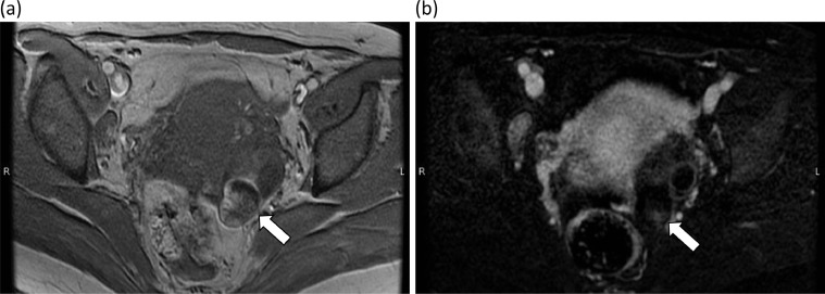
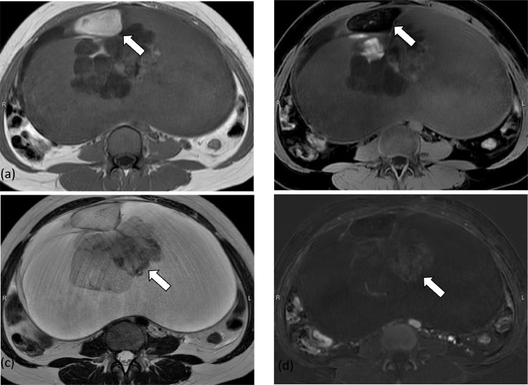
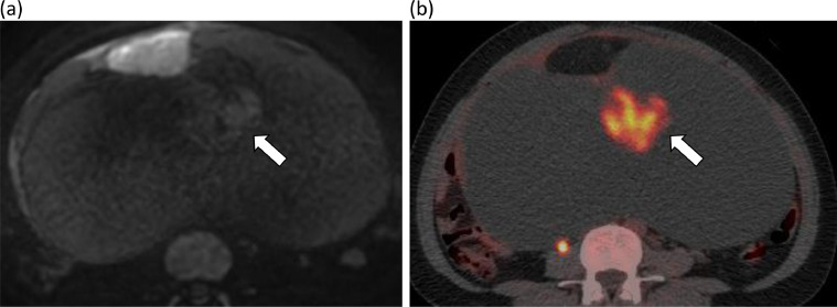
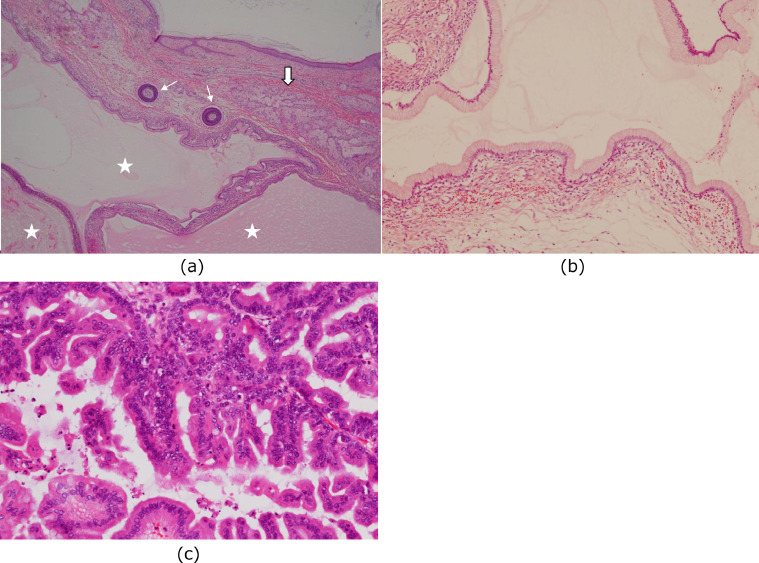
Ovarian mature teratomas are benign, but malignant transformation can occur infrequently, especially in women of advanced age. The tissue that undergoes malignant change is mostly squamous cell carcinoma, although adenocarcinoma has been reported in a small number of cases. The immunostaining results of adenocarcinoma usually show a cytokeratin (CK)7-/CK20+ expression profile, corresponding to lower gastrointestinal tract origin. In this report, we describe a case of mucinous carcinoma arising from an ovarian mature teratoma showing a CK7+/CK20+ profile and discuss its imaging features. A 40-year-old woman presented to her primary care physician with abdominal distension and poor oral intake, and she was referred to our hospital. She had been diagnosed with an ovarian mature teratoma at our institution 3 years earlier. At the current presentation, pelvic magnetic resonance imaging showed a large multilocular cystic mass with adipose tissue extending into the upper abdomen. Densely packed cysts were observed inside the mass, which showed weak contrast enhancement on contrast-enhanced imaging and a mildly high signal on diffusion-weighted imaging. A portion of the cysts also showed abnormal 18F-fluorodeoxyglucose uptake (maximum standardized uptake value, 13.2) on positron emission tomography/computed tomography. The patient was subsequently diagnosed with mucinous carcinoma showing a CK7+/CK20+ profile arising from a mature teratoma by pathologic examination. This mucinous carcinoma arising from a mature teratoma showed a CK7+/CK20+ profile and took the form of densely packed multilocular cysts. In this respect, it was similar to primary ovarian epithelial mucinous carcinoma on both magnetic resonance imaging and pathologic examination despite showing a much higher maximum standardized uptake value than that of primary ovarian mucinous carcinoma. When a large ovarian teratoma contains a large multilocular cyst, the presence of densely packed multilocular cysts should not be missed even in a mass without solid components. Clinicians should consider the possibility of mucinous carcinoma showing a CK7+/CK20+ profile arising from a mature teratoma in such cases.
Keywords: Adenocarcinoma; MRI; Mucinous; Ovary; PET-CT scan; Teratoma.
© 2024 The Authors. Published by Elsevier Inc. on behalf of University of Washington.
Figures
References
-
- Shaaban AM, Menias CO, Rezvani M, Tubay MS, El Sayed RF, Woodward PJ. 2nd ed. Elsevier; Philadelphia: 2015. Gynecology.
-
- Ueda G, Fujita M, Ogawa H, Sawada M, Inoue M, Tanizawa O. Adenocarcinoma in a benign cystic teratoma of the ovary: report of a case with a long survival period. Gynecol Oncol. 1993;48(2):259–263. - PubMed
-
- Kikkawa F, Nawa A, Tamakoshi K, Ishikawa H, Kuzuya K, Suganuma N, et al. Diagnosis of squamous cell carcinoma arising from mature cystic teratoma of the ovary. Cancer. 1998;82(11):2249–2255. - PubMed
-
- Mori Y, Nishii H, Takabe K, Shinozaki H, Matsumoto N, Suzuki K, et al. Preoperative diagnosis of malignant transformation arising from mature cystic teratoma of the ovary. Gynecol Oncol. 2003;90(2):338–341. - PubMed
Publication types
LinkOut - more resources
Full Text Sources
Research Materials
