[Preprint]. 2024 Jan 21:2024.01.20.576458.
doi: 10.1101/2024.01.20.576458.

Lecanemab Blocks the Effects of the Aβ/Fibrinogen Complex on Blood Clots and Synapse Toxicity in Organotypic Culture

Affiliations

Lecanemab Blocks the Effects of the Aβ/Fibrinogen Complex on Blood Clots and Synapse Toxicity in Organotypic Culture

Pradeep Kumar Singh et al. bioRxiv. .

Update in

Abstract

Proteinaceous brain inclusions, neuroinflammation, and vascular dysfunction are common pathologies in Alzheimer's disease (AD). Vascular deficits include a compromised blood-brain barrier, which can lead to extravasation of blood proteins like fibrinogen into the brain. Fibrinogen's interaction with the amyloid-beta (Aβ) peptide is known to worsen thrombotic and cerebrovascular pathways in AD. Lecanemab, an FDA-approved antibody therapy for AD, shows promising results in facilitating reduction of Aβ from the brain and slowing cognitive decline. Here we show that lecanemab blocks fibrinogen's binding to Aβ protofibrils, normalizing Aβ/fibrinogen-mediated delayed fibrinolysis and clot abnormalities in vitro and in human plasma. Additionally, we show that lecanemab dissociates the Aβ/fibrinogen complex and prevents fibrinogen from exacerbating Aβ-induced synaptotoxicity in mouse organotypic hippocampal cultures. These findings reveal a possible protective mechanism by which lecanemab may slow disease progression in AD.

Keywords: ARIA; Alzheimer’s disease; Biological Sciences/Neuroscience; NAS section: Biochemistry; amyloid-beta; fibrinogen; fibrinolysis; lecanemab.


Conflict of interest statement

Competing Interests: The authors have no competing interests.

Figures
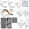
Figure 1.
Figure 1.. Lecanemab restores Aβ42 protofibril-induced delayed fibrinolysis and clot abnormalities by inhibiting the Aβ42/fibrinogen interaction in vitro.
(A) Biotinylated Aβ42 protofibrils (B-Aβ42) bind to fibrinogen-coated wells. (B) Lecanemab dose-dependently blocked the binding of B-Aβ42 to fibrinogen, while human IgG control (IgG) had no effect. (C) Quantification of B (at equimolar ratio). A-C: Data from 3 independent experiments, n=8-16/group. (D) Transmission electron micrograph of Aβ42 protofibrils. Scale bar, 200 nm. (E) Turbidity assay shows clotting and clot lysis phases. Lecanemab, but not control IgG, corrected the Aβ42-induced delayed fibrinolysis. (F) Quantification of clot lysis rate in E (slopes between vertical dotted lines). (G) Quantification of maximum clot turbidity in E. E-G: Data from 6 independent experiments, n=6/group. (H) Representative scanning electron micrographs of fibrin clots from purified fibrinogen with different treatments. Scale bar, 10 μm. (I, J) Quantification of fibrin diameter and total area of abnormal fibrin clumps/clusters in clot images. Data from three independent experiments. Vehicle constitutes PBS+DMSO. Comparisons among multiple groups were performed using one-way ANOVA followed by Newman-Keuls multiple comparison test. Data are presented as mean ± SEM. ****p<0.0001, ***p<0.001; ns, not significant.
Figure 2.
Figure 2.. Lecanemab blocks Aβ42 protofibril-induced clot abnormalities and delays fibrinolysis in normal human plasma (NHP) and Aβ42/fibrinogen-mediated synaptotoxicity in mouse organotypic hippocampal culture (OHC).
(A) Biotinylated Aβ protofibrils (B-Aβ42) immunoprecipitated fibrinogen from NHP but did not in the presence of lecanemab. Control IgG had no effect. (B) Quantification of A. Data from 3 independent experiments; n=8/group. (C) Clotting and fibrinolysis in NHP using turbidity assay. Lecanemab restored the Aβ42-induced delayed fibrinolysis in NHP. (D) Quantification of clot lysis rate in C (slopes between vertical dotted lines). (E) Quantification of maximum clot turbidity in C. C-E: Data from 7 experiments, n=7/group. (F) Representative scanning electron micrographs of clots formed from NHP with different treatments. Scale bar, 5 μm. (G, H) Analyses of scanning electron micrographs showing quantifications of fibrin diameter and total area of abnormal fibrin clumps/clusters. Data from 3 independent experiments. (I) Western blotting shows Aβ42+fibrinogen treatment reduced SYP and PSD- 95 levels in OHC. However, in the presence of lecanemab, but not control IgG, the Aβ42/fibrinogen- mediated reduction in PSD-95 and SYP was minimized. (J, K) Quantification of SYP and PSD-95. Data from 4 experiments. Changes in synaptic markers were not due to cell death as determined by propidium iodide staining. (L) B-Aβ42 was added to NHP and incubated for one hour to form complexes with fibrinogen as shown in A. Lecanemab, buffer, or control IgG was then added. Western blot analysis of immunoprecipitation shows that lecanemab dissociated Aβ42/fibrinogen complexes. (M) Quantification of L. Data is from 3 independent experiments. (N) Western blotting shows preformed [Aβ42+fibrinogen] complex treatment reduced SYP and PSD-95 levels in OHC. However, treatment of preformed [Aβ42+fibrinogen] complex with lecanemab mitigated its synaptotoxicity. (O, P) Quantification of SYP and PSD-95. Data from 3 independent experiments. Vehicle constitutes PBS+DMSO. Comparisons among multiple groups were performed using one-way ANOVA followed by Newman-Keuls multiple comparison test. Data are presented as mean ± SEM. ****p<0.0001, ***p<0.001, **p<0.01; ns, not significant).

References

    1. Strickland S., Blood will out: vascular contributions to Alzheimer’s disease. J Clin Invest 128, 556–563 (2018). - PMC - PubMed
    1. Karran E., De Strooper B., The amyloid hypothesis in Alzheimer disease: new insights from new therapeutics. Nat Rev Drug Discov 21, 306–318 (2022). - PubMed
    1. Ryu J. K., McLarnon J. G., A leaky blood-brain barrier, fibrinogen infiltration and microglial reactivity in inflamed Alzheimer’s disease brain. J Cell Mol Med 13, 2911–2925 (2009). - PMC - PubMed
    1. Merlini M. et al. Fibrinogen Induces Microglia-Mediated Spine Elimination and Cognitive Impairment in an Alzheimer’s Disease Model. Neuron 101, 1099–1108 e1096 (2019). - PMC - PubMed
    1. Zamolodchikov D. et al. Biochemical and structural analysis of the interaction between beta- amyloid and fibrinogen. Blood 128, 1144–1151 (2016). - PMC - PubMed

Publication types