[Preprint]. 2024 Feb 2:2024.01.16.575894.
doi: 10.1101/2024.01.16.575894.

MAIT Cells Modulate Innate Immune Cells and Inhibit Colon Cancer Growth

Affiliations

MAIT Cells Modulate Innate Immune Cells and Inhibit Colon Cancer Growth

Olivia J Cheng et al. bioRxiv. .

Update in

Abstract

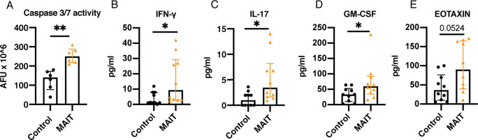
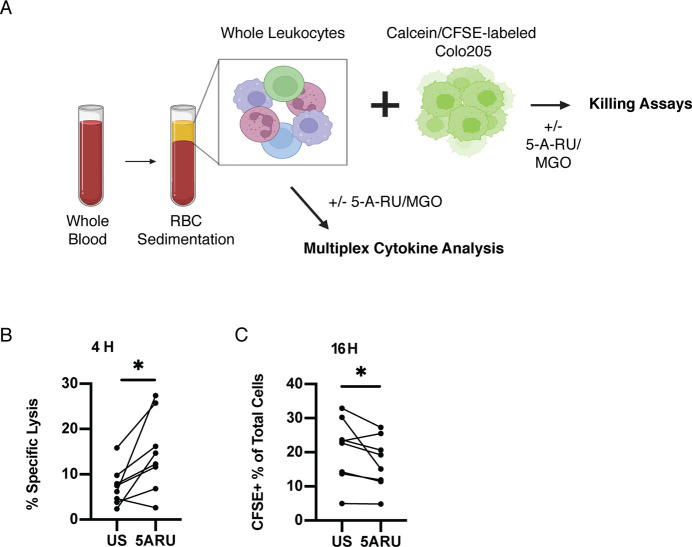
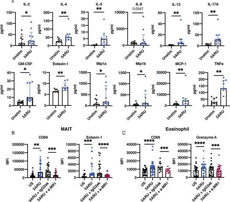
Mucosal-associated invariant T (MAIT) cells are innate-like T cells that can be activated by microbial antigens and cytokines and are abundant in mucosal tissues including the colon. MAIT cells have cytotoxic and pro-inflammatory functions and have potentials for use as adoptive cell therapy. However, studies into their anti-cancer activity, including their role in colon cancer, are limited. Using an animal model of colon cancer, we show that peritumoral injection of in vivo-expanded MAIT cells into RAG1-/- mice with MC38-derived tumors inhibits tumor growth compared to control. Multiplex cytokine analyses show that tumors from the MAIT cell-treated group have higher expression of markers for eosinophil-activating cytokines, suggesting an association between eosinophil recruitment and tumor inhibition. In a human peripheral leukocyte co-culture model, we show that leukocytes stimulated with MAIT ligand show an increase in eotaxin-1 production and activation of eosinophils, associated with increased cancer cell killing. In conclusion, we show that MAIT cells have a protective role in a murine colon cancer model, associated with modulation of the immune response to cancer, potentially involving eosinophil-associated mechanisms. Our results highlight the potential of MAIT cells for non-donor restricted colon cancer immunotherapy.


Conflict of interest statement

The authors have declared that no conflict of interest exists.

Figures

Figure 1.
Figure 1.. MAIT cell peritumoral injection leads to inhibition of MC38 cells-derived tumor growth.
A) Experimental setup of MAIT cell expansion in C57BL/6J mice and MAIT cell peritumoral injection in MC38-bearing RAG1−/− mice. MAIT cells were expanded in C57BL/6J mice for 7 days and sorted MAIT cells were injected peritumorally in RAG1−/− mice 1 day after MC38 cell injection. Control: n = 11 males. MAIT: n = 10 (9 males and 1 female). Tumor volume was monitored during the course of the experiment. Tumors were extracted at the end of the experiment for weight measurement and Luminex multiplex analysis. B) MC38 cells-derived tumor volume in mm3 over time from 2 independent experiments. C-D) Tumor measurements. Each dot represents a tumor sample and data is shown as median with interquartile range. C) Volume of extracted tumors in mm3 and D) Weight of extracted tumors in mg. *p<0.05 by two-tailed Mann-Whitney U test. E) Representative photos of extracted tumors from each experimental group. Figure created using BioRender.com
Figure 2.
Figure 2.. MAIT cell peritumoral injection induces changes in cytokines produced within tumors.
Comparison of (A) caspase activity and (B – E) concentration of various cytokines and chemokines released from extracted tumors from Figure 1 between control group and MAIT group. A) Caspase activity is measured in arbitrary fluorescent unit (AFU). B) GM-CSF, C) eotaxin, D) IFN-γ, and E) IL-17. 25 μL of supernatants from 18-hour culture of 8 mg tumor pieces were analyzed using Luminex multiplex assay. Each dot represents an individual sample of supernatant. Data was collected from 2 independent experiments and is shown as median with interquartile range. *p<0.05 **p<0.01 by two-tailed Mann-Whitney U test.
Figure 3.
Figure 3.. Activation of MAIT cells enhance killing against colon cancer cells.
A) Experimental setup of human whole leukocyte isolation and killing assay co-culture with COLO 205. B) Percent specific lysis of COLO 205 co-cultured with human whole leukocytes with or without 5-A-RU/MGO stimulation using a 4-hour Calcein Release Assay. C) Frequency of CFSE+ cells after overnight co-culture of human whole leukocytes with COLO 205 with or without 5-A-RU/MGO stimulation for 16 hours by flow cytometry. Each dot represents a sample from an individual healthy donor and data was collected from 2 independent experiments. Figure created using BioRender.com. *p<0.05 by Wilcoxon ranked test.
Figure 4.
Figure 4.. Stimulation of human peripheral leukocytes with MAIT ligand induces eotaxin-1 production in MAIT cells and activation of eosinophils in an MR-1 dependent manner.
A) Supernatant concentration of various cytokines and chemokines detected in overnight cultures of human whole leukocytes with or without 5-A-RU/MGO stimulation analyzed by Luminex multiplex assay. Each dot represents an individual sample of supernatant from a healthy donor. Data was collected from 2 independent experiment and is shown as median with interquartile range. B) Flow cytometry analysis of CD69 and eotaxin-1 expression in MAIT cells. C) Flow cytometry analysis of CD69 and granzyme A expression in eosinophils. (B and C) Expression of each marker is shown as MFI (median fluorescence intensity). Each dot represents a sample from an individual healthy donor. Data was collected from 4 independent experiments. **p<0.01 *** p<0.001 **** p<0.0001 by Wilcoxon ranked test.

Similar articles

References

    1. Treiner E, Duban L, Bahram S, Radosavljevic R, Wanner V, Tilloy F, Affaticati P, Gilfillan S, Lantz O. Selection of evolutionarily conserved mucosal-associated invariant T cells by MR1. Nature. 2003;422. - PubMed
    1. Le Bourhis L, Martin E, Peguillet I, Guihot A, Froux N, Core M, Levy E, Dusseaux M, Meyssonnier V, Premel V, Ngo C, Riteau B, Duban L, Robert D, Huang S, Rottman M, Soudais C, Lantz O. Antimicrobial activity of mucosal-associated invariant T cells. Nat Immunol. 2010;11(8):701–8. - PubMed
    1. Mak JYW, Liu L, Fairlie DP. Chemical Modulators of Mucosal Associated Invariant T Cells. Acc Chem Res. 2021;54(17):3462–75. - PMC - PubMed
    1. Hagel J, Garner L, Bilton M, Mehta H, Leng T, Hackstein C, Phalora P, Amini A, Akther H, Provine N, Edmans M, Willberg C, Klenerman P. Human MAIT Cell Activation. Methods in Moelcular Biology. 2020;2098:97–124. - PubMed
    1. Ussher JE, Bilton M, Attwod E, Shadwell J, Richardson R, de Lara C, Mettke E, Kurioka A, Hansen TH, Klenerman P, Willberg CB. CD161++ CD8+ T cells, including the MAIT cell subset, are specifically activated by IL-12+IL-18 in a TCR-independent manner. Eur J Immunol. 2014;44(1):195–203. - PMC - PubMed

Publication types