

Postmortem chest computed tomography in COVID-19: A minimally invasive autopsy method
- PMID: 38293283
- PMCID: PMC10825618
- DOI: 10.1016/j.ejro.2024.100546
Postmortem chest computed tomography in COVID-19: A minimally invasive autopsy method
Abstract
Objectives: Performing autopsies in a pandemic scenario is challenging, as the need to understand pathophysiology must be balanced with the contamination risk. A minimally invasive autopsy might be a solution. We present a model that combines radiology and pathology to evaluate postmortem CT lung findings and their correlation with histopathology.
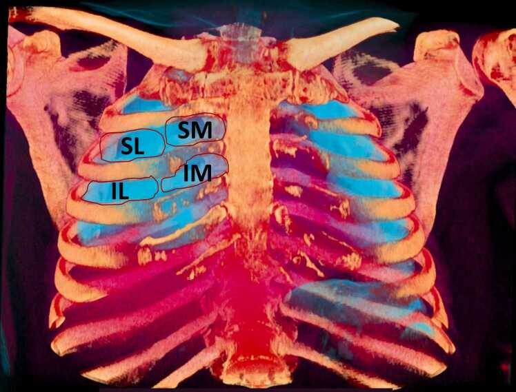
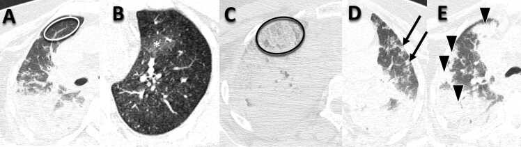
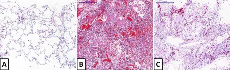
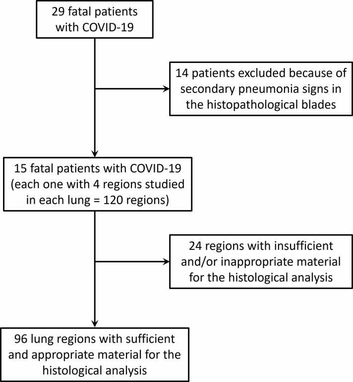
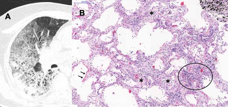
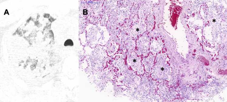
Methods: Twenty-nine patients with fatal COVID-19 underwent postmortem chest CT, and multiple lung tissue samples were collected. The chest CT scans were analyzed and quantified according to lung involvement in five categories: normal, ground-glass opacities, crazy-paving, small consolidations, and large or lobar consolidations. The lung tissue samples were examined and quantified in three categories: normal lung, exudative diffuse alveolar damage (DAD), and fibroproliferative DAD. A linear index was used to estimate the global severity of involvement by CT and histopathological analysis.
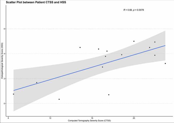
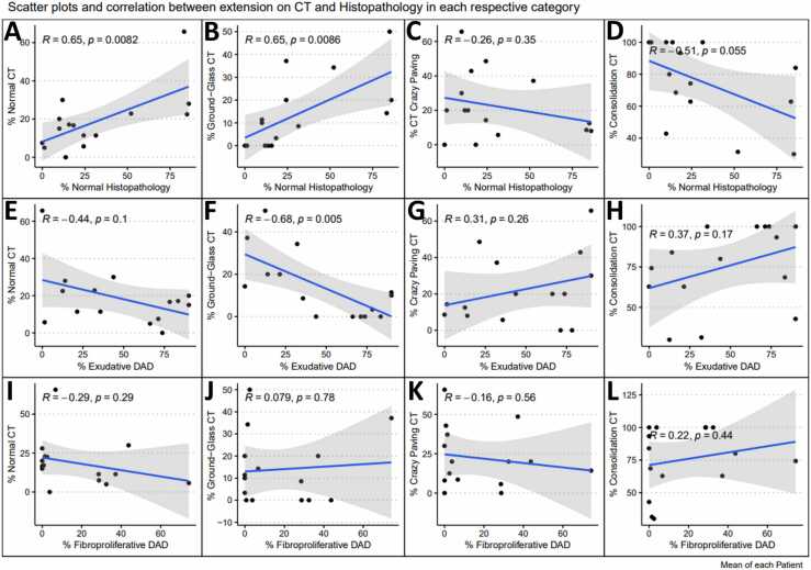
Results: There was a positive correlation between patient mean CT and histopathological severity score indexes - Pearson correlation coefficient (R) = 0.66 (p = 0.0078). When analyzing the mean lung involvement percentage of each finding, positive correlations were found between the normal lung percentage between postmortem CT and histopathology (R=0.65, p = 0.0082), as well as between ground-glass opacities in postmortem CT and normal lungs in histopathology (R=0.65, p = 0.0086), but negative correlations were observed between ground-glass opacities extension and exudative diffuse alveolar damage in histological slides (R=-0.68, p = 0.005). Additionally, it was found is a trend toward a decrease in the percentage of normal lung tissue on the histological slides as the percentage of consolidations in postmortem CT scans increased (R =-0.51, p = 0.055). The analysis of the other correlations between the percentage of each finding did not show any significant correlation or correlation trends (p ≥ 0.10).
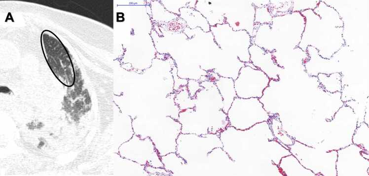
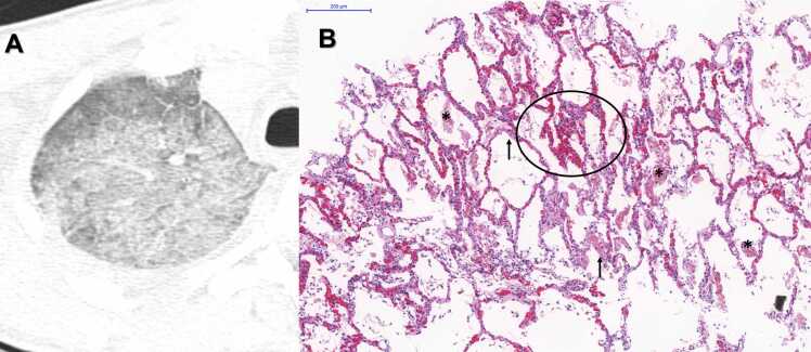
Conclusions: A minimally invasive autopsy is valid. As the severity of involvement is increased in CT, more advanced disease is seen on histopathology. However, we cannot state that one specific radiological category represents a specific pathological correspondent. Ground-glass opacities, in the postmortem stage, must be interpreted with caution, as expiratory lungs may overestimate disease.
Keywords: Autopsy; Pathology; Thorax; Tomography.
© 2024 The Authors.
Conflict of interest statement
The authors declare the following financial interests/personal relationships which may be considered as potential competing interests: Marisa Dolhnikoff reports financial support was provided by Bill and Melinda Gates Foundation. Marisa Dolhnikoff reports financial support was provided by Conselho Nacional de Desenvolvimento Científico e Tecnológico. Thais Mauad reports financial support was provided by Conselho Nacional de Desenvolvimento Científico e Tecnológico. Marisa Dolhnikoff reports financial support was provided by Fundação de Amparo a Pesquisa do Estado de São Paulo (FAPESP). Ellison Fernando Cardoso reports financial support was provided by University of Sao Paulo Hospital of Clinics. Luiz Fernando Ferraz da Silva reports financial support was provided by University of Sao Paulo Hospital of Clinics.
Figures
References
-
- Hanley B., Lucas S.B., Youd E., Swift B., Osborn M. Autopsy in suspected COVID-19 cases. J. Clin. Pathol. 2020;73(5):239–242. - PubMed
-
- CDC. Center for Prevention and Disease Control-Coronavirus Disease 2019 (COVID-19). Collection and Submission of Postmortem Specimens from Deceased Persons with Known or Suspected COVID-19 〈https://www.cdc.gov/coronavirus/2019-ncov/hcp/guidance-postmortem-speci... p. Accessed on 05–31-2020.
-
-
Sao Paulo State Secretary of Heath. SS-32 Resolution. State of Sao Paulo Official Diary - Executive Power - Seccion I20th March 2020.
-
LinkOut - more resources
Full Text Sources
Research Materials
