. 2024 Feb 20;98(2):e0197523.
doi: 10.1128/jvi.01975-23. Epub 2024 Jan 31.

Alternative translation contributes to the generation of a cytoplasmic subpopulation of the Junín virus nucleoprotein that inhibits caspase activation and innate immunity

Affiliations

Alternative translation contributes to the generation of a cytoplasmic subpopulation of the Junín virus nucleoprotein that inhibits caspase activation and innate immunity

Linus Bostedt et al. J Virol. .

Abstract

The highly pathogenic arenavirus, Junín virus (JUNV), expresses three truncated alternative isoforms of its nucleoprotein (NP), i.e., NP53kD, NP47kD, and NP40kD. While both NP47kD and NP40kD have been previously shown to be products of caspase cleavage, here, we show that expression of the third isoform NP53kD is due to alternative in-frame translation from M80. Based on this information, we were able to generate recombinant JUNVs lacking each of these isoforms. Infection with these mutants revealed that, while all three isoforms contribute to the efficient control of caspase activation, NP40kD plays the predominant role. In contrast to full-length NP (i.e., NP65kD), which is localized to inclusion bodies, where viral RNA synthesis takes place, the loss of portions of the N-terminal coiled-coil region in these isoforms leads to a diffuse cytoplasmic distribution and a loss of function in viral RNA synthesis. Nonetheless, NP53kD, NP47kD, and NP40kD all retain robust interferon antagonistic and 3'-5' exonuclease activities. We suggest that the altered localization of these NP isoforms allows them to be more efficiently targeted by activated caspases for cleavage as decoy substrates, and to be better positioned to degrade viral double-stranded (ds)RNA species that accumulate in the cytoplasm during virus infection and/or interact with cytosolic RNA sensors, thereby limiting dsRNA-mediated innate immune responses. Taken together, this work provides insight into the mechanism by which JUNV leverages apoptosis during infection to generate biologically distinct pools of NP and contributes to our understanding of the expression and biological relevance of alternative protein isoforms during virus infection.IMPORTANCEA limited coding capacity means that RNA viruses need strategies to diversify their proteome. The nucleoprotein (NP) of the highly pathogenic arenavirus Junín virus (JUNV) produces three N-terminally truncated isoforms: two (NP47kD and NP40kD) are known to be produced by caspase cleavage, while, here, we show that NP53kD is produced by alternative translation initiation. Recombinant JUNVs lacking individual NP isoforms revealed that all three isoforms contribute to inhibiting caspase activation during infection, but cleavage to generate NP40kD makes the biggest contribution. Importantly, all three isoforms retain their ability to digest double-stranded (ds)RNA and inhibit interferon promoter activation but have a diffuse cytoplasmic distribution. Given the cytoplasmic localization of both aberrant viral dsRNAs, as well as dsRNA sensors and many other cellular components of innate immune activation pathways, we suggest that the generation of NP isoforms not only contributes to evasion of apoptosis but also robust control of the antiviral response.

Keywords: Junín virus; alternative translation; arenavirus; caspase activation; exonuclease activity; interferon antagonism; nucleoprotein.


Conflict of interest statement

The authors declare no conflict of interest.

Figures

Fig 1
Fig 1
JUNV NP53kD is not the product of canonical caspase cleavage. (A) Location of putative caspase cleavage motifs within JUNV NP. Putative caspase cleavage motifs found within the region of NP expected to be associated with NP53kD formation (i.e., amino acids D30–D160) are shown underlined and with the associated aspartate (D) residue in bold. Sites for which an impact on NP53kD formation had already been examined in a previous study (20) are shown boxed, and the 160DVKD163 identified as being responsible for the production of NP47kD is shown boxed and in bold. (B) Expression of NP53kD after elimination of the putative caspase cleavage motifs. Vero76 cells were transfected with C-terminal FLAG-tagged versions of JUNV NPWT or mutants in which the indicated D residues [corresponding to the P1 position of the putative caspase sites shown in (A)] were replaced with asparagine (N). Samples were collected 96 h after transfection, and expression of NP was analyzed in Western blot using an anti-FLAG antibody. The positions of full-length NP65kD and the NP53kD isoform are indicated by arrowheads.
Fig 2
Fig 2
JUNV NP53kD is the product of alternative translation starting at position M80. (A) Schematic of the sequence region associated with NP53kD expression. The relevant region was divided into blocks of 20 amino acids (blocks 1–6) or 10 amino acids (block 7), which were substituted against a corresponding number of alanine (A) residues in the respective NP expression constructs. (B) Expression of alanine-scanning mutants of NP. Vero76 cells were transfected with constructs encoding either JUNV NPWT or JUNV NP containing the substitutions described in (A), as indicated. All constructs contained a C-terminal FLAG tag for detection. After 96 h, cell lysates were collected, and NP expression was analyzed by Western blot using an anti-FLAG antibody. Positions of full-length NP65kD and NP53kD are indicated by arrowheads. (C)Schematic of methionine residues in the region from amino acids 70 to 110 in JUNV NP. Three methionine residues in-frame with the canonical NP start codon (i.e., M1) were found within or in proximity to the region responsible for NP53kD expression (i.e., M78, M80, and M100). (D) Expression of NP with mutations in putative alternative in-frame start codons. Vero76 cells were transfected with C-terminal FLAG tag containing constructs encoding either JUNV NPWT or JUNV NP containing point mutations in the alternative in-frame methionine residues described in (C), as indicated. Samples were analyzed as described in (B).
Fig 3
Fig 3
NP isoforms show a lack of function in viral RNA synthesis. (A) Schematic overview of motifs related to viral RNA synthesis. The relative positions of key motifs within JUNV NP that are associated with viral RNA synthesis are shown. In particular, an N-terminal coiled-coil motif required for NP homo-oligomerization is shown, as are the locations of basic RNA-binding amino acids (i.e., K248–R324, S242, and Y205) required for viral RNA synthesis. The regions within the C-terminus responsible for interaction of NP with the matrix protein Z and polymerase (L) are also shown. The N-termini for the NP53kD, NP47kD, and NP40kD isoforms are indicated. (B) Ability of JUNV NP isoforms to support viral RNA synthesis. BSR-T7/5 cells were transfected with the indicated JUNV NP constructs, in combination with the other plasmids required for a minigenome assay (i.e., JUNV L, T7 polymerase, and a NanoLuc luciferase-encoding minigenome). After 72 h, lysates were collected, and reporter activity was measured. Values were normalized to Firefly luciferase activity (transfection control). Omitting NP served as a negative control (-NP). Means and standard deviations for each sample are shown based on the results of five independent experiments. Statistical analyses were based on a one-way ANOVA with Dunnett’s post hoc test, comparing each sample with the negative control (-NP) (n.s., not significant; ****, P > 0.0001). (C) Impact of JUNV NP isoforms on viral RNA synthesis mediated by full-length NP. JUNV minigenome assays were performed as described in (B) with the exception that wild type (NPWT) was expressed in addition to each of the three other NP isoforms, as indicated. Means and standard deviations are based on data from at least three independent experiments. Statistical analyses were based on a one-way ANOVA with Dunnett’s post hoc test, with each sample compared to the NPWT control (n.s., not significant; ****, P > 0.0001). (D) Subcellular localization of JUNV NP isoforms. Huh7 cells were transfected with C-terminally FLAG-tagged constructs expressing JUNV NPWT or one of the truncated NP isoforms, as indicated. After 48 h, cells were fixed and stained with an anti-FLAG antibody to visualize the distribution of NP (green). DAPI (blue) was used as a nuclear counterstain. The scale bars indicate a distance of 10 µm. (E) Impact of NP isoforms on inhibition of viral RNA synthesis by Z. JUNV minigenome assays were performed as described in (C) but with additional transfection of 1 or 2.5 ng of plasmid encoding the matrix protein (Z), as indicated. The averages and standard deviations were calculated based on at least three independent experiments. Statistical analyses were performed using a one-way ANOVA with Dunnett’s post hoc test comparing each sample to the corresponding NPWT + Z control (n.s., not significant).
Fig 4
Fig 4
NP isoforms exhibit both robust 3'−5' exonuclease activity and antagonism of IFN-β promoter activation. (A) Schematic overview of motifs related to innate immune regulation found within the C-terminal domain of NP. Regions of NP that interact with IKKε to inhibit IFN-β promoter activation, as well as residues involved in forming the 3'−5' exonuclease domain, are indicated. (B) Exonuclease activity of JUNV NP isoforms. HEK293T cells were transfected with plasmids encoding a FLAG-tagged version of TCRV NP (positive control) or a catalytically inactive mutant (E388A, negative control), JUNV NPWT or its isoforms (i.e., NP53kD, NP47kD, and NP40kD), as indicated. Cell lysates were harvested 24 h post-transfection and incubated with poly(I:C) for 24 h. RNase activity was quantified based on absorbance at 260 nm (A260nm) with subtraction of background values from empty vector (pCAGGS) transfected control samples as well as the sample-matched 0-h values. Values are shown as means and standard deviations of three independent experiments. Statistical differences to the negative control [i.e., TCRV NP (E388A) were determined by ordinary one-way ANOVA with Dunnett’s post hoc test (***, P > 0.001)]. (C) Inhibition of IFN-β promoter activation by JUNV NP isoforms. HEK293T cells were transfected with a plasmid encoding Firefly luciferase under the control of a minimal human IFN-β promotor, as well as plasmids for either Flag-tagged JUNV NPWT, or its isoforms (i.e., NP53kD, NP47kD, and NP40kD), as indicated. After 24 h, cells were either infected with Sendai virus (MOI = 200), or mock infected. At 24 h post-infection, the cells were lysed, and luciferase activity was measured. Values were normalized to Renilla luciferase activity (transfection control). Omitting NP served as a positive control (-NP). Promoter activity is shown as fold induction compared to the matched uninfected samples. Values are shown as means and standard deviations of three independent experiments. Statistical analyses of differences to the JUNV NPWT control were based on a one-way ANOVA with Dunnett’s post hoc test (n.s., not significant; ****, P > 0.0001).
Fig 5
Fig 5
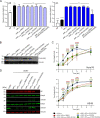
Alternative translation and caspase cleavage of JUNV NP both contribute to inhibition of apoptosis during infection. (A) Activity of NP mutants lacking specific NP isoforms in the viral lifecycle. trVLP assays were performed by transfecting BSR-T7/5 cells (p0 cells) with a bicistronic minigenome plasmid encoding the viral matrix protein Z and a glycoprotein-NanoLuc luciferase fusion. In addition, cells were transfected with plasmids encoding JUNV L, the T7 polymerase, and either JUNV NPWT or one of the JUNV NP mutants lacking specific NP isoforms, as indicated. Omission of NP (-NP) was used as a negative control in both p0 and p1 cells. Alternatively, a minigenome in which the Z open reading frame was not present (-Z) was used as a negative control for p1. Lysates of p0 cells were harvested after 72 h, and NanoLuc reporter activity was measured. Reporter levels were normalized to Firefly luciferase (transfection control). Furthermore, the supernatants of p0 cells were used to infect Huh7 cells that had been previously transfected with plasmids expressing JUNV NPWT and L. After 72 h of incubation, the p1 cells were lysed, and NanoLuc reporter activity was again determined. Means and standard deviations were calculated based on two independent experiments. One-way ANOVA with Dunnett’s post hoc test was used to determine significance relative to the NPWT positive control (n.s., not significant; **, P < 0.01; ****, P > 0.0001). (B) Expression of JUNV NP mutants containing isoform deletions. HEK293T cells were transfected with C-terminal FLAG-tagged versions of JUNV NPWT or mutants lacking specific NP isoforms, as indicated. After 96 h, cell lysates were collected, and NP expression was analyzed by Western blot using antibodies specific for FLAG or vinculin (as a loading control). (C) Growth kinetics of rJUNV mutants. Vero76 cells (IFN deficient, top panel) or A549 cells (IFN competent, bottom panel) were infected at an MOI of 0.005 using either recombinant JUNVWT or recombinant JUNV lacking specific NP isoforms, as indicated [i.e., ΔNP53kD (M80A); ΔNP53/52kD (M80L/M100A); ΔNP47kD (D160/163N); ΔNP40kD (D222N); ΔNP47/40kD (D160/163/222N)]. Supernatants were collected daily for 4 days and again after 7 days. Titers were determined by plaque assay. Data reflect the means and standard deviations of three independent experiments. Statistical analyses are based on a two-way ANOVA combined with Dunnett’s post hoc test, with comparisons to rJUNVWT at each time point (****, P > 0.0001; ***, P > 0.001; **, P < 0.01; *, P > 0.1). (D) Caspase activation during rJUNV infection. Vero76 cells were mock infected or infected (MOI = 0.005) with TCRVWT (positive control), rJUNVWT, or the indicated rJUNV mutants. After 4 days, cell lysates were analyzed for caspase activation by Western blot using antibodies specific for caspase 9, caspase 3, a cross-reactive guinea pig anti-JUNV NP antibody, or vinculin (as a loading control).
Fig 6
Fig 6
Proposed model for the biological functions of JUNV NP and its isoforms during JUNV infection. Translation of the NP mRNA primarily occurs at the canonically described AUG and leads to the production of the full-length form of NP65kD (step 1), which localizes to inclusion bodies where it participates in viral RNA synthesis. However, in some cases, the ribosome bypasses this AUG and initiates translation at a downstream in-frame start codon 238AUG240 (corresponding to the methionine at position 80), leading to the production of NP53kD (step 2). In contrast to the full-length NP65kD, NP53kD lacks domains essential for NP homo-oligomerization and, therefore, remains exclusively in the cytoplasm. Full-length NP65kD and the NP53kD isoform are both able to function as decoy substrates for cleavage by activated caspases at the motifs 160DVKD163 and 219QEHD222, and this process inhibits further caspase activation and subsequent apoptotic cell death (step 3). Caspase cleavage at these sites also gives rise to NP47kD and NP40kD. Full-length NP65kD as well as all three smaller NP isoforms contain an intact C-terminal domain, which encodes functions involved in the regulation of the innate immune response. Specifically, the 3′−5′ exonuclease activity is preserved and digests viral dsRNA, that is produced as a by-product of viral RNA synthesis during virus infection (step 4), and which has been shown to activate both RIG-I-like receptors and PKR during virus infection. Furthermore, the motifs involved in IKKε binding (preventing IRF3 activation), and thus inhibition of IFN-β upregulation, are also maintained, allowing these isoforms to serve as IFN pathway antagonists. Finally, NP40kD is also able to enter the nucleus where it may have other as-yet unknown functions and/or interactions (step 5).

Similar articles

Cited by

References

    1. Buchmeier M, Peters CJ, d.l.T.J . 2007. Arenaviridae: the viruses and their replication, p 1791–1828. In H.P. e, Knipe DM (ed), Fields Virology. Lippincott Williams & Wilkins Philadelphia.
    1. Hallam SJ, Koma T, Maruyama J, Paessler S. 2018. Review of mammarenavirus biology and replication. Front Microbiol 9:1751. doi:10.3389/fmicb.2018.01751 - DOI - PMC - PubMed
    1. Radoshitzky SR, Bào Y, Buchmeier MJ, Charrel RN, Clawson AN, Clegg CS, DeRisi JL, Emonet S, Gonzalez J-P, Kuhn JH, Lukashevich IS, Peters CJ, Romanowski V, Salvato MS, Stenglein MD, de la Torre JC. 2015. Past, present, and future of arenavirus taxonomy. Arch Virol 160:1851–1874. doi:10.1007/s00705-015-2418-y - DOI - PubMed
    1. McCormick JB, Fisher-Hoch SP. 2002. Lassa fever. Curr Top Microbiol Immunol 262:75–109. doi:10.1007/978-3-642-56029-3_4 - DOI - PubMed
    1. Paweska JT, Sewlall NH, Ksiazek TG, Blumberg LH, Hale MJ, Lipkin WI, Weyer J, Nichol ST, Rollin PE, McMullan LK, et al. . 2009. Nosocomial outbreak of novel arenavirus infection, Southern Africa. Emerg Infect Dis 15:1598–1602. doi:10.3201/eid1510.090211 - DOI - PMC - PubMed

Publication types

MeSH terms

LinkOut - more resources