Case Reports
. 2024 Sep 1;63(17):2415-2420.
doi: 10.2169/internalmedicine.2989-23. Epub 2024 Feb 1.

Acute Liver Injury and Bilateral Pulmonary Artery Thrombosis Due to Hypereosinophilic Syndrome

Affiliations
Case Reports

Acute Liver Injury and Bilateral Pulmonary Artery Thrombosis Due to Hypereosinophilic Syndrome

Keishi Ouchi et al. Intern Med. .

Abstract

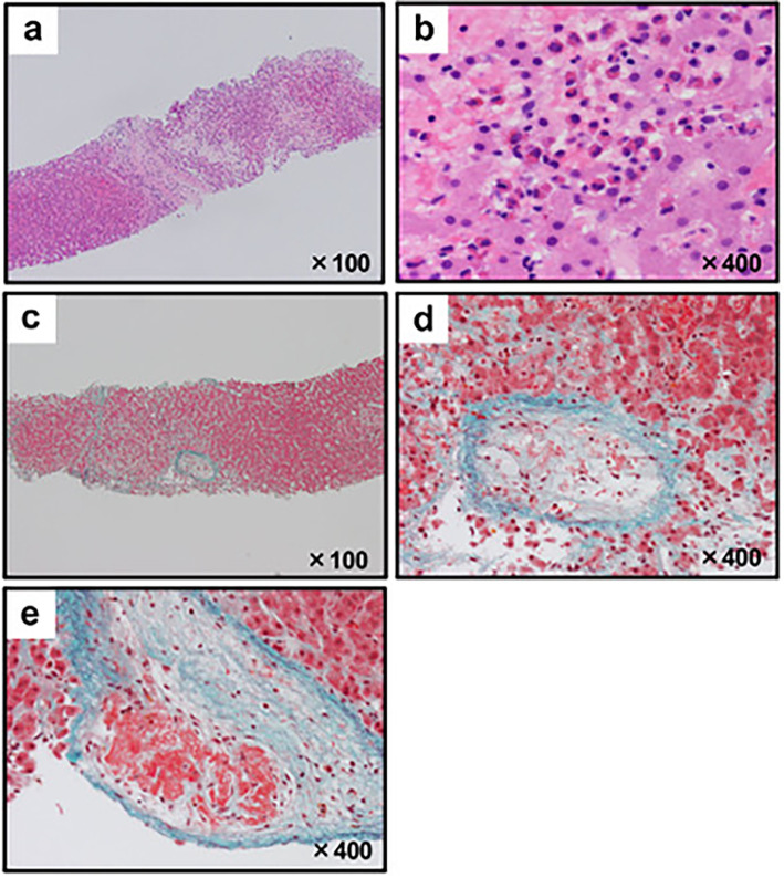
A 46-year-old Japanese man was referred to our hospital because of a marked increase in his eosinophil count (22,870/μL) and elevated liver enzyme levels. Computed tomography (CT) showed thrombi measuring approximately 8 cm in both femoral veins. A liver biopsy revealed eosinophilic infiltration, hepatocyte necrosis, fibrosis, and multiple thrombi. We suspected acute liver injury and deep vein thrombosis associated with hypereosinophilic syndrome and initiated steroids and heparin treatment. Four days after starting treatment, the patient experienced sudden chest pain and cardiopulmonary arrest. CT revealed bilateral pulmonary artery thrombosis, and despite administration of a tissue plasminogen activator, the patient died.

Keywords: acute liver injury; hypereosinophilic syndrome; pulmonary embolism.


Conflict of interest statement

The authors state that they have no Conflict of Interest (COI).

Figures

Figure 1.
Figure 1.
Images of abdominal contrast-enhanced computed tomography (a, b), esophagogastroduodenoscopy (c, d) and Hematoxylin and Eosin staining from a bone marrow puncture (e, f) on admission. (a) Atrophy of the left hepatic lobe, enlargement of the right hepatic lobe, and an extensive irregular area with contrast enhancement in the liver were found. No bile duct obstruction or periportal collar sign was observed. (b) High-density thrombi approximately 8 cm in length in both femoral veins were found (arrows). Lymphadenopathy at the hilum of liver and that from bilateral iliac regions to the groin were found. (c) No esophagitis was observed. (d) Redness and erosion of the mucosa throughout the stomach were found. A biopsy from the stomach revealed few eosinophils. (e, f) The bone marrow was slightly hyperplastic, and the morphology of the eosinophilia was normal. No numerical or morphological findings in megakaryocytes and no chromosomal abnormalities were found. (e) Low magnification. (f) High magnification.
Figure 2.
Figure 2.
Microphotographs from the liver biopsy. (a, b) Hematoxylin and Eosin staining showed the infiltration of eosinophils, ischemic changes in hepatocytes, fibrosis and loss of hepatocytes due to necrosis. (a) Low magnification. (b) High magnification. (c-e) Elastica-Masson staining with high magnification showed fibrosis and thrombus formation in several central veins. (c) Low magnification. (d, e) High magnification.
Figure 3.
Figure 3.
Computed tomography images at the onset of pulmonary embolism. Arrows indicate thrombi in both pulmonary arteries. (a) Right pulmonary artery. (b) Left pulmonary artery.
Figure 4.
Figure 4.
Clinical course during hospitalization. WBC: white blood cells, Eo: eosinophils, ALT: alanine transaminase, PSL: prednisolone

Similar articles

References

    1. Shomali W, Gotlib J. World Health Organization-defined eosinophilic disorders: 2022 update on diagnosis, risk stratification, and management. Am J Hematol 97: 129-148, 2022. - PubMed
    1. Li W, Ed. In: Leukemia. The 5(th) Edition of the World Health Organization Classification of Hematolymphoid Tumors. Exon Publications, Brisbane, 2022 [Internet]. [cited 2023 Sep 29]. Available from: https://doi.org/10.36255/exon-publications-leukemia. - PubMed
    1. Liu Y, Meng X, Feng J, Zhou X, Zhu H. Hypereosinophilia with concurrent venous thromboembolism: clinical features, potential risk factors, and short-term outcomes in a Chinese cohort. Sci Rep 10: 8359, 2020. - PMC - PubMed
    1. Kawamura T, Hiraoka A, Toshimori A, et al. . A possible case of hepatitis due to hypereosinophilic syndrome. Intern Med 55: 1453-1458, 2016. - PubMed
    1. Ogbogu PU, Rosing DR, Horne MK. Cardiovascular manifestations of hypereosinophilic syndromes. Immunol Allergy Clin 27: 457-475, 2007. - PMC - PubMed

Publication types