. 2024 Feb 1;15(1):954.
doi: 10.1038/s41467-024-44968-y.

Hydrogel dressings with intrinsic antibiofilm and antioxidative dual functionalities accelerate infected diabetic wound healing

Affiliations

Hydrogel dressings with intrinsic antibiofilm and antioxidative dual functionalities accelerate infected diabetic wound healing

Dicky Pranantyo et al. Nat Commun. .

Abstract

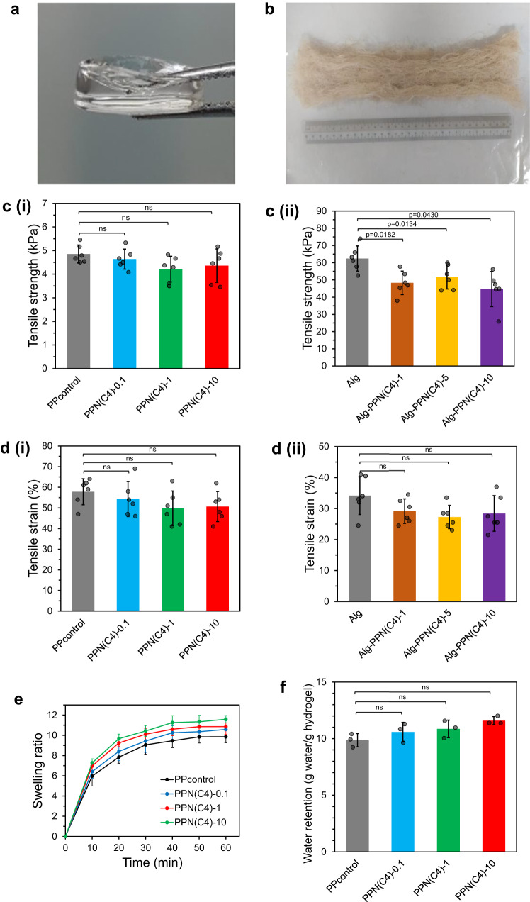
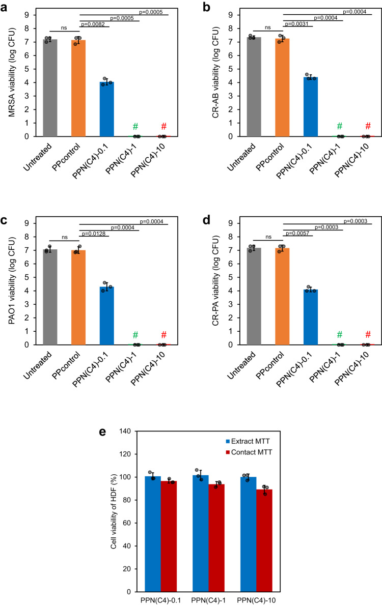
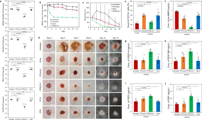
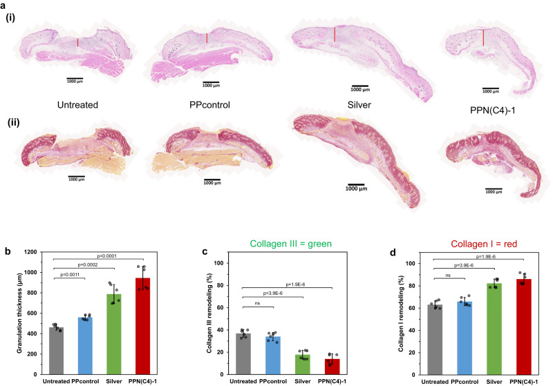
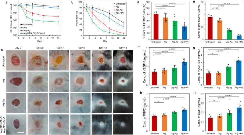
Chronic wounds are often infected with biofilm bacteria and characterized by high oxidative stress. Current dressings that promote chronic wound healing either require additional processes such as photothermal irradiation or leave behind gross amounts of undesirable residues. We report a dual-functionality hydrogel dressing with intrinsic antibiofilm and antioxidative properties that are synergistic and low-leaching. The hydrogel is a crosslinked network with tethered antibacterial cationic polyimidazolium and antioxidative N-acetylcysteine. In a murine diabetic wound model, the hydrogel accelerates the closure of wounds infected with methicillin-resistant Staphylococcus aureus or carbapenem-resistant Pseudomonas aeruginosa biofilm. Furthermore, a three-dimensional ex vivo human skin equivalent model shows that N-acetylcysteine promotes the keratinocyte differentiation and accelerates the re-epithelialization process. Our hydrogel dressing can be made into different formats for the healing of both flat and deep infected chronic wounds without contamination of the wound or needing other modalities such as photothermal irradiation.


Conflict of interest statement

D.P., C.K.Y., Y.W., X.X., N.S.T., and M.B.C.-P. have filed the Singapore provisional patents for this work under the patent numbers 10202250243U and 10202301624S. C.K.Y., S.H.M., N.S.T., and M.B.C.-P. have filed a patent for this work under the patent number US20210244846A1. D.P., C.K.Y, Y.W., X.X., S.H.M, N.S.T, and M.B.C.-P. declare no other competing interests. C.F., Y.S.Y., M.I.G.V., P.L.K.L., L.Y., P.T.H., and D.I.L. declare no competing interests.

Figures

Fig. 1
Fig. 1. Preparation schemes of the film and fiber hydrogel wound dressings.
a Structures of the main components in hydrogel film and fiber dressings. Syntheses of (b) PPN hydrogel film and (c) Alg-PPN hydrogel fiber dressings.
Fig. 2
Fig. 2. Physical characterizations of the film and fiber hydrogel wound dressings.
Visual appearance of the wound dressings investigated: (a) PPN(C4)−1 film hydrogel and (b) Alg-PPN(C8)-5 alginate fiber hydrogel. Tensile strength of the (c(i)) film hydrogels and (c(ii)) alginate fiber hydrogels (n = 6 independent experiments, two-tailed Student’s t test). Tensile strain (elongation) of the (d(i)) film hydrogels and (d(ii)) alginate fiber hydrogels (n = 6 independent experiments, two-tailed Student’s t test). e Swelling kinetics (mass increase/initial mass versus time) of the film hydrogels (n = 3 independent experiments). f Water retention capacity of the film hydrogels after 1 day immersion in water (n = 3 independent experiments, two-tailed Student’s t test). Data are presented as mean values ± SD.
Fig. 3
Fig. 3. Film hydrogels exhibited in vitro antibacterial activity and cytocompatibility.
ad Antibacterial efficacy of PPN(C4) film hydrogels assessed via contact testing for 1 h. # denotes that no bacterial colonies were observed on the agar plate (n = 3 biologically independent samples, two-tailed Student’s t test). e Cell viability of human dermal fibroblasts (HDFs) after incubation with the extracts (blue) or direct contact immersion (red) of PPN(C4) film hydrogels at 37 °C for 24 h (n = 3 cells examined over 3 independent experiments). Data are presented as mean values ± SD.
Fig. 4
Fig. 4. Ex vivo human skin equivalent (HSE) model of the film hydrogels.
a Representative images of MTT staining for wound healing. b Quantitative measurement of the MTT assay at the day 4 and day 7 time points (triplicated; n = 9 biologically independent samples, two-tailed Student’s t test, data are presented as mean values ± SEM). c Representative immunostaining images of H&E, p63, Keratin 10 (K10), and Keratin 14 (K14) after treatment with the PPN(C4)−1 hydrogel and its variants (PP-N is without PIM(C4)−1, and PPN- is without NAC). Scale bar = 100 μm.
Fig. 5
Fig. 5. Mouse in vivo diabetic wound infection model with hydrogel films treatment beginning 24 h post-infection.
a Bacterial counts of MRSA USA300, PA01, CR-AB, and CR-PA on various control and treated wounds after 24 h of treatment (n = 6 mice, two-tailed Student’s t test). bd Full wound healing study. b Bacterial counts of MRSA USA300 on untreated control, silver dressing-, PPcontrol- and PPN(C4)−1 hydrogel-treated wounds on days 0, 1, 3, 5, 7, 9, 12 and 14 post-treatment (n = 6 mice). c Wound sizes of untreated control, silver dressing-, PPcontrol- and PPN(C4)−1 hydrogel-treated wounds on various days as a percentage of the initial wound size (n = 6 mice). d Visual appearance of representative untreated control-, silver dressing-, PPcontrol- and PPN(C4)−1 hydrogel-treated wounds between dressing changes. Scale bar = 5 mm. ej Characterization of wound tissues. Measurements in MRSA USA300-infected diabetic mice (n = 6 mice, two-tailed Student’s t test) on day 2 post-treatment: e Percentage of CD11b+ cells in wounds. The percentage of CD11b+ cells is directly proportional to the extent of inflammation in the skin. f Concentration of pro-MMP9 in wounds. Concentrations of wound healing factors (g) VEGF-A, (h) PDGF-BB, (i) FGF-2 and (j) EGF in wounds. Data are presented as mean values ± SD.
Fig. 6
Fig. 6. Wound histology of diabetic mouse model treated 24 h post-infection of MRSA USA300 with hydrogel films.
a Representative images depicting (i) H&E and (ii) picrosirius red stained tissue sections of untreated control, silver dressing, PPcontrol, and PPN(C4)−1 hydrogel-treated wounds on day 7 post-treatment. Scale bar = 1 mm. b Quantification of granulation tissue formation thickness based on histological wound samples harvested on day 7 post-treatment. Quantification of (c) collagen III (immature collagen, stained green) and (d) collagen I (native collagen, stained red) deposition and remodeling in the histological wound samples harvested on day 7 post-treatment. (n = 6 mice, two-tailed Student’s t test, data are presented as mean values ± SD).
Fig. 7
Fig. 7. Antibacterial and antibiofilm activities of the fiber hydrogels.
Viability of (ad) planktonic bacteria and (eh) biofilms of (a, e) MRSA, (b, f) CR-AB, (c, g) PAO1, and (d, h) CR-PA after contact incubation with the surface of Alg-PPN(Cn)-0.1 (blue), Alg-PPN(Cn)−1 (green), Alg-PPN(Cn)-5 (orange), and Alg-PPN(Cn)−10 (red) fibers at 37 °C (n = 3 biologically independent samples, two-tailed Student’s t test, p values denote significant difference compared to untreated controls, data are presented as mean values ± SD). The contact periods were 1 h for planktonic bacteria and 24 h for biofilms.
Fig. 8
Fig. 8. Full wound healing study of murine infected diabetic wounds with hydrogel fibers treatment beginning 24 h post-infection.
a Bacterial counts of CR-PA on untreated control, Alg, Alg-Ag, and Alg-PPN fibers-treated wounds on days 0, 1, 3, 5, 7, 9, 12 and 14 post-treatment (n = 6 mice). b Wound sizes of untreated control, Alg, Alg-Ag, and Alg-PPN fibers-treated wounds on various days as a percentage of the initial wound size (n = 6 mice). c Visual appearance of representative untreated control, Alg, Alg-Ag, and Alg-PPN fibers-treated wounds between dressing changes. Scale bar = 5 mm. d–i Characterization of wound tissues. Measurements in CR-PA-infected diabetic mice (n = 6 mice, two-tailed Student’s t test) on day 2 post-treatment: d Percentage of CD11b+ cells in wounds. The percentage of CD11b+ cells is directly proportional to the extent of inflammation in the skin. e Concentration of pro-MMP9 in wounds. Concentrations of wound healing factors (f) VEGF-A, (g) PDGF-BB, (h) FGF-2 and (i) EGF in wounds. Data are presented as mean values ± SD.

Similar articles

Cited by

References

    1. Singer AJ, Clark RAF. Cutaneous wound healing. N. Engl. J. Med. 1999;341:738–746. - PubMed
    1. Han G, Ceilley R. Chronic wound healing: A review of current management and treatments. Adv. Ther. 2017;34:599–610. - PMC - PubMed
    1. Simoes D, et al. Recent advances on antimicrobial wound dressing: A review. Eur. J. Pharm. Biopharm. 2018;127:130–141. - PubMed
    1. Eming SA, Hammerschmidt M, Krieg T, Roers A. Interrelation of immunity and tissue repair or regeneration. Semin. Cell Dev. Biol. 2009;20:517–527. - PubMed
    1. Frykberg RG, Banks J. Challenges in the treatment of chronic wounds. Adv. Wound Care. 2015;4:560–582. - PMC - PubMed