. 2024 Jan 31;15(1):919.
doi: 10.1038/s41467-024-44957-1.

DENND6A links Arl8b to a Rab34/RILP/dynein complex, regulating lysosomal positioning and autophagy

Affiliations

DENND6A links Arl8b to a Rab34/RILP/dynein complex, regulating lysosomal positioning and autophagy

Rahul Kumar et al. Nat Commun. .

Abstract

Lysosomes help maintain cellular proteostasis, and defects in lysosomal positioning and function can cause disease, including neurodegenerative disorders. The spatiotemporal distribution of lysosomes is regulated by small GTPases including Rabs, which are activated by guanine nucleotide exchange factors (GEFs). DENN domain proteins are the largest family of Rab GEFs. Using a cell-based assay, we screened DENND6A, a member of the DENN domain protein family against all known Rabs and identified it as a potential GEF for 20 Rabs, including Rab34. Here, we demonstrate that DENND6A activates Rab34, which recruits a RILP/dynein complex to lysosomes, promoting lysosome retrograde transport. Further, we identify DENND6A as an effector of Arl8b, a major regulatory GTPase on lysosomes. We demonstrate that Arl8b recruits DENND6A to peripheral lysosomes to activate Rab34 and initiate retrograde transport, regulating nutrient-dependent lysosomal juxtanuclear repositioning. Loss of DENND6A impairs autophagic flux. Our findings support a model whereby Arl8b/DENND6A/Rab34-dependent lysosomal retrograde trafficking controls autophagy.


Conflict of interest statement

The authors declare no competing interests.

Figures
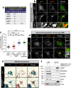
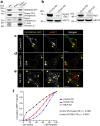
Fig. 1
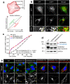
Fig. 1. DENND6A-GFP localizes to lysosomes.
a BioID interactome of DENND6A, taken from human cell map. b HeLa cells expressing either GFP alone or DENND6A-GFP were fixed, stained with LAMP1 antibody and imaged using confocal microscopy (Leica SP8). The cell periphery is outlined by a white dotted line. Scale bar = 10 µm for low magnification images; 2.8 µm and 2.2 µm for high magnification images corresponding to inset 1 and 2. The yellow arrow indicates colocalization between DENND6A-GFP and LAMP1. c Quantification of the Pearson correlation coefficient for the co-localization of GFP with indicated markers (LAMP1, EEA1); means ± SEM; One-way ANOVA (**** P ≤ 0.0001; ns = not significant; n = 30 cells for all conditions from 3 replicates). d HeLa cells expressing either GFP alone or DENND6A-GFP were fixed, stained with LAMP1 antibody. 3D-SIM images were acquired using LSM880-Elyra PS1 super-resolution microscopy. The cell periphery is outlined by a white dotted line. Scale bar = 10 µm for low magnification images; 4.18 µm and 4.14 µm for high magnification images corresponding to insets from GFP or DENND6A-GFP expressing cells. e 3D reconstruction of SIM imaging performed in (d). Scale bar = 10 µm for low magnification images; 3 µm for high magnification images. f Lysates from HEK-293 cells expressing the Tmem192-3xHA (HA-Lyso cells) or the Tmem192-2xFlag (Control-Lyso cells) were prepared as per the protocol. Lysosomes were immunoprecipitated using anti-HA magnetic beads and analyzed by immunoblot. SM stands for starting material and IP stands for immunoprecipitation. Red arrow indicates specific band corresponding to TMEM192-FLAG.
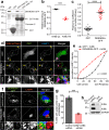
Fig. 2
Fig. 2. DENND6A promotes juxtanuclear clustering of lysosomes.
a Schematic representation of the quantification of cumulative intensity distribution method for lysosomal distribution (adapted from). b Unstarved HeLa cells expressing either GFP alone or DENND6A-GFP were fixed, stained with LAMP1 antibody and imaged using confocal microscopy (Leica SP8). The cell periphery is outlined by a white dotted line. Scale bar = 10 µm for low magnification images; 3.4 µm and 2.1 µm for high magnification images corresponding to inset 1 and 2. c Quantification of cumulative distribution of LAMP1 intensity; mean ± SEM; two-tailed extra sum of F-squares test following nonlinear regression and curve fitting; n = 30 cells from 3 replicates. d Immunoblot showing the protein levels in WT and DENND6A KO HeLa cells. Immunoblot probed with anti-DENND6A and anti-GAPDH antibodies. e WT and DENND6A KO unstarved HeLa cells were fixed, stained with LAMP1 antibody and DAPI, and imaged using confocal microscopy (Leica SP8). The cell periphery is outlined by a white dotted line. Scale bar = 10 µm. Yellow arrow indicates peripheral lysosomes.
Fig. 3
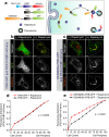
Fig. 3. DENND6A is sufficient to promote juxtanuclear clustering.
a Schematic representation of rapamycin-induced relocalization of DENND6A to the peroxisome. FKBP = FK506-binding protein domain; FRB = FKBP-rapamycin–binding domain (adapted from). HeLa cells were co-transfected with (b) FRB-GFP and PEX3-FKBP-mCherry, c DENND6A-FRB-GFP and PEX3-FKBP-mCherry and treated with or without rapamycin for 1 h. Following rapamycin treatment, cells were fixed and imaged. The cell periphery is outlined by a white dotted line. Scale bars = 10 μm. d–e Quantification of cumulative peroxisomal distribution (mCherry intensity) in experiments performed in (b) and (c); mean ± SEM; two-tailed extra sum of F-squares test following nonlinear regression and curve fitting; n [(GFP-FRB + Rapamycin), (GFP-FRB - Rapamycin), (DENND6A-GFP-FRB + Rapamycin), (DENND6A-GFP-FRB - Rapamycin)] = (31, 30, 30, 29) cells from 3 replicates.
Fig. 4
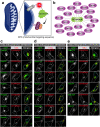
Fig. 4. DENN6A targeted to the mitochondria recruits corresponding Rab partners.
a Schematic model of the cell-based assay (adapted from). b List of Rabs recruited by DENND6A at the mitochondria. c–e HeLa cells co-transfected with GFP-Rabs and mito-mSc-DENND6A were fixed and imaged. The nucleus and cell periphery are outlined by blue and white dotted line respectively. Scale bar = 10 µm.
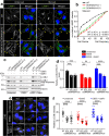
Fig. 5
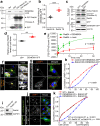
Fig. 5. DENND6A promotes juxtanuclear clustering of lysosomes via activation of Rab34.
a HEK-293T cell lysates expressing DENND6A-GFP were incubated with indicated purified proteins. Bound proteins were detected by immunoblot with anti-GFP antibody. Starting material (SM). b Quantification from (a); means ± SEM; two-tailed unpaired t test (*** P ≤ 0.0005; n = 4 from three replicates). c GFP or DENND6A-GFP expressing HEK-293T cell lysates were incubated with purified T7-RILP. Bound proteins were detected by anti-Rab34, anti-GFP and anti-HSC70 antibody. d Quantification from c; means ± SEM; two-tailed unpaired t test (**P ≤ 0.0025; n = 3 from three replicates). e In vitro GEF assays using purified Rab34 with or without DENND6A as indicated. Relative incorporation of [35S]GTPγS on Rab34 is plotted over time; data represent mean ± SEM; two-tailed extra sum of F-squares test following nonlinear regression one-phase association curve fit, n = 3 from three replicates. f HeLa cells co-expressing DENND6A-GFP and mCherry-Rab34 were fixed/stained with LAMP1 antibody. Cell periphery outlined by a white dotted line. Scale bar = 10 and 3.13 µm for low and high magnification images. g HeLa cells treated with control or Rab34 siRNA were transfected with DENND6A-GFP. Post transfection, cells were fixed and stained with LAMP1 antibody. Cell periphery is outlined by a white dotted line. Scale bar = 10 µm. h Quantification of cumulative LAMP1 distribution from g; mean ± SEM; two-tailed extra sum of F-squares test following nonlinear regression and curve fitting; n = 30 cells from 3 replicates. i Immunoblot showing the Rab34 protein levels in control and Rab34 siRNA treated HeLa cells. Immunoblot probed with anti-Rab34 and anti-GAPDH antibodies. j WT and DENND6A KO HeLa cells were transfected with GFP-Rab34 or GFP-Rab34 QL. Post transfection, cells were fixed and stained with LAMP1 antibody. Cell periphery is outlined by a white dotted line. Scale bar = 10 µm. k Quantification of cumulative LAMP1 distribution from (j); mean ± SEM; two-tailed extra sum of F-squares test following nonlinear regression and curve fitting; n = 27, 30 and 30 cells from 3 replicates corresponding to the three conditions Rab34+WT; Rab34 + KO1; Rab34 QL + KO1.
Fig. 6
Fig. 6. DENND6A mediated juxtanuclear lysosomal clustering depends on RILP and dynein.
a Lysates from HEK-293T cells expressing DENND6A-GFP were incubated with wither beads covalently linked to T7 antibody or T7 linked beads coupled to T7-RILP protein. Specifically bound proteins were detected by immunoblot with anti-GFP antibody, anti- dynein intermediate chain (DIC) antibody and anti-Rab34 antibody. The starting material (SM) was run in parallel to detect the total DENND6A-GFP, DIC and Rab34. b Immunoblot showing DIC protein levels in control and dynein siRNA treated HeLa cells. Immunoblot probed with anti-DIC and anti-HSC70 antibodies. Immunoblot showing RILP protein levels in control and RILP siRNA treated HeLa cells. Immunoblot probed with anti-RILP and anti-HSC70 antibodies. HeLa cells were treated with (c) control siRNA or (d) dynein siRNA or (e) RILP siRNA and transfected with DENND6A-GFP. 16 h post transfection, cells were fixed and stained with LAMP1 antibody. The cell periphery is outlined by a white dotted line. The yellow arrow indicates the presence of lysosomes corresponding to DENND6-GFP. Scale bars = 10 μm. f Quantification of cumulative distribution of LAMP1 intensity in experiments performed in (c)(e); mean ± SEM; two-tailed extra sum of F-squares test following nonlinear regression and curve fitting; n = 29, 30, 30 cells from 3 replicates corresponding to control, dynein, and RILP siRNA treated cells.
Fig. 7
Fig. 7. DENND6A is an Arl8b effector, and their interaction promotes juxtanuclear lysosomal clustering.
a Purified GST, GST-Arl8b QL and GST-Arl8b TN were incubated with lysates from HEK-293T cells expressing DENND6A-GFP. Bound proteins were detected by immunoblot with anti-GFP antibody. The starting material (SM) was run in parallel to detect the total DENND6A-GFP. b Quantification of experiment in (a); means ± SEM; two-tailed unpaired t test (*** P ≤ 0.0005; n = 3 from three replicates). c Graphical representation of the Pearson correlation coefficient for the co-localization of GFP or DENND6A-GFP with Arl8b-mCherry from experiments performed in (d); means ± SEM; two-tailed unpaired t test (****P ≤ 0.0001; n = 29 and 30 corresponding to GFP and DENND6A-GFP, from 3 replicates). d HeLa cells co-expressing either GFP alone or DENND6A-GFP along with Arl8b-mCherry were fixed/stained with LAMP1 antibody and imaged (confocal microscopy). Cell periphery is outlined by a white dotted line. The yellow arrow indicates the presence of lysosomes corresponding to Arl8b-cherry in the presence of either GFP or DENND6-GFP. Scale bar = 10 µm for low magnification images; 5.22 µm and 2.12 µm for high magnification images corresponding to inset 1 and 2. e Quantification of cumulative distribution of LAMP1 intensity in (d); mean ± SEM; two-tailed extra sum of F-squares test following nonlinear regression and curve fitting; n = 25 and 26 cells, corresponding to GFP and DENND6A-GFP, from 3 replicates. f Control or Arl8 siRNA treated HeLa cells were transfected with DENND6A-GFP. 16 h Post transfection, cells were fixed/stained with LAMP1 antibody and imaged (confocal microscopy). The cell periphery is outlined by a white dotted line. Scale bar = 10 µm for low magnification images; 3.79 µm and 4.97 µm for high magnification images corresponding to inset 1 and 2. g Quantification of experiment in (f); means ± SEM; two-tailed unpaired t test (***P = 0.0004; n = 42 and 47, corresponding to control and Arl8 siRNA, from 3 replicates). h Immunoblot showing Arl8a and Arl8b protein levels in control and Arl8(a + b) siRNA treated HeLa cells. Immunoblot probed with anti-Arl8a, anti-Arl8b and anti-GAPDH antibodies.
Fig. 8
Fig. 8. Loss of DENND6A impairs nutrient dependent lysosomal positioning and autophagic flux.
a Unstarved or Earle’s Balanced Salt Solution (EBSS) starved HeLa cells were fixed, stained with LAMP1 antibody and imaged using confocal microscopy (Leica SP8). The cell periphery is outlined by a white dotted line. Scale bar = 10 µm. Yellow arrows indicate the presence of peripheral lysosomes. b Quantification of cumulative distribution of LAMP1 intensity in experiments performed in (a) (under starvation condition); mean ± SEM; two-tailed extra sum of F-squares test following nonlinear regression and curve fitting; n = 28, 29, 30 cells, corresponding to WT, DENND6A KO1 and DENND6A KO2, from 3 replicates. c Immunoblot showing LC3B-II protein levels under various conditions (unstarved; EBSS starved; and EBSS starved + Bafilomycin A1 (BafA1)) Immunoblot probed with anti-LC3B-II, anti-p62 and anti-HSC70 antibodies. d Quantification of experiment in (c); means ± SEM; two-way ANOVA (**P ≤ 0.0025; ***P ≤ 0.0005; ****P ≤ 0.0001; n = 3 from three replicates). e HeLa WT and DENND6A KOs cells from c were fixed and stained with LC3B-II antibody and DAPI. The cell periphery is outlined by a white dotted line. Scale bar = 25 µm. f Quantification of experiment in (e); means ± SEM; Kruskal-Wallis test ****P ≤ 0.0001; n = (32, 43, 37); (36, 34, 42); (34, 41, 34) cells, corresponding to WT, DENND6A KO1 and DENND6A KO2 in unstarved; EBSS; EBSS + BafA1, from 3 replicates).
Fig. 9
Fig. 9. Schematic representation of Arl8b/DENND6A/Rab34 mediated retrograde lysosomal trafficking.
We propose that DENND6A functions as an effector of Arl8b. Furthermore, we hypothesize the presence of an additional factor (marked by ‘?’) that may assist in DENND6A’s recruitment to lysosomes. The BORC complex recruits Arl8b to peripheral lysosomes (adapted from), where Arl8b in turn recruits DENND6A. Once recruited, DENND6A activates Rab34, and the activated Rab34 then recruits its own effector, RILP, which is a proposed dynein adapter. Consequently, DENND6A links Arl8b and Rab34, facilitating retrograde lysosomal transport, from the plus to minus ends of the microtubules.

References

    1. Ballabio A, Bonifacino JS. Lysosomes as dynamic regulators of cell and organismal homeostasis. Nat. Rev. Mol. Cell Biol. 2019;21:101–118. doi: 10.1038/s41580-019-0185-4. - DOI - PubMed
    1. Pu J, Guardia CM, Keren-Kaplan T, Bonifacino JS. Mechanisms and functions of lysosome positioning. J. Cell Sci. 2016;129:4329. - PMC - PubMed
    1. Bright NA, Davis LJ, Luzio JP. Endolysosomes Are the Principal Intracellular Sites of Acid Hydrolase Activity. Curr. Biol. 2016;26:2233–2245. doi: 10.1016/j.cub.2016.06.046. - DOI - PMC - PubMed
    1. Bissig C, Hurbain I, Raposo G, van Niel G. PIKfyve activity regulates reformation of terminal storage lysosomes from endolysosomes. Traffic. 2017;18:747–757. doi: 10.1111/tra.12525. - DOI - PubMed
    1. Cabukusta B, Neefjes J. Mechanisms of lysosomal positioning and movement. Traffic. 2018;19:761. doi: 10.1111/tra.12587. - DOI - PMC - PubMed

MeSH terms