. 2024 Jan 31;14(1):2572.
doi: 10.1038/s41598-024-51787-0.

Molecular insights into the dynamic modulation of bacterial ClpP function and oligomerization by peptidomimetic boronate compounds

Affiliations

Molecular insights into the dynamic modulation of bacterial ClpP function and oligomerization by peptidomimetic boronate compounds

Bruno Alves França et al. Sci Rep. .

Abstract

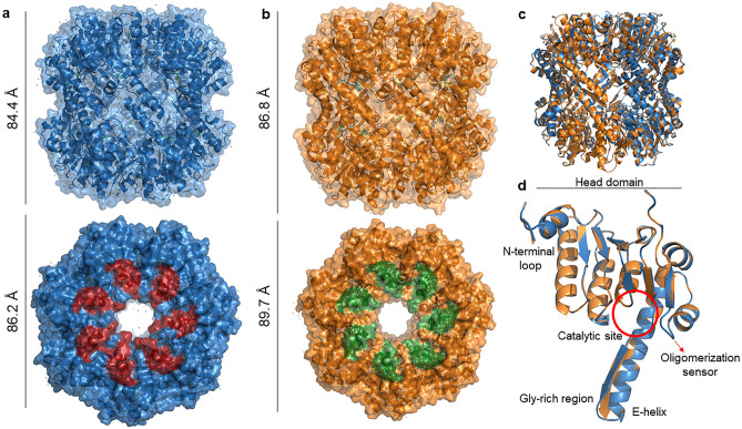
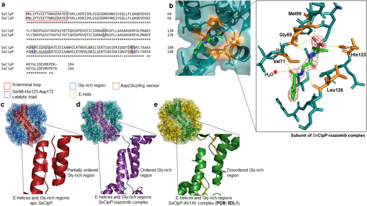
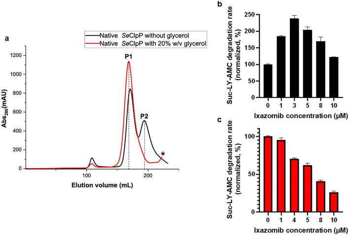
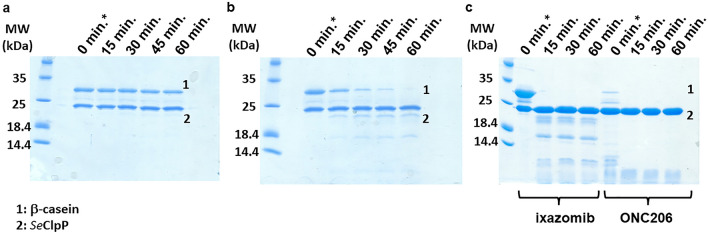
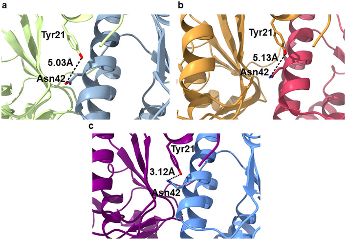
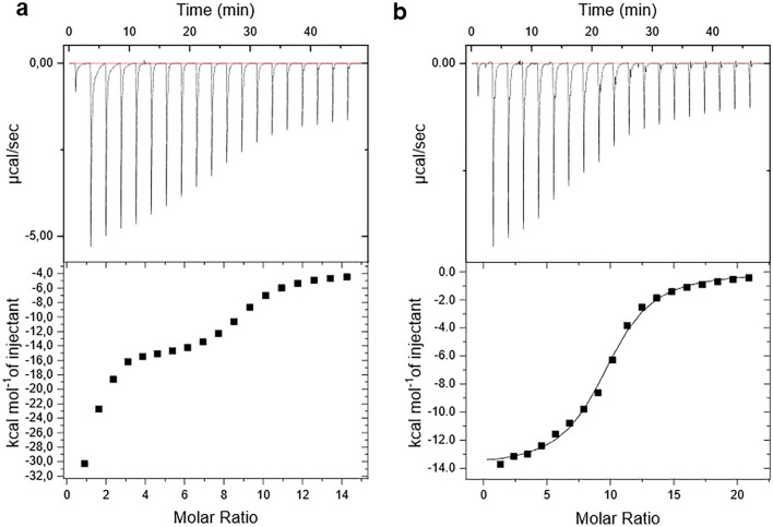
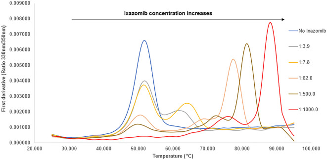
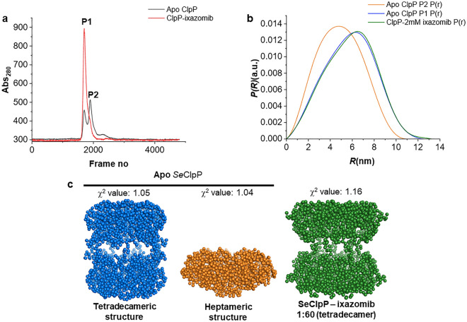
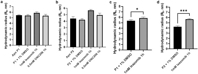
Bacterial caseinolytic protease P subunit (ClpP) is important and vital for cell survival and infectivity. Recent publications describe and discuss the complex structure-function relationship of ClpP and its processive activity mediated by 14 catalytic sites. Even so, there are several aspects yet to be further elucidated, such as the paradoxical allosteric modulation of ClpP by peptidomimetic boronates. These compounds bind to all catalytic sites, and in specific conditions, they stimulate a dysregulated degradation of peptides and globular proteins, instead of inhibiting the enzymatic activity, as expected for serine proteases in general. Aiming to explore and explain this paradoxical effect, we solved and refined the crystal structure of native ClpP from Staphylococcus epidermidis (Se), an opportunistic pathogen involved in nosocomial infections, as well as ClpP in complex with ixazomib at 1.90 Å and 2.33 Å resolution, respectively. The interpretation of the crystal structures, in combination with complementary biochemical and biophysical data, shed light on how ixazomib affects the ClpP conformational state and activity. Moreover, SEC-SAXS and DLS measurements show, for the first time, that a peptidomimetic boronate compound also induces the assembly of the tetradecameric structure from isolated homomeric heptameric rings of a gram-positive organism.


Conflict of interest statement

The authors declare no competing interests.

Figures

Figure 1
Figure 1
Crystal structures of ClpP from Staphylococcus epidermidis: apo SeClpP and SeClpP-ixazomib complex. The structures consist of two stacked heptameric rings that contain 14 identical catalytic sites in a central chamber. (a) Front and top views of apo SeClpP; (b) front and top views of SeClpP-ixazomib complex. In both cases, the protein dimensions are displayed. (c) The superposition of both tetradecamers demonstrates that there is not any significant three-dimensional difference between them. (d) Superimposition of monomers from apo SeClpP (blue) and SeClpP (orange) and their main parts.
Figure 2
Figure 2
(a) ClustalW sequence alignment with SeClpP and S. aureus ClpP (SaClpP) amino acid sequences in FASTA format; (b) catalytic site of a monomer from the SeClpP-ixazomib complex and interactions between ixazomib and residues in the catalytic cleft. The amino acids that form hydrogen bonds with the ligand are labeled and colored in orange. Ixazomib is shown with the 2Fo-Fc electron density at 1.5σ. All the ClpP monomers bound to the ligand are shown in Supplementary Fig. S1. Comparison between the Gly-rich regions of different crystal structures of ClpP: (c) apo SeClpP with its partially disordered Gly-rich region; (d) SeClpP-ixazomib complex with its ordered Gly-rich region (two antiparallel beta-strands); (e) SaClpP-AV145 complex with its disordered, or unstructured, Gly-rich region.
Figure 3
Figure 3
(a) Chromatograms after size-exclusion chromatography (SEC) with SeClpP sample free from glycerol (black curve) and supplemented with 20% w/v glycerol (red curve). As seen here, the use of the triol avoids the formation of heptamers (P2). When glycerol is not added to the protein sample, both tetradecameric (P1) and heptameric (P2) species are present. Suc-LY-AMC degradation rate vs. ixazomib concentration using reaction mixtures without glycerol (black bars) ( b) and with 20% w/v glycerol (red bars) (c). All assays were performed with experimental triplicates. In Supplementary Fig. S2, curves of peptidase activity (fluorescence unit) vs. time, at different ixazomib concentrations, are presented. * Only a low amount of small oligomers of ClpP can be seen in the protein sample containing 20% w/v glycerol.
Figure 4
Figure 4
15% SDS-PAGE after reactions with β-casein monitored for 60 minutes. (a) ixazomib and SeClpP concentrations: 200μM and 10μM, respectively; (b) ixazomib and SeClpP concentrations: 500μM and 10μM, respectively; (c) Comparative experiment with ixazomib (1mM) and ONC206 (10μM), where it is noticeable that the molecular weight (MW) of product fragments vary, according to the ligand used. A possible explanation for this difference is that ixazomib occupies the catalytic sites, affecting the processive degradation of the substrate. ONC206 only binds to the allosteric regions. Degradation products of low molecular weight can be seen in (b) and (c). *t= 0 min corresponds to the time point with no incubation, but until the complete denaturation of ClpP before the SDS-PAGE, β-casein degradation happened in the quick reaction with ONC206. The entire image of each gel is found in Supplementary Fig. S3.
Figure 5
Figure 5
Illustration of the conformation of the Asn42 sidechain in different crystal structures of ClpP: (a) SeClpP-ixazomib complex, (b) active mutant (SaClpP Y63A) (PDB ID: 5C90) of ClpP from Staphylococcus aureus (Sa), and (c) native SeClpP. In (a) and (b), Asn42 is in a “down” position, with open pores (active for proteolysis). In contrast, in (c), the same amino acid residue is found in the “up” position, characteristic of closed ClpP.
Figure 6
Figure 6
ITC measurements with SeClpP and ixazomib, at different molar ratios, (a) in a glycerol-free environment and (b) with 20% w/v glycerol, both at 36°C (the same temperature set for the enzymatic assays). In the presence of glycerol, the ITC data could be fitted, and the following parameters were calculated: N = 9.53 ±0.0993 sites, K = 1.86E5 ±1.99E4 M−1 , ΔH= -1.390E4 ±198.0 cal/mol, and ΔS= -20.8 cal/mol/deg.
Figure 7
Figure 7
Thermal stability analysis of SeClpP based on the first derivatives calculated from nanoDSF measurements. In Supplementary Fig. S4, a graph with more curves is presented, including a scan with the fluorescence intensity of samples that contain only ixazomib (at 1 mM) and buffer to discard any influence of this compound on the measurements.
Figure 8
Figure 8
SEC-SAXS data with SeClpP samples composed of heptamers and tetradecamers, as expected for protein samples without glycerol. (a) Chromatogram with apo SeClpP (black) and SeClpP sample incubated with 1 mM ixazomib (1:60 protein-to-ligand ratio; red). (b) Inter-atomic pair distribution functions of native SeClpP P1 (blue), native SeClpP P2 (orange), and SeClpP-ixazomib complex (green). (c) Ab initio low-resolution models calculated from SAXS scattering curves of native SeClpP P1 (blue), native SeClpP P2 (orange), and SeClpP-ixazomib complex (green). In Supplementary Table 2, there are experimental data of SAXS measurements. Guinier plots with scattering intensity graphs (I(q) vs q) are available in Supplementary Fig. S5.
Figure 9
Figure 9
DLS measurements with apo SeClpP from samples P1 (a) and P2 (b). Apo P1/P2 and P1/P2 supplemented with 1% v/v DMSO were selected as controls. 1 mM ixazomib and 0.5 mM ONC206 were incubated with the protein for 1 h, at 36 °C prior to the measurements. In (c,d), an unpaired t-test was applied to analyze how significant is the difference that ixazomib causes to RH values. In both cases, the change was significant (p < 0.05): p = 0.0363 (c) and p = 0.0004 (d). The last p-value shows that a higher alteration occurs when P2 is incubated with the peptidomimetic boronate.

Similar articles

Cited by

References

    1. Alexopoulos JA, Guarné A, Ortega J. ClpP: A structurally dynamic protease regulated by AAA+ proteins. J. Struct. Biol. 2012;179:202–210. doi: 10.1016/j.jsb.2012.05.003. - DOI - PubMed
    1. Gersch M, et al. AAA+ chaperones and acyldepsipeptides activate the ClpP protease via conformational control. Nat. Commun. 2015;6:122. doi: 10.1038/ncomms7320. - DOI - PubMed
    1. Moreno-Cinos C, et al. ClpP protease, a promising antimicrobial target. Int. J. Mol. Sci. 2019 doi: 10.3390/ijms20092232. - DOI - PMC - PubMed
    1. Dougan DA. Chemical activators of ClpP: Turning jekyll into hyde. Chem. Biol. 2011;18:1072–1074. doi: 10.1016/j.chembiol.2011.09.003. - DOI - PubMed
    1. Zheng D, Xu Y, Yuan G, Wu X, Li Q. Bacterial ClpP protease is a potential target for methyl gallate. Front. Microbiol. 2021;11:31. doi: 10.3389/fmicb.2020.598692. - DOI - PMC - PubMed

MeSH terms