. 2024 Jan 31;25(1):40.
doi: 10.1186/s13059-024-03175-0.

BORIS/CTCFL epigenetically reprograms clustered CTCF binding sites into alternative transcriptional start sites

Affiliations

BORIS/CTCFL epigenetically reprograms clustered CTCF binding sites into alternative transcriptional start sites

Elena M Pugacheva et al. Genome Biol. .

Abstract

Background: Pervasive usage of alternative promoters leads to the deregulation of gene expression in carcinogenesis and may drive the emergence of new genes in spermatogenesis. However, little is known regarding the mechanisms underpinning the activation of alternative promoters.

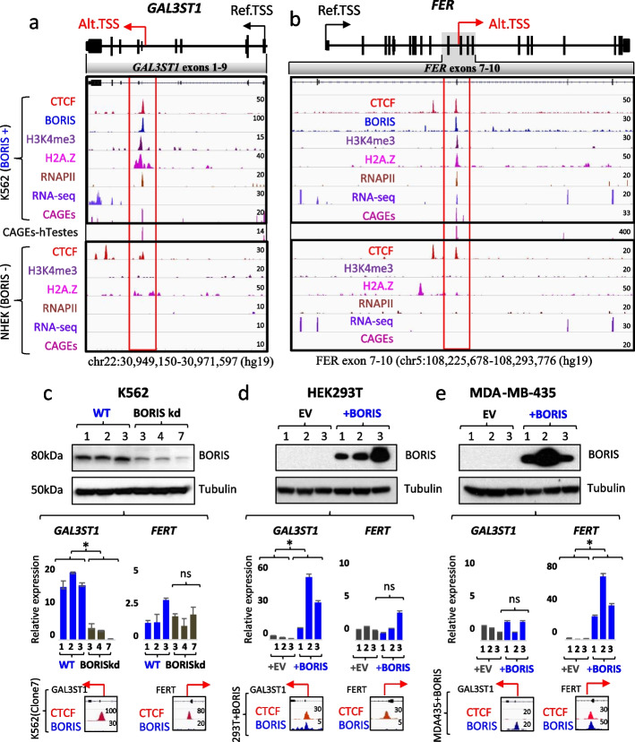
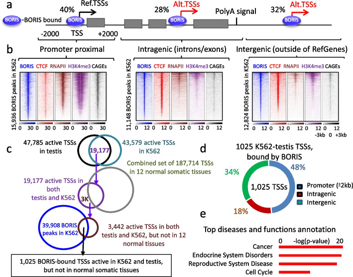
Results: Here we describe how alternative cancer-testis-specific transcription is activated. We show that intergenic and intronic CTCF binding sites, which are transcriptionally inert in normal somatic cells, could be epigenetically reprogrammed into active de novo promoters in germ and cancer cells. BORIS/CTCFL, the testis-specific paralog of the ubiquitously expressed CTCF, triggers the epigenetic reprogramming of CTCF sites into units of active transcription. BORIS binding initiates the recruitment of the chromatin remodeling factor, SRCAP, followed by the replacement of H2A histone with H2A.Z, resulting in a more relaxed chromatin state in the nucleosomes flanking the CTCF binding sites. The relaxation of chromatin around CTCF binding sites facilitates the recruitment of multiple additional transcription factors, thereby activating transcription from a given binding site. We demonstrate that the epigenetically reprogrammed CTCF binding sites can drive the expression of cancer-testis genes, long noncoding RNAs, retro-pseudogenes, and dormant transposable elements.

Conclusions: Thus, BORIS functions as a transcription factor that epigenetically reprograms clustered CTCF binding sites into transcriptional start sites, promoting transcription from alternative promoters in both germ cells and cancer cells.

Keywords: Alternative transcription; CTCF; CTCFL/BORIS; Cancer; Germ cells.


Conflict of interest statement

B.R. is a co-founder and consultant of Arima Genomics Inc. and co-founder of Epigenome Technologies, Inc. The other authors declare that they have no competing interests.

Figures

Fig. 1
Fig. 1
CTCF and BORIS co-binding to intronic regions of GAL3ST1 and FER genes associated with cancer-testis-specific transcription. a,b Above: Schematic representation of the gene structure of GAL3ST1 (a) and FER (b). Arrows denote somatic (black) and testis-specific (red) TSSs. Below: In the upper part, ChIP-seq peaks illustrate CTCF (red) and BORIS (blue) co-binding in BORIS-positive (BORIS +) K562 cells across GAL3ST1 and FER (exon 7-10). The co-binding coincides with the enrichment of active histones/marks H3K4me3 (purple), H2A.Z (magenta), and RNAPII (brown). RNA-seq peaks (indigo) and CAGE-seq peaks (pink) highlight alternative transcription in K562 cells. In the lower part, CTCF binding alone in BORIS-negative (BORIS −) NHEK cells does not activate testis-specific promoters. CAGE-seq for human testes (pink) is shown between the two panels, with red boxes highlighting testis-specific TSSs. c–e In the upper part, Western blots from whole cell lysates show BORIS protein detection in c K562 wild-type (WT1-total culture, WT2 and WT3 – single-cell wild-type clones transfected with control RNA) versus BORIS knockdown (kd) K562 single-cell clones (#3,4,7), obtained by zinc finger nuclease (ZFN) treatment. d HEK293T and e MDA-MB-435 cells transfected with empty (EV) or BORIS-expressing vector. Tubulin is used as a loading control. Numbers (1,2,3) indicate different single-cell clones. The middle part displays RT-qPCR results indicating the relative expression of GAL3ST1 and FERT in K562, HEK293T, and MDA-MB-435 cell lines. Statistical analysis was performed using two-tailed Student’s t test (*, p < 0.0005). Error bars indicate mean ± SD (n = 3), ns – non-significant. In the bottom part, ChIP-seq peaks show CTCF and BORIS occupancy in K562 (clone#7) BORIS kd cells, HEK293T, and MDA-MB-435 cells. Abbreviations; Ref. TSS (reference transcriptional start site), Alt. TSS (alternative transcriptional start site)
Fig. 2
Fig. 2
Intragenic and intergenic BORIS binding sites are associated with active transcription in K562 and male germ cells. a Schematic representation of ChIP-seq data illustrating the distribution of BORIS binding sites in K562 cells relative to a reference gene structure. Among the identified binding sites, 40, 28, and 32% are located around TSSs, inside introns or exons, and outside of RefSeqGenes, respectively. Canonical and alternative TSSs are denoted by black and red arrows, respectively, based on NGS data shown in panel b. b Heatmaps illustrating the enrichment profiles of BORIS, CTCF, RNAPII, H3K4me3 ChIP-seq signals, and CAGE-seq reads within a 6-kilobase (kb) window centered on BORIS binding sites in K562 cells. These heatmaps correspond to the genomic distribution pattern depicted in panel a. c Schematic representation of the strategy used to map TSSs associated with K562-testis-specific transcription driven from BORIS sites in K562 cells. d Genomic distribution of 1025 TSSs, active in both testis and K562 cells, relative to their genomic location with respect to RefSeqGenes. e Top diseases or function annotation for the 1025 TSSs identified using the strategy illustrated in panel c
Fig. 3
Fig. 3
Ectopic BORIS expression transforms NIH3T3 cells and deregulates gene transcription. a Representative images of soft agar colonies. b Quantification of the number of soft agar colonies. Two-tailed Student’s t test (***—p < 0.0001). c Western blot analysis of BORIS expression in nuclear extracts isolated from four single-cell clones of NIH3T3 cells, recovered from a soft agar. K562—positive control. Parental (WT) and EV-expressing NIH3T3 cells were used as a negative control for BORIS expression. RAD21 Abs were used as a loading control. d RT-qPCR results displaying the relative expression levels of testis-specific Gal3St1 in NIH3T3 + EV, including EV-total culture (#1), and single-cell clones with EV (#2, #3), compared to NIH3T3 + BORIS clones recovered from soft agar (#1, 2, 3, 4). Two-tailed Student’s t test ( *—p < 0.005). Error bars indicate mean ± SD (n = 3). e Heatmap depicting BORIS occupancy in three BORIS-expressing clones (#2,3,4) compared to EV. f Quantification of the number of upregulated (red) and downregulated (blue) RefSeqGenes, lncRNAs, and TEs in three BORIS-expressing clones (#2,3,4), compared to EV, with log2 fold change > 1.3 and p-value (padj) < 0.05. g–k NIH3T3 cells were stably transfected with either the doxycycline-inducible empty vector (pBIGi-EV) or BORIS (pBIGi-BORIS). The cells were cultured under different conditions: in the absence of doxycycline (No Dox), induced with doxycycline (Dox) for a specified number of hours, and doxycycline removal (Wash Off) for an indicated number of hours. g Western blot confirms BORIS activation by dox. h Heatmap of BORIS occupancy in doxycycline-treated cells compared to EV and cells without doxycycline treatment. i Quantification of upregulated (red) and downregulated (blue) RefSeqGenes in doxycycline-induced EV or BORIS cells compared to EV (no dox) or EV (dox 12 h), respectively, with log2 > 1.3, p-value < 0.001. k The heatmap depicts the expression profile of 1377 genes (RNA-seq data, 2 replicates for each condition), which were significantly deregulated in BORIS-expressing cells treated with dox for 24 h, comparing the expression in EV versus BORIS-induced cells. l GSEA of RNA-seq data for BORIS-expressing cells isolated from soft agar, compared to EV
Fig. 4
Fig. 4
BORIS binding epigenetically reprograms transcriptionally inert CTCF binding sites into active promoters. a Genome browser view of ChIP-seq data and RNA-seq data for NIH3T3 + EV versus NIH3T3 + BORIS (clone#2, soft-agar derived) cells. CTCF (red) and BORIS (blue) co-occupancy in NIH3T3 + BORIS cells leads to the activation of Oct4 (Pou5f1) gene expression from an alternative intronic promoter (highlighted by red arrow and open box) in all three BORIS-expressing clones (RNA-seq data), compared to no expression in NIH3T3 + EV cells. b Upper panel shows the exon/intron structure of Pou5f1. Black and red arrows indicate reference and alternative promoters of Pou5f1, respectively. Black and white boxes represent coding exons specific to isoforms 4A and 4B, respectively; grey boxes represent exons shared by both isoforms. Lower panel compares the relative expression of Pou5f1 isoforms in three BORIS-expressing soft-agar-derived NIH3T3 clones to NIH3T3 + EV cells, revealing upregulation of only isoform 4B upon BORIS expression. c,d Genome browser view showing that CTCF and BORIS co-binding in NIH3T3 + BORIS (clone#2) cells is associated with the enrichment of active histones/marks (H2A.Z, H3K4me3, H3K27ac), RNAPII, CAGE-seq reads and the activation of alternative transcription (RNA-seq) from Slc6a19 (c) and Hck (d) genes. The alternative promoters are silent under CTCF-only occupancy in NIH3T3 + EV cells. e–g Left panel shows scatter plots of normalized read counts (log10) for H2A.Z e, H3K4me3 f, and CAGE g occupancy at BORIS-bound sites in NIH3T3 + BORIS (clone#2) cells compared to the same genomic sites in NIH3T3 + EV cells. BORIS sites with gain or loss of active histone marks or CAGEs are highlighted by red or green colors, respectively. The right panel displays a heatmap of H2A.Z e, H3K4me3 f, and CAGE g occupancy at BORIS-bound sites that gained active marks in NIH3T3 + BORIS (clone#2) cells compared to NIH3T3 + EV cells. Red arrows connect the left and right panels, indicating the number of gained active histone marks at BORIS-bound sites. h,i Heatmap representation of CTCF (red), BORIS (blue), RNAPII (pink), H2A.Z (yellow), H3K4me3 (purple), and H3K27ac (green) occupancy in NIH3T3 + EV cells (h) compared to NIH3T3 + BORIS (clone#2) cells (i) at the 5871 CTCF/BORIS binding sites, which gained occupancy of at least one active mark of transcription
Fig. 5
Fig. 5
BORIS recruits SRCAP, which drives H2A.Z occupancy at its target sites. a Co-IP: BORIS directly interacts with SRCAP in NIH3T3 + BORIS (clone#2) cells. Benzonase-treated nuclear cell extracts were immunoprecipitated (IP) with either mouse IgG, anti-BORIS Abs, or anti-SRCAP Abs. Western blot analysis with indicated antibodies confirms the interaction. b Average plot of SRCAP occupancy (ChIP-seq data) at BORIS-bound sites in NIH3T3 + BORIS (clone#2) cells compared to NIH3T3 + EV cells. The p-value is calculated by Kolmogorov–Smirnov test. c Heatmap displaying BORIS (left panel, blue), SRCAP (middle panel, purple), and H2A.Z (right panel, brown) occupancy at the 4654 BORIS-bound sites in NIH3T3 + BORIS (clone#2) cells compared to NIH3T3 + EV cells. Arrows atop the heatmaps indicate the proposed sequence of events: BORIS binding to chromatin leads to SRCAP recruitment, resulting in the gain of H2A.Z occupancy around BORIS sites. d Heatmap displaying the correlation between H2A.Z occupancy and BORIS binding in both doxycycline-inducible empty vector (EV) and BORIS-expressing cells. The cells were subjected to treatment with or without doxycycline for a specified duration of time, as indicated
Fig. 6
Fig. 6
BORIS expression is associated with a significant increase in CTCF occupancy and a more open chromatin state around CTCF sites. a CTCF occupancy mapped in NIH3T3 + EV versus NIH3T3 + BORIS (clone#2) cells, depicted through a violin plot. b Scatter plot shows normalized read counts (log10) for CTCF occupancy at the combined set of CTCF binding sites in NIH3T3 + BORIS (clone#2) cells compared to NIH3T3 + EV cells. CTCF sites with increased or decreased occupancy by more than threefold are highlighted in red and green, respectively. c Heatmap illustrating CTCF (red) and BORIS (blue) occupancy at the 6521 CTCF sites from panel b (connected by red arrow). d Scatter plot of normalized read counts (log10) for ATAC-seq tag density at the combined set of ATAC-seq genomic sites mapped in both NIH3T3 + EV and NIH3T3 + BORIS (clone#2) cells. Genomic sites with increased or decreased occupancy by more than twofold are highlighted in red and green, respectively. e Heatmaps combining ATAC-seq (black) accessibility with CTCF, BORIS, H2A.Z, H3K4me3, and H3K27ac occupancy at genomic regions from panel d (connected by red and green arrows). Upper and lower panels display data for increased and decreased ATAC-seq sites, respectively. f Average plot of chromatin accessibility (ATAC-seq) at 4654 BORIS binding sites that gained H2A.Z occupancy in NIH3T3 + BORIS (blue) cells compared to NIH3T3 + EV cells (black). P-value calculated by the Kolmogorov–Smirnov test. g Genome browser view of two alternative intronic promoters in Snx31 and Hsf3 genes activated by BORIS (blue) binding to CTCF (red) sites in NIH3T3 + BORIS (clone#2) cells. Black arrows indicate increased chromatin accessibility (ATAC-seq, black) beyond CTCF and BORIS ChIP-seq peaks. Red arrows show alternative transcription initiated from a CTCF binding site with BORIS recruitment
Fig. 7
Fig. 7
Opening of chromatin by BORIS facilitates binding of other transcriptional factors. a Scatter plot displaying the enrichment of 190 TF motifs at CTCF/BORIS binding sites that reprogrammed into active promoters, compared to transcriptionally silent CTCF/BORIS sites in NIH3T3 + BORIS (clone#2) cells. b MAZ and MXI1 motifs are significantly enriched at CTCF/BORIS binding sites converted into active promoters. c Genome browser view illustrates the recruitment of TBP, HCFC1, MXI1, and MAZ proteins at the CTCF site within the Rbpjl promoter, activated by BORIS binding in NIH3T3 + BORIS (clone#2) cells. The activated promoter is highlighted by a red open box. d Left panel: Scatter plot of normalized read counts (log10) for HCFC1 occupancy at the combined set of HCFC1 binding sites (46,287) in NIH3T3 + BORIS (clone#2) cells compared to the same genomic sites in EV cells. Right panel: Heatmap of CTCF (red), BORIS (blue), and HCFC1 (brown) occupancy at the 19,519 HCFC1 sites from the left panel (connected by red arrow). e TF motifs enriched at HCFC1 peaks (60 bp around the summit of peak) in NIH3T3 + EV versus NIH3T3 + BORIS (clone#2) cells. f Heatmap of BORIS (blue), TBP (purple), HCFC1 (brown), MXI1 (orange), and MAZ (green) occupancy at the 5871 CTCF/BORIS binding sites converted into active promoters in NIH3T3 + BORIS (clone#2) cells compared to NIH3T3 + EV cells from Fig. 4h. g Summary of epigenetic reprogramming: BORIS binding recruits SRCAP, which replaces H2A histone with H2A.Z, leading to the opening of chromatin around CTCF sites. This, in turn, attracts other TFs to bind and stimulate transcription, resulting in the conversion of transcriptionally inert CTCF sites into active promoters

References

    1. Lin S, Lin Y, Nery JR, Urich MA, Breschi A, Davis CA, Dobin A, Zaleski C, Beer MA, Chapman WC, et al. Comparison of the transcriptional landscapes between human and mouse tissues. Proc Natl Acad Sci U S A. 2014;111:17224–17229. doi: 10.1073/pnas.1413624111. - DOI - PMC - PubMed
    1. Schmidt EE. Transcriptional promiscuity in testes. Curr Biol. 1996;6:768–769. doi: 10.1016/S0960-9822(02)00589-4. - DOI - PubMed
    1. Larouche JD, Trofimov A, Hesnard L, Ehx G, Zhao Q, Vincent K, Durette C, Gendron P, Laverdure JP, Bonneil E, et al. Widespread and tissue-specific expression of endogenous retroelements in human somatic tissues. Genome Med. 2020;12:40. doi: 10.1186/s13073-020-00740-7. - DOI - PMC - PubMed
    1. Sun J, Lin Y, Wu J. Long non-coding RNA expression profiling of mouse testis during postnatal development. PLoS ONE. 2013;8:e75750. doi: 10.1371/journal.pone.0075750. - DOI - PMC - PubMed
    1. Soumillon M, Necsulea A, Weier M, Brawand D, Zhang X, Gu H, Barthes P, Kokkinaki M, Nef S, Gnirke A, et al. Cellular source and mechanisms of high transcriptome complexity in the mammalian testis. Cell Rep. 2013;3:2179–2190. doi: 10.1016/j.celrep.2013.05.031. - DOI - PubMed

Publication types