. 2023 Dec;38(4):e2023028-0.
doi: 10.5620/eaht.2023028. Epub 2023 Dec 29.

Antagonistic effectiveness of Anacardium occidentale leaf extract on lead-acetate exposure-induced hepatorenal toxicity in rats

Affiliations

Antagonistic effectiveness of Anacardium occidentale leaf extract on lead-acetate exposure-induced hepatorenal toxicity in rats

Aisha Aminu et al. Environ Anal Health Toxicol. 2023 Dec.

Abstract

Lead (Pb) poisoning is an environmental substance that accumulates in the hepato-renal tissue, which is hazardous to health, while Anacardium occidentale L. is a tropical herb used to treat oxidative stress and inflammatory diseases. The aim of this study was to investigate the antagonistic effect of Anacardium occidentale leaf extract on lead acetate exposure-induced hepatorenal toxicity in rats. Thirty-six adult Wistar rats were split into six equal groups (n = 6). Group I served as a control, and groups II and III were administered lead acetate (50 mg/kg) and Anacardium occidentale leaf extract (400 mg/kg), respectively, while rats in groups IV-VI were administered Anacardium occidentale (L) extract (200 mg/kg and 400 mg/kg) and 10 mg/kg of Succimer, respectively, and were then administered lead acetate (50 mg/kg). When compared to the group I, rats administered lead acetate showed an increase in hepatic enzymes, urea, creatinine, MDA, TNF-α, and IL-1β (p < 0.001) levels and decreased levels of SOD, CAT, and GSH, whereas Anacardium occidentale prevented the increase in hepatorenal function parameters, oxidative stress, and inflammatory markers (TNF-α and IL-1β) induced by lead acetate. Rats administered only lead acetate had a marked increase in hepatic Pb concentration, severe hepatic steatosis, and renal glomerulus degeneration. However, treatment with Anacardium occidentale extract and succimer decreases the Pb concentration, oxidative stress, and inflammation, and also reduces histological liver steatosis and glomerular cytoarchitecture deterioration in the kidney. The results of this study revealed that Anacardium occidentale extract protects against lead acetate-induced liver and kidney toxicity by decreasing oxidative stress and inflammation.

Keywords: Anacardium occidentale; Kidney; Lead acetate; Liver; Oxidative stress; Succimer.


Conflict of interest statement

The authors declare there are no competing interests.

The research was approved by University of Maiduguri Directorate of Academic Planning and Monitoring (Approval No: (UM/HA/UGR20.21-0740067) and was conducted according to the ARRIVE Guidelines.

Figures
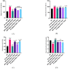
Figure 1.
Figure 1.
After 28 days of the experiment, a bar chart of the liver enzyme parameters (A) AST, (B) AST, (C) ALT, and (D) Total protein, ALP = alkaline phosphatase, ALT = alanine aminotransferase, AST = aspartate aminotransferase. #Significant difference compared with PbAc control #P<0.05; ##P<0.002; ###P<0.0001. *Significant difference compared with control *P<0.033; **P<0.002; ***P<0.0001. n=5
Figure 2.
Figure 2.
Bar charts of kidney function parameters (A) creatinine (B) Uric Acid, and (C) Urea and (D) Pb concentration after 28 days of the experiment. #Significant difference compared with PbAc control #P<0.05; ##P<0.002; ###P<0.0001. *Significant difference compared with control *P<0.033; **P<0.002; ***P<0.0001. n=5.
Figure 3.
Figure 3.
Bar charts of oxidative stress parameters ((A) MDA, (B) SOD, (C) CAT and (D) GSH) after 28 days of the experiment. GSH = glutathione reductase, CAT = catalase, MDA = malondialdehyde, SOD = superoxide dismutase #Significant difference compared with PbAc control #P<0.05; ##P<0.002; ###P<0.0001. *Significant difference compared with control *P<0.033; **P<0.002; ***P<0.0001. n=5.
Figure 4.
Figure 4.
Bar charts of Inflammatory markers (A) TNF-α and (B) IL-6. TNF-α =, Tumour Necrosis Factor Alpha.; IL-1 β = interleukin 1β #Significant difference compared with PbAc control #P<0.05; ##P<0.002; ###P<0.0001. *Significant difference compared with control (NC) *P<0.033; **P<0.002; ***P<0.0001. n=5.
Figure 5.
Figure 5.
Control liver photomicrograph (A) and AOLE treated liver (C) showed with normal hepatocytes (green arrow) and central vein (CV); PbAc treated liver photomicrograph (B) with remarkable degenerating hepatocytes and fat hepatocellular vacuoles (blue arrow). AOLE 200/400mg/kg + 50 mg/kg PbAc (D&E) demonstrated mild inflamed hepatocyte (green arrow) and normal hepatocyte; Succimer + 50 mg/kg PbAc treated rats (F) demonstrated mild vacuolated hepatocyte (blue arrow). H and E staining at X200 magnification.
Figure 6.
Figure 6.
(A) Composite photomicrographs of control (A) and liver of rats treated with only AOLE (C), showing normal distribution cytoarchitecture of the liver; PbAc treated liver photomicrograph (B) showing excess accumulation of PbAc residue within the liver cytoarchitecture (blue arrow). AOLE 200/400mg/kg + 50 mg/kg PbAc (D&E) demonstrated mild accumulation of PbAc within the hepatocyte (blue arrow) in a dose dependent manner, the hight dose the PbAc concentration is very minimal; Succimer + 50 mg/kg PbAc treated rats (F) demonstrated mild deposit of the PbAc (red arrow). sodium rhodizonate staining at X200 magnification.
Figure 7.
Figure 7.
Composite photomicrographs of the kidney from the control group (A) and AOLE (C) show a normal glomerulus (G) and renal tubules; the PbAc-treated micrograph (B) shows spontaneous lipid vacuolation of the glomerulus (G); the AOLE 200mg/kg + 50 mg/kg PbAc micrograph (D) displays mildly degenerated and spontaneous lipid vacuolation of the glomerulus; the AOLE 400mg/kg + 50 mg/kg PbAc micrograph (E) showing very normal rental tubules and normal glomerulus (G), Succimer treated group (E), exhibited mild obliterative form of glomerulus (G). H and E staining at X200 magnification.

Similar articles

References

    1. Wu X, Cobbina SJ, Mao G, Xu H, Zhang Z, Yang L. A review of toxicity and mechanisms of individual and mixtures of heavy metals in the environment. Environ Sci Pollut Res Int. 2016;23(9):8244–8259. doi: 10.1007/s11356-016-6333-x. - DOI - PubMed
    1. Obeng-Gyasi E. Sources of lead exposure in various countries. Rev Environ Health. 2019;34(1):25–34. doi: 10.1515/reveh-2018-0037. - DOI - PubMed
    1. Mudipalli A. Lead hepatotoxicity & potential health effects. Indian J Med Res. 2007;126(6):518–527. - PubMed
    1. Al-Megrin WA, Alkhuriji AF, Yousef AOS, Metwally DM, Habotta OA, Kassab RB, et al. Antagonistic efficacy of luteolin against lead acetate exposure-associated with hepatotoxicity is mediated via antioxidant, anti-Inflammatory, and antiapoptotic activities. Antioxidants (Basel) 2019;9(1):10. doi: 10.3390/antiox9010010. - DOI - PMC - PubMed
    1. Mujaibel LM, Kilarkaje N. Mitogen-activated protein kinase signaling and its association with oxidative stress and apoptosis in lead-exposed hepatocytes. Environ Toxicol. 2015;30(5):513–529. doi: 10.1002/tox.21928. - DOI - PubMed

LinkOut - more resources