. 2023 Nov:106:41-94.
doi: 10.3114/sim.2023.106.02. Epub 2023 Jul 19.

Diversity of colacosome-interacting mycoparasites expands the understanding of the evolution and ecology of Microbotryomycetes

Affiliations

Diversity of colacosome-interacting mycoparasites expands the understanding of the evolution and ecology of Microbotryomycetes

N Schoutteten et al. Stud Mycol. 2023 Nov.

Abstract

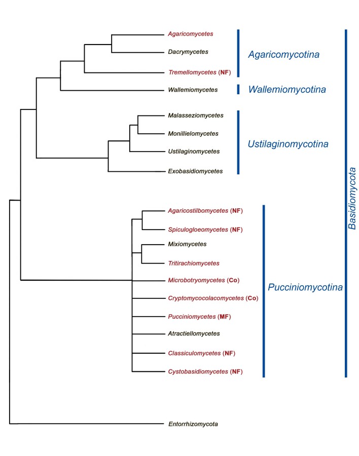
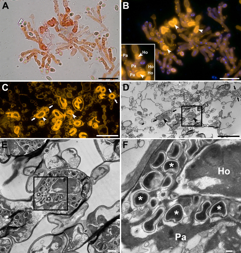
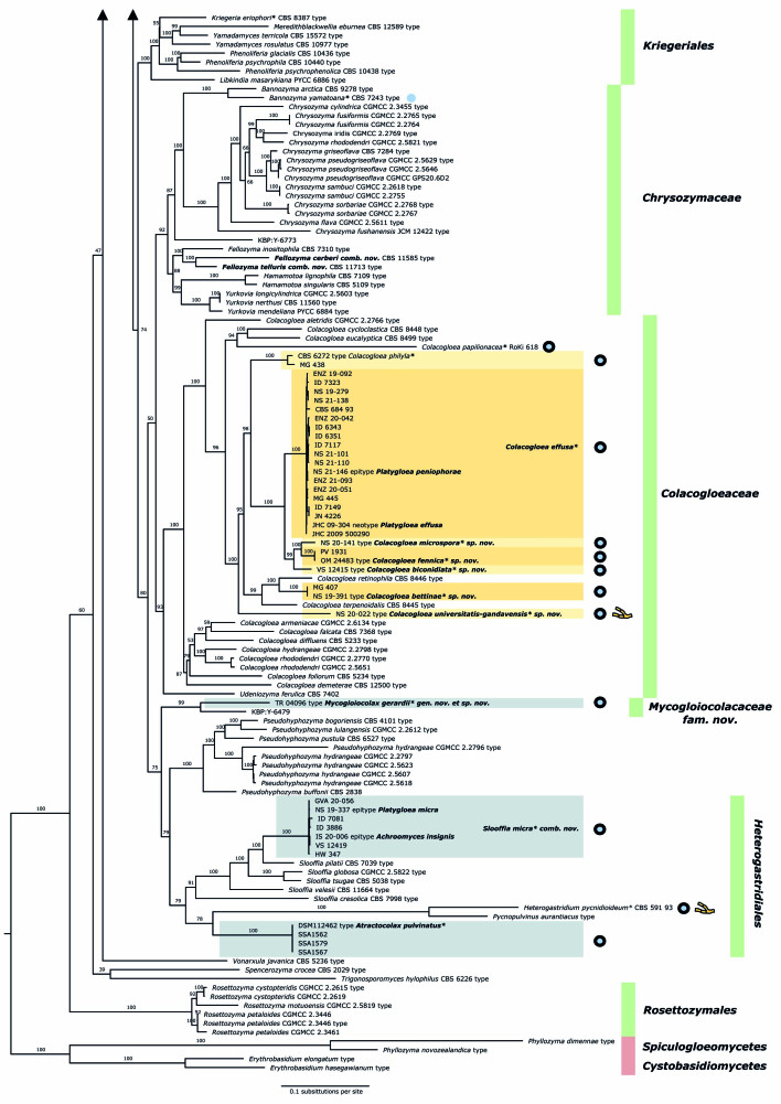
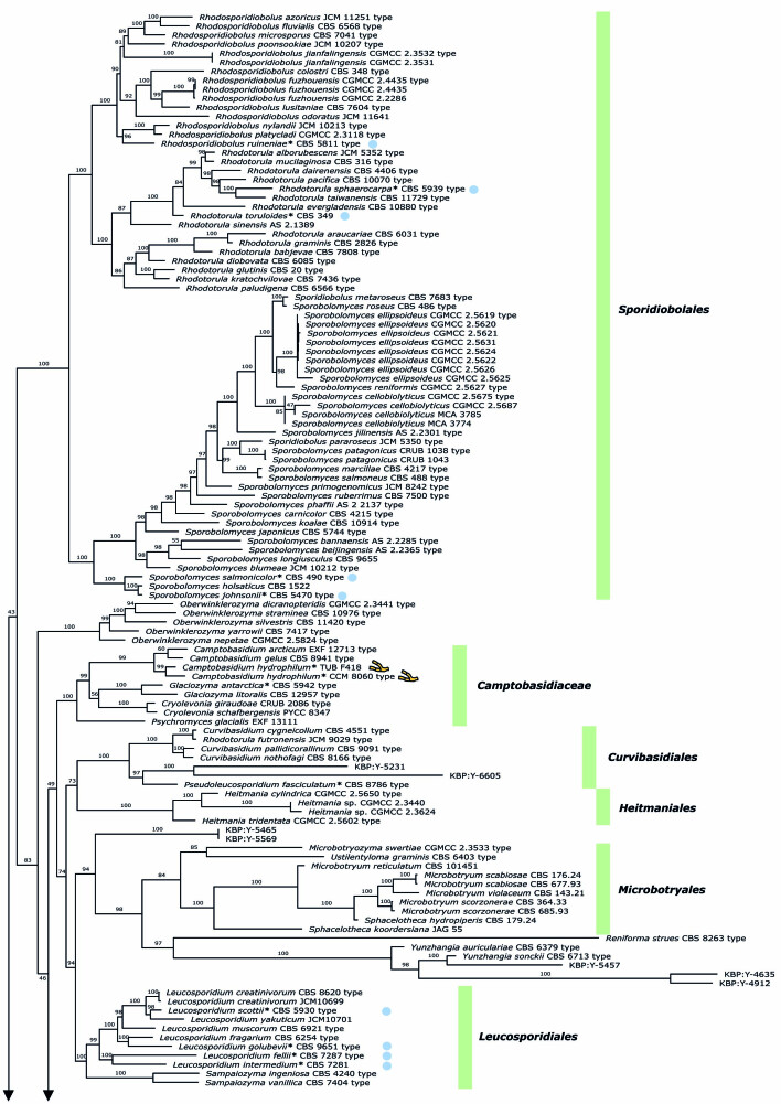
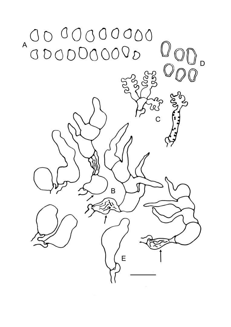
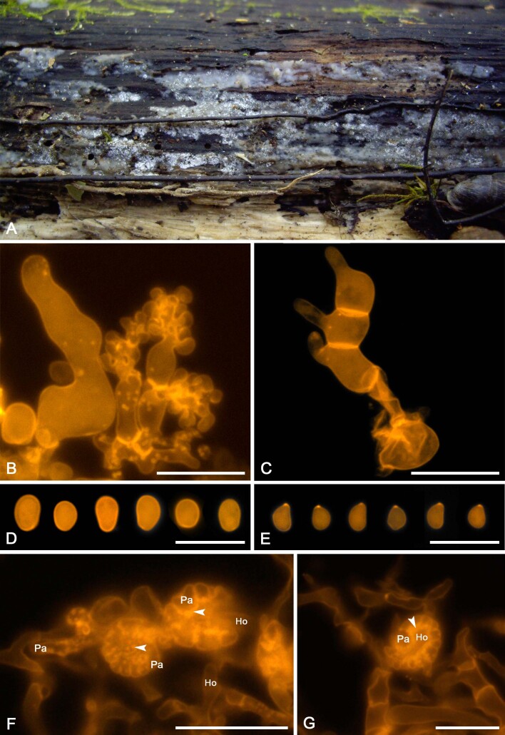
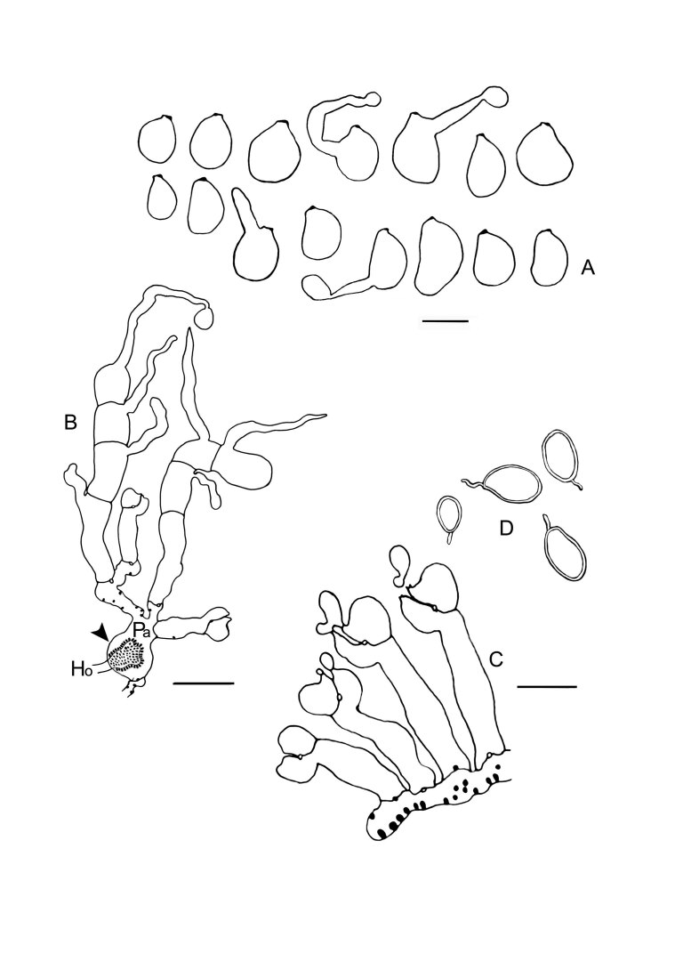
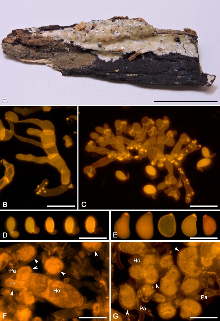
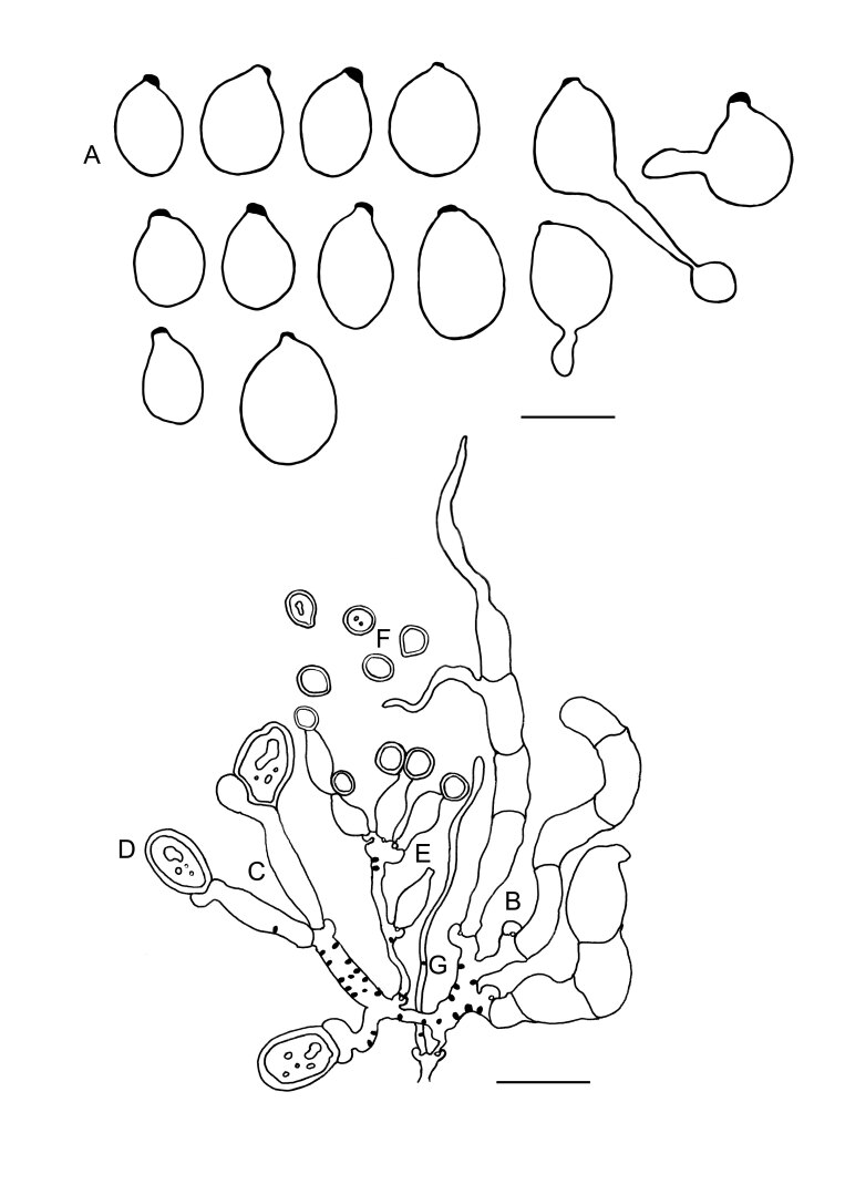
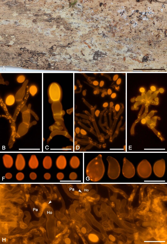
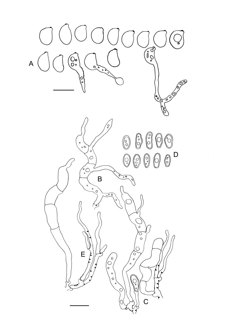
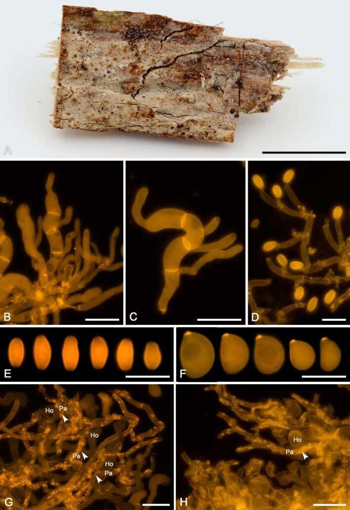
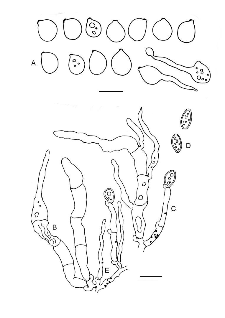
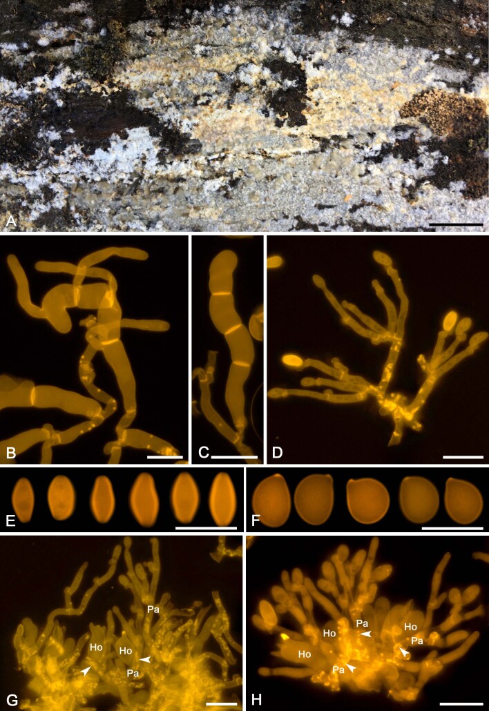
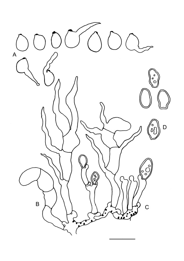
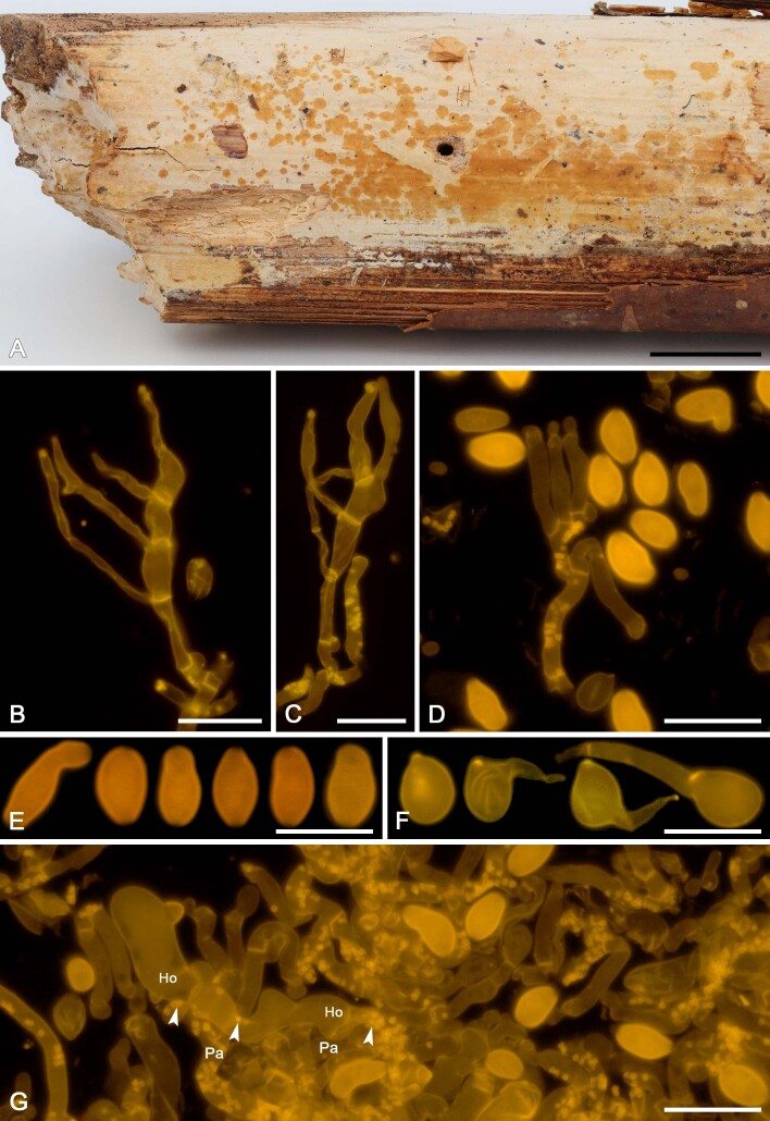
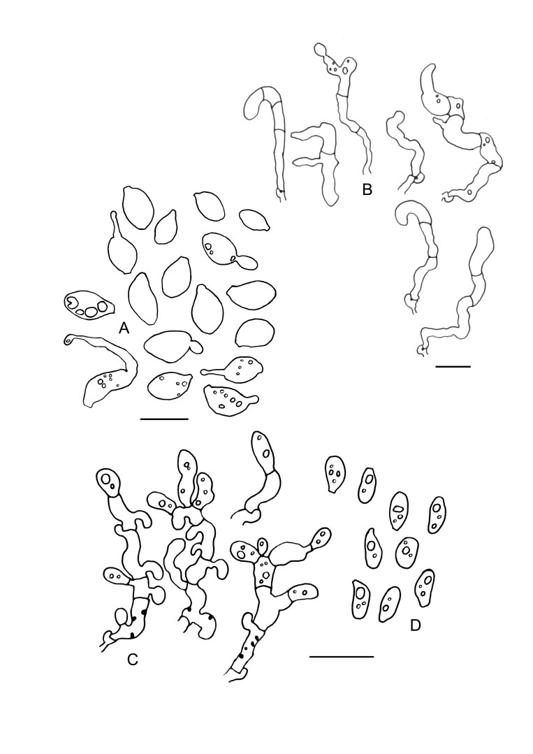
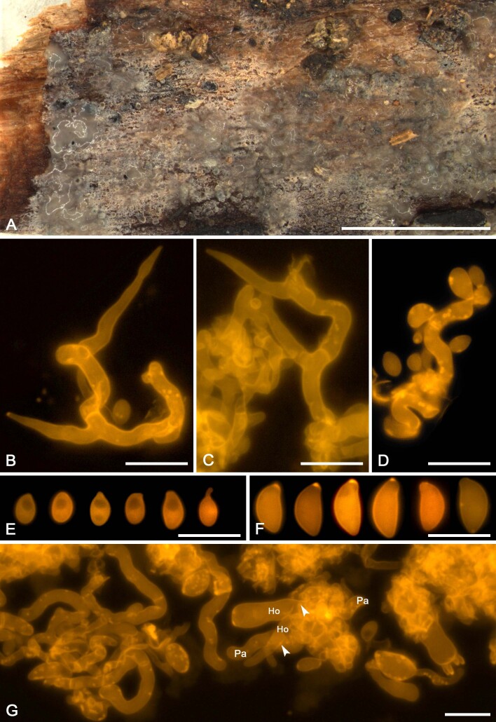
Mycoparasites in Basidiomycota comprise a diverse group of fungi, both morphologically and phylogenetically. They interact with their hosts through either fusion-interaction or colacosome-interaction. Colacosomes are subcellular structures formed by the mycoparasite at the host-parasite interface, which penetrate the parasite and host cell walls. Previously, these structures were detected in 19 fungal species, usually by means of transmission electron microscopy. Most colacosome-forming species have been assigned to Microbotryomycetes (Pucciniomycotina, Basidiomycota), a highly diverse class, comprising saprobic yeasts, mycoparasites, and phytoparasites. In general, these myco- and phytoparasites are dimorphic organisms, with a parasitic filamentous morph and saprobic yeast morph. We investigated colacosome-forming mycoparasites based on fungarium material, freshly collected specimens, and cultures of yeast morphs. We characterised the micromorphology of filamentous morphs, the physiological characteristics of yeast morphs, and inferred phylogenetic relationships based on DNA sequence data from seven loci. We outline and employ an epifluorescence-based microscopic method to assess the presence and organisation of colacosomes. We describe five new species in the genus Colacogloea, the novel dimorphic mycoparasite Mycogloiocolax gerardii, and provide the first report of a sexual, mycoparasitic morph in Colacogloea philyla and in the genus Slooffia. We detected colacosomes in eight fungal species, which brings the total number of known colacosome-forming fungi to 27. Finally, we revealed three distinct types of colacosome organisation in Microbotryomycetes. Taxonomic novelties and typifications: New family: Mycogloiocolacaeae Schoutteten & Yurkov; New genus: Mycogloiocolax Schoutteten & Rödel; New species: Colacogloea bettinae Schoutteten & Begerow, C. biconidiata Schoutteten, C. fennica Schoutteten & Miettinen, C. microspora Schoutteten, C. universitatis-gandavensis Schoutteten & Verbeken, Mycogloiocolax gerardii Schoutteten & Rödel; New combinations: Slooffia micra (Bourdot & Galzin) Schoutteten, Fellozyma cerberi (A.M. Yurkov et al.) Schoutteten & Yurkov, Fellozyma telluris (A.M. Yurkov et al.) Schoutteten & Yurkov; Epitypifications (basionyms): Achroomyces insignis Hauerslev, Platygloea micra Bourdot & Galzin, Platygloea peniophorae Bourdot & Galzin; Lectotypification (basionym): Platygloea peniophorae Bourdot & Galzin Citation: Schoutteten N, Yurkov A, Leroux O, Haelewaters D, Van Der Straeten D, Miettinen O, Boekhout T, Begerow D, Verbeken A (2023). Diversity of colacosome-interacting mycoparasites expands the understanding of the evolution and ecology of Microbotryomycetes. Studies in Mycology 106: 41-94. doi: 10.3114/sim.2022.106.02.

Keywords: Basidiomycota; Pucciniomycotina; Transmission Electron Microscopy; epifluorescence microscopy; molecular phylogeny; new taxa; systematics; yeasts.


Conflict of interest statement

The authors declare that there is no conflict of interest.

Figures

Fig. 1
Fig. 1
Phylogram of Basidiomycota, interpretation based on of different previously published phylogenetic reconstructions of this phylum (Aime et al. 2006, Bauer et al. 2006, Schell et al. 2011, Wang et al. 2015a, Zhao et al. 2017, He et al. 2019). Names of classes indicated in red represent those comprising mycoparasitic species. Colacosome-interacting (Co) mycoparasites belong to Cryptomycocolacomycetes and Microbotryomycetes. Nanopore fusioninteracting (NF) mycoparasites belong to Agaricostilbomycetes, Classiculomycetes, Cystobasidiomycetes, Spiculogloeomycetes and Tremellomycetes. Micropore fusion-interacting (MF) mycoparasites belong to Pucciniomycetes
Fig. 2
Fig. 2
Brightfield, epifluorescence and transmission electron microscopy (TEM) of Colacogloea Universitatis-gandavensis sp. nov. A, B. Whole-mount preparation, stained with Congo red and DAPI, visualised using brightfield (A) and epifluorescence (B) microscopy. Epifluorescence microscopy facilitates fast detection of colacosomes as they exhibit bright fluorescence signals. Inset shows the intricate host–parasite (Ho-Pa) interface. Arrowheads indicate regions of colacosome clustering. Note the occurrence of individual colacosomes in parasite tissue (bright spots). C, D. Serial sections of a Spurr-embedded sample, showing the same region. Corresponding structures are indicated with arrows. (C) Section stained with Congo red and visualised using epifluorescence microscopy. (D) Equivalent serial section of the same region as in (C), visualised using TEM. E, F. High-magnification details of colacosome clusters (arrowheads), composed of many individual colacosomes (asterisks), arranged in parasitic hyphae (Pa) along the host–parasite interface (Ho-Pa), showing their typical electron dense cores. Scale bars: A–D = 20 μm, E = 10 μm, F = 200 nm.
Fig. 3
Fig. 3
Phylogenetic relationships of colacosome-forming species in Microbotryomycetes based on a seven-locus ML tree inference. Species names in bold indicate taxonomic novelties. Species which were explicitly investigated for the presence of colacosomes are indicated with a * symbol behind the species name. Species for which the presence of colacosomes was positively assessed are indicated by blue-filled circles. Blue circles with black outline indicate species which have been isolated as a mycoparasite and for which an interaction with a host was reported. Blue circles without outline indicate species which were only reported to form colacosomes in pure culture. Species for which currently only a filamentous morph was observed are indicated by a branching hyphae icon, for all other species in the tree, at least a yeast morph is known. Clades investigated in detail in this study are indicated with boxes. Boxes in yellow tones represent the Colacogloea effusa complex. Green vertical lines represent the highest described taxon available for species in the tree (family or order). Numbers on branches indicate ultrafast bootstrap values. Cystobasidiomycetes and Spiculogloeomycetes are used as outgroup.
Fig. 3
Fig. 3
Phylogenetic relationships of colacosome-forming species in Microbotryomycetes based on a seven-locus ML tree inference. Species names in bold indicate taxonomic novelties. Species which were explicitly investigated for the presence of colacosomes are indicated with a * symbol behind the species name. Species for which the presence of colacosomes was positively assessed are indicated by blue-filled circles. Blue circles with black outline indicate species which have been isolated as a mycoparasite and for which an interaction with a host was reported. Blue circles without outline indicate species which were only reported to form colacosomes in pure culture. Species for which currently only a filamentous morph was observed are indicated by a branching hyphae icon, for all other species in the tree, at least a yeast morph is known. Clades investigated in detail in this study are indicated with boxes. Boxes in yellow tones represent the Colacogloea effusa complex. Green vertical lines represent the highest described taxon available for species in the tree (family or order). Numbers on branches indicate ultrafast bootstrap values. Cystobasidiomycetes and Spiculogloeomycetes are used as outgroup.
Fig. 4
Fig. 4
Slooffia micra comb. nov. (KH7222) line drawings. A. Basidiospores. B. Basidia. C. Cluster of conidiophores. D. Conidia. E. Basidioles, arrows indicate probasidia. Black dots represent colacosomes. Scale bar = 10 μm.
Fig. 5
Fig. 5
Slooffia micra comb. nov. (KH7222). A. Basidiome (VS 12419). B. Basidiole (left) and cluster of conidiophores (right), note colacosomes in hyphae and conidiophores. C. Three-septate basidium with three sterigmata, the first cell of the basidium and the probasidium are collapsed. D. Conidia. E. Basidiospores. F. Host–parasite interface, Pa = parasite hyphae, Ho = host hyphae, arrowheads indicate some positions of colacosomes. Scale bars = 10 μm.
Fig. 6
Fig. 6
Colacogloea bettinae sp. nov. (NS 19-391) line drawings. A. Basidiospores and germinating basidiospores with secondary spores. B. Cluster of basidia and conidiophores. C. Cluster of conidiophores, showing subsequent stages of conidiogenesis. Each conidiophore consists of two conidiogenous cells. Each cell produces a conidium, of which one grows larger than the other. Subsequently the two daughter conidia fuse, and the cellular content of the smaller conidium is transferred to the larger conidium, after which the zygoconidium is abscised. The cell wall of the smeller conidium remains attached to the larger conidium. D. Conidia. E. Gall-like cell of the parasite (Pa) enveloping a host hyphae (Ho). Black dots represent colacosomes. Note the different distribution of colacosomes in the gall-like cell and the hyphae. Scale bars = 10 μm.
Fig. 7
Fig. 7
Colacogloea bettinae sp. nov. (NS 19-391). A. Basidiome. B. Three-septate basidium with four sterigmata, note one attached basidiospore. C. Cluster of conidiophores and conidia. D. Conidia. E. Basidiospores. F, G. Host–parasite interface, Pa = parasite cell, Ho = host cell, arrowheads indicate gall-like cells of the parasite enveloping host hyphae, colacosomes are formed along the contact interface within these galls. Scale bars: A = 1 cm; B–G = 10 μm.
Fig. 8
Fig. 8
Colacogloea biconidiata sp. nov. (VS 12415) line drawings. A. Basidiospores and germinating basidiospores by hyphae and secondary spores. B. Cluster of basidia and basidioles. C. Type-1 conidiophores. D. Type-1 conidia with basal clamps. E. Type-2 conidiophores. F. Type-2 conidia. G. Hyphidium. Black dots represent colacosomes. Scale bars = 10 μm.
Fig. 9
Fig. 9
Colacogloea biconidiata sp. nov. (VS 12415). A. Basidiome.. B. Three-septate basidium with four sterigmata, note hyphae with numerous colacosomes and three well stained conidia. C. Type-1 conidiophore and attached conidium with basal clamp. D. Cluster of type-1 conidiophores and conidia. E. Cluster of type-2 conidiophores. F. Upper row represent type-1 conidia, lower row represent type-2 conidia. G. Basidiospores. H. Host–parasite interface, Pa = parasite cell, Ho = host cell, arrowheads indicate some positions of colacosomes. Scale bars: A = 1 cm; B–G = 10 μm.
Fig. 10
Fig. 10
Colacogloea effusa (NS 21-146) line drawings. A. Basidiospores and germinating basidiospores by hyphae and secondary spores. B. Basidia. C. Conidiophore. D. Conidia. E. Hyphidia. Black dots represent colacosomes. Scale bars = 10 μm.
Fig. 11
Fig. 11
Colacogloea effusa (NS 21-146). A. Basidiome. B. Cluster of basidium, basidiole and hyphidia. C. Three-septate basidium with four sterigmata. D. Conidiophores. E. Conidia. F. Basidiospores. G, H. Host–parasite interface, Pa = parasite cell, Ho = host cell, arrowheads indicate some positions of colacosomes. Scale bars: A = 1 cm; B–H = 10 μm.
Fig. 12
Fig. 12
Colacogloea fennica sp. nov. (OM 22483) line drawings. A. Basidiospores and germinating basidiospores by secondary spores. B. Basidium and basidiole. C. Conidiophore and basidium. D. Conidia, note the clamp at the base of conidia. E. Hyphidium. Black dots represent colacosomes. Scale bar = 10 μm.
Fig. 13
Fig. 13
Colacogloea fennica sp. nov. (OM 22483). A. Basidiome. B. Three-septate basidium with four sterigmata, note colacosomes in hyphae bearing the basidium but not in the basidium. C. Basidiole. D. Cluster of conidiophores and conidia, note colacosomes in hyphae but not in conidiophores. E. Conidia. F. Basidiospores. G, H. Host–parasite interface, Pa = parasite cell, Ho = host cell, arrowheads indicate some positions of colacosomes. Scale bars: A = 1 cm; B–G = 10 μm.
Fig. 14
Fig. 14
Colacogloea microspora sp. nov. (NS 20-141) line drawings. A. Basidiospores and germinating basidiospores by hyphae and secondary spores. B. Two basidia and basidiole. C. Conidiophore. D. Conidia. Black dots represent colacosomes. Scale bar = 10 μm.
Fig. 15
Fig. 15
Colacogloea microspora sp. nov. (NS 20-141). A. Basidiome. B, C. Three-septate basidia with sterigmata, note colacosomes in hyphae. D. Cluster of conidiophores and conidia, note colacosomes in hyphae. E. Conidia. F. Basidiospores. G. Host–parasite interface, Pa = parasite cell, Ho = host cell, arrowheads indicate some positions of colacosomes. Scale bars: A = 1 cm; B–G = 10 μm.
Fig. 16
Fig. 16
Colacogloea philyla (MG 438) line drawings. A. Basidiospores and germinating basidiospores by hyphae and secondary spores. B. Basidium. C. Conidiophore. D. Conidia. E. Hyphidium. Black dots represent colacosomes. Scale bars = 10 μm.
Fig. 17
Fig. 17
Colacogloea philyla (MG 438). A. Basidiome. B. Cluster of three-septate basidia, basidioles and hyphidia. C. Two-septate basidium with apical sterigma. D. Cluster of conidiophores and conidia, note the colacosomes in the hyphae but not in the conidiophores. E. Conidia. F. Basidiospores. G, H. Host–parasite interface, Pa = parasite cell, Ho = host cell, arrowheads indicate some positions of colacosomes. Scale bars: A = 1 cm; B–H = 10 μm.
Fig. 18
Fig. 18
Colacogloea universitatis-gandavensis sp. nov. (H6086094) line drawings. A. Basidiospores and germinating basidiospores by secondary spores. B. Cluster of basidia and conidiophores. C. Conidiophore. D. Conidiophores showing subsequent stages of conidiogenesis and conidia. E. Gall-like cell of the parasite (Pa) enveloping a host hyphae (Ho). Black dots represent colacosomes. Scale bars = 10 μm.
Fig. 19
Fig. 19
Colacogloea universitatis-gandavensis sp. nov. (NS 21-013). A. Basidiome. B. Cluster of three-septate basidium with four sterigmata and conidiophores, note one attached basidiospore. C. Cluster of basidium, basidiole and conidiophores. D. Cluster of conidiophores, note the colacosomes in hyphae. E. Conidia. F. Basidiospores. G. Host–parasite interface, Pa = parasite cell, Ho = host cell, arrowheads indicate some gall-like cells of the parasite enveloping host hyphae, colacosomes are formed along the contact interface within these galls. Scale bars: A = 1 cm; B–G = 10 μm.
Fig. 20
Fig. 20
Mycogloiocolax gerardii sp. nov. (TR 04096) line drawings. A. Basidiospores and germinating basidiospores by budding, hyphae and secondary spores. B. Basidia and basidioles. C. Conidiophores. D. Conidia. Black dots represent colacosomes. Scale bars = 10 μm.
Fig. 21
Fig. 21
Mycogloiocolax gerardii sp. nov. (TR 04096). A. Basidiome. B, C. One-septate basidia with two sterigmata. D. Conidiophore and conidia. E. Conidia. F. Basidiospores. G. Host–parasite interface, Pa = parasite hyphae, Ho = host hyphae, arrowheads indicate some positions of colacosomes. Scale bar: A = cm; B–G = 10 μm.

References

    1. Aime MC, Castlebury LA, Abbasi M. et al. (2018). Competing sexual and asexual generic names in Pucciniomycotina and Ustilaginomycotina (Basidiomycota) and recommendations for use. IMA Fungus 9: 75–89. - PMC - PubMed
    1. Aime MC, Matheny PB, Henk DA. et al. (2006).An overview of the higher-level classification of Pucciniomycotina based on combined analyses of nuclear large and small subunit rDNA sequences. Mycologia 98: 896–905. - PubMed
    1. Aime MC, Toome M, McLaughlin DJ. (2014). Pucciniomycotina. In: The Mycota VII. Systematics and evolution. Part A. (McLaughlin DJ, Spatafora JW eds). Springer, Germany: 271–294.
    1. Ali SS, Wu J, Xie R. et al. (2017). Screening and characterizing of xylanolytic and xylose-fermenting yeasts isolated from the wood-feeding termite, Reticulitermes chinensis. PLoS One 12: e0181141. - PMC - PubMed
    1. Bandoni RJ. (1956). A preliminary survey of the genus Platygloea. Mycologia 48: 821–840.

LinkOut - more resources