

Spatial normalization for voxel-based lesion symptom mapping: impact of registration approaches
- PMID: 38298911
- PMCID: PMC10828036
- DOI: 10.3389/fnins.2024.1296357
Spatial normalization for voxel-based lesion symptom mapping: impact of registration approaches
Abstract
Background: Voxel-based lesion symptom mapping (VLSM) assesses the relation of lesion location at a voxel level with a specific clinical or functional outcome measure at a population level. Spatial normalization, that is, mapping the patient images into an atlas coordinate system, is an essential pre-processing step of VLSM. However, no consensus exists on the optimal registration approach to compute the transformation nor are downstream effects on VLSM statistics explored. In this work, we evaluate four registration approaches commonly used in VLSM pipelines: affine (AR), nonlinear (NLR), nonlinear with cost function masking (CFM), and enantiomorphic registration (ENR). The evaluation is based on a standard VLSM scenario: the analysis of statistical relations of brain voxels and regions in imaging data acquired early after stroke onset with follow-up modified Rankin Scale (mRS) values.
Materials and methods: Fluid-attenuated inversion recovery (FLAIR) MRI data from 122 acute ischemic stroke patients acquired between 2 and 3 days after stroke onset and corresponding lesion segmentations, and 30 days mRS values from a European multicenter stroke imaging study (I-KNOW) were available and used in this study. The relation of the voxel location with follow-up mRS was assessed by uni- as well as multi-variate statistical testing based on the lesion segmentations registered using the four different methods (AR, NLR, CFM, ENR; implementation based on the ANTs toolkit).
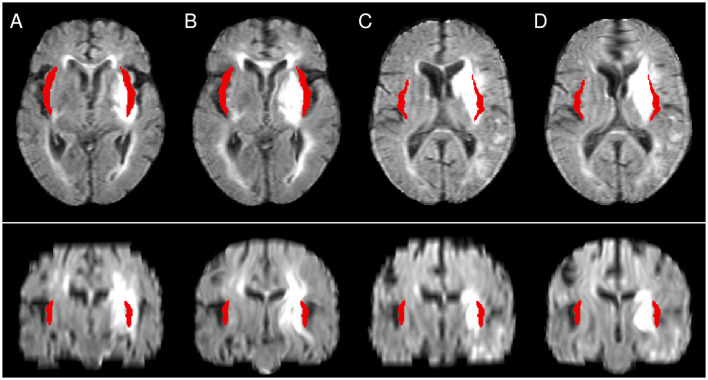
Results: The brain areas evaluated as important for follow-up mRS were largely consistent across the registration approaches. However, NLR, CFM, and ENR led to distortions in the patient images after the corresponding nonlinear transformations were applied. In addition, local structures (for instance the lateral ventricles) and adjacent brain areas remained insufficiently aligned with corresponding atlas structures even after nonlinear registration.
Conclusions: For VLSM study designs and imaging data similar to the present work, an additional benefit of nonlinear registration variants for spatial normalization seems questionable. Related distortions in the normalized images lead to uncertainties in the VLSM analyses and may offset the theoretical benefits of nonlinear registration.
Keywords: VLSM; image registration; neuroimaging; spatial normalization; stroke.
Copyright © 2024 Jühling, Rajashekar, Cheng, Hilgetag, Forkert and Werner.
Conflict of interest statement
The authors declare that the research was conducted in the absence of any commercial or financial relationships that could be construed as a potential conflict of interest.
Figures


Similar articles
-
Improved accuracy of lesion to symptom mapping with multivariate sparse canonical correlations.Neuropsychologia. 2018 Jul 1;115:154-166. doi: 10.1016/j.neuropsychologia.2017.08.027. Epub 2017 Sep 5. Neuropsychologia. 2018. PMID: 28882479
-
Is VLSM a valid tool for determining the functional anatomy of the brain? Usefulness of additional Bayesian network analysis.Neuropsychologia. 2018 Dec;121:69-78. doi: 10.1016/j.neuropsychologia.2018.10.003. Epub 2018 Oct 25. Neuropsychologia. 2018. PMID: 30449718
-
Multiclass Support Vector Machine-Based Lesion Mapping Predicts Functional Outcome in Ischemic Stroke Patients.PLoS One. 2015 Jun 22;10(6):e0129569. doi: 10.1371/journal.pone.0129569. eCollection 2015. PLoS One. 2015. PMID: 26098418 Free PMC article.
-
Connectome-based lesion-symptom mapping (CLSM): A novel approach to map neurological function.Neuroimage Clin. 2017 Aug 24;16:461-467. doi: 10.1016/j.nicl.2017.08.018. eCollection 2017. Neuroimage Clin. 2017. PMID: 28884073 Free PMC article. Review.
-
On the validity of lesion-behaviour mapping methods.Neuropsychologia. 2018 Jul 1;115:17-24. doi: 10.1016/j.neuropsychologia.2017.07.035. Epub 2017 Aug 3. Neuropsychologia. 2018. PMID: 28782546 Review.
Cited by
-
Current Clinical Applications of Structural MRI in Neurological Disorders.J Clin Neurol. 2025 Jul;21(4):277-293. doi: 10.3988/jcn.2025.0185. J Clin Neurol. 2025. PMID: 40635533 Free PMC article. Review.
References
-
- Avants B. B., Libon D. J., Rascovsky K., Boller A., McMillan C. T., Massimo L., et al. . (2014). Sparse canonical correlation analysis relates network-level atrophy to multivariate cognitive measures in a neurodegenerative population. Neuroimage 84, 698–711. 10.1016/j.neuroimage.2013.09.048 - DOI - PMC - PubMed
-
- Avants B. B., Tustison N., Song G. (2009). Advanced normalization tools (ANTS). Insight J. 2, 1–35. 10.54294/uvnhin - DOI
LinkOut - more resources
Full Text Sources
Research Materials