. 2024 Feb 20;98(2):e0165223.
doi: 10.1128/jvi.01652-23. Epub 2024 Feb 1.

SHIV-C109p5 NHP induces rapid disease progression in elderly macaques with extensive GI viral replication

Affiliations

SHIV-C109p5 NHP induces rapid disease progression in elderly macaques with extensive GI viral replication

Deepanwita Bose et al. J Virol. .

Abstract

CCR5-tropic simian/human immunodeficiency viruses (SHIV) with clade C transmitted/founder envelopes represent a critical tool for the investigation of HIV experimental vaccines and microbicides in nonhuman primates, although many such isolates lead to spontaneous viral control post infection. Here, we generated a high-titer stock of pathogenic SHIV-C109p5 by serial passage in two rhesus macaques (RM) and tested its virulence in aged monkeys. The co-receptor usage was confirmed before infecting five geriatric rhesus macaques (four female and one male). Plasma viral loads were monitored by reverse transcriptase-quantitative PCR (RT-qPCR), cytokines by multiplex analysis, and biomarkers of gastrointestinal damage by enzyme-linked immunosorbent assay. Antibodies and cell-mediated responses were also measured. Viral dissemination into tissues was determined by RNAscope. Intravenous SHIV-C109p5 infection of aged RMs leads to high plasma viremia and rapid disease progression; rapid decrease in CD4+ T cells, CD4+CD8+ T cells, and plasmacytoid dendritic cells; and wasting necessitating euthanasia between 3 and 12 weeks post infection. Virus-specific cellular immune responses were detected only in the two monkeys that survived 4 weeks post infection. These were Gag-specific TNFα+CD8+, MIP1β+CD4+, Env-specific IFN-γ+CD4+, and CD107a+ T cell responses. Four out of five monkeys had elevated intestinal fatty acid binding protein levels at the viral peak, while regenerating islet-derived protein 3α showed marked increases at later time points in the three animals surviving the longest, suggesting gut antimicrobial peptide production in response to microbial translocation post infection. Plasma levels of monocyte chemoattractant protein-1, interleukin-15, and interleukin-12/23 were also elevated. Viral replication in gut and secondary lymphoid tissues was extensive.IMPORTANCESimian/human immunodeficiency viruses (SHIV) are important reagents to study prevention of virus acquisition in nonhuman primate models of HIV infection, especially those representing transmitted/founder (T/F) viruses. However, many R5-tropic SHIV have limited fitness in vivo leading to many monkeys spontaneously controlling the virus post acute infection. Here, we report the generation of a pathogenic SHIV clade C T/F stock by in vivo passage leading to sustained viral load set points, a necessity to study pathogenicity. Unexpectedly, administration of this SHIV to elderly rhesus macaques led to extensive viral replication and fast disease progression, despite maintenance of a strict R5 tropism. Such age-dependent rapid disease progression had previously been reported for simian immunodeficiency virus but not for R5-tropic SHIV infections.

Keywords: AIDS; SHIV; aging.


Conflict of interest statement

The authors declare no conflict of interest.

Figures

Fig 1
Fig 1
In vivo adaptation and characterization of SHIVC109. (A) Schematics of in vivo adaptation of SHIVC109P3/EL78 to generate the SHIV-C109p5. An in vitro stock was generated initially from SHIVC109P3/EL78 in rhesus activated PBMCs, which was titrated intrarectally (IR) in three macaques. Next, the same stock mixed with the original SHIVC109P3/EL78 were used to infect rhesus monkey 56C to perform in vivo passage 4 adaptation. Monkey 56C was euthanized on day 14 p.i., and SHIV-C109p4 was generated from PBMCs, lymph node cells, and splenocytes activated and cultured ex vivo. This stock was similarly passaged in vivo in monkey 00-09, which was also euthanized at day 14 p.i., and SHIVC109p5 generated ex vivo cultures of activated PBMCs, lymph node cells, and splenocytes. (B) Replication kinetics of the virus in plasma (viral loads) of different passages. (C) Ex vivo viral production from 00 to 09 splenocyte, lymph node cell, and PBMC cultures. Supernatants with p27 values >300 ng were pooled to generate the high-titer SHIV-C109p5 stock. (D) SHIV-C109p5 stock virus tropism was verification by infection of Ghost (3) CXCR4+ cells and Ghost (3) CCR5+(Hi5) cells along with different viruses (SHIV CH505, SHIV-RT, HIV1 TYBE hu PBL). (E) Amino acid sequence comparison of the gp120 V1–V5 region of SHIVC109P3/EL78 and SHIV-C109p5. The six N-linked glycosylation sites are based on the ZM109F.PB4 sequence marked in red boxes. (F) Neutralization of SHIV-C109p5 was performed with a panel of monoclonal antibodies (mAbs) on TZM-bl cell line. The 50% inhibitory concentration (IC50) was determined based on the mAb dilution that caused 50% reduction of luciferase activity (RLU) compared to virus control (no mAb).
Fig 2
Fig 2
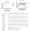
Viral replication and evolution of SHIV-C109p5 in vivo. (A) Plasma viral loads (viral copies/mL) in the five geriatric rhesus macaques infected I.V. “†” represents the time point of euthanasia. (B) Plasma viral loads of 16 young adult rhesus macaques infected with SHIV-C109p5 IV. (C) Envelope sequence (V1–V5) alignment of SHIV-C109p5 virus stock and proviral DNA sequences derived from PBMCS of all the five elderly monkeys obtained on day 14 p.i. Mismatched amino acids are indicated in red.
Fig 3
Fig 3
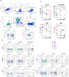
Phenotype analyses of elderly monkey PBMCs on days 0 (pre infection), 14 (acute phase), and at necropsy. (A) Gating schematics of T cell panel used to identify CD4+ and CD8+ T cells and memory subsets. Subsets analyzed were CD4+, CD8+, CD4+CD8+, and CD4CD8 populations within CD3+ gates. Further categorization was performed into central memory (CD28+CD95+), effector memory (CD28CD95+), and naïve (CD28CD95) subsets. (B) Percentage loss of CD4+ and CD4+/CD8+ cells with days post infection relative to initial percentage of these cells calculated as (%CD4+ cell on day 14) × 100/(%CD4+ cells on day 0). (C) Percentage of CD4+, CD8+, CD4+CD8+ double-positive, and CD4CD8 double-negative populations based on total CD3+ cells. (D) Percentages of CD4+ and CD8+ memory subsets based on total CD4+ and CD8+ T cells, respectively. (E) Percentages of CD4+CD8+ and CD4CD8 memory subsets based on total CD4+CD8+ and CD4CD8 T cells, respectively. Statistical differences between two groups (day 14 or necropsy vs day 0, or day 14 vs necropsy) were evaluated by the Wilcoxon matched-pairs signed rank test. The level of significance is indicated by P-values as follows: *P < 0.05; **P < 0.01; ns, not significant.
Fig 4
Fig 4
Analysis of myeloid and B cells on all five elderly monkey PBMCs from day 0 (pre infection), day 14 (acute phase), and necropsy. (A) Gating of myeloid and B cell populations. CD20CD3HLADR+ cells were gated for pDC and mDC, while the CD20+ cells were gated for mature and memory B cells. (B) Gating and percentages for pDC and mDC for all five elderly monkey for day 0 baseline, day 14, and necropsy. (C) Percentages of B cells gated on lymphocytes, monocytes gated on lymphocytes-monocytes, mDCs and pDCs gated on Lin−HLA-DR+ in all five elderly monkey. Statistical differences between two groups (day 14 or necropsy vs day 0, or day 14 vs necropsy) were evaluated by the Wilcoxon matched-pairs signed rank test. The level of significance is indicated by P-values as follows: *P < 0.05; ns, not significant.
Fig 5
Fig 5
HIV-1 Env clade C binding IgG titers in sequential plasma samples from the five elderly monkeys. Curves shown are representative of duplicate experiments. Mean titer values are summarized at each indicated time point. Symbol (*) represents each animal at necropsy.
Fig 6
Fig 6
SHIV-specific CD4+ and CD8+ T cell immune responses during SHIV-C109p5 infection. Only the two elderly macaques surviving the acute phase, BV16 (days 28, 55, and 87) and X388A (days 28 and 55) were included, since responses from the other three acute animals were negative. PBMCs were stimulated with either HIV1 consensus clade C Env N terminal or C terminal peptide pools or SIVmac239 Gag peptide pool (2 mg/mL) or dimethyl sulfoxide (DMSO) (negative control). Net frequencies of antigen-specific CD4+ and CD8+ T cell responses are presented after subtraction of the negative DMSO stimulated values.
Fig 7
Fig 7
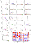
Multiplex evaluation of plasma biomarkers. (A) Pro-inflammatory immune biomarkers in plasma from five elderly monkey were analyzed by multiplex assay for different time points post infection. (B) REG3α and I-FABP levels were determined by ELISA from the plasma of all five elderly macaques at different time points post infection. (C) Fold changes in analyte levels over time in plasma samples from SHIV-C109p5-infected elderly monkeys.
Fig 8
Fig 8
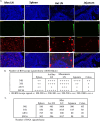
Evaluation of SHIV-C109p5 expression in tissues using RNAscope. (A) Representative images of axillary lymph node (Axi LN), mesenteric lymph node (Mes LN), spleen, and jejunum (40×) from monkey 34K (day 21 p.i.) showing abundant vRNA+ cells in lymphoid tissues and gut. Images were acquired on an Olympus IX71 microscope using Cell Sens software. Shown are DAPI nucleus in blue, FITC for verification of background autofluorescence, and TRITC SHIV-specific RNA signals, and the last row shows merged images. (B and C) Quantitation of SHIV mRNA signals in the various tissues analyzed in a single frame of 280 × 160 µm tissue areas counted for each tissue.

References

    1. Novitsky V, Smith UR, Gilbert P, McLane MF, Chigwedere P, Williamson C, Ndung’u T, Klein I, Chang SY, Peter T, Thior I, Foley BT, Gaolekwe S, Rybak N, Gaseitsiwe S, Vannberg F, Marlink R, Lee TH, Essex M. 2002. Human immunodeficiency virus type 1 subtype C molecular phylogeny: consensus sequence for an AIDS vaccine design? J Virol 76:5435–5451. doi:10.1128/JVI.76.11.5435-5451.2002 - DOI - PMC - PubMed
    1. Evans DT, Silvestri G. 2013. Nonhuman primate models in AIDS research. Curr Opin HIV AIDS 8:255–261. doi:10.1097/COH.0b013e328361cee8 - DOI - PMC - PubMed
    1. Del Prete GQ, Lifson JD, Keele BF. 2016. Nonhuman primate models for the evaluation of HIV-1 preventive vaccine strategies: model parameter considerations and consequences. Curr Opin HIV AIDS 11:546–554. doi:10.1097/COH.0000000000000311 - DOI - PMC - PubMed
    1. Hatziioannou T, Evans DT. 2012. Animal models for HIV/AIDS research. Nat Rev Microbiol 10:852–867. doi:10.1038/nrmicro2911 - DOI - PMC - PubMed
    1. Johnson PR, Hirsch VM. 1992. SIV infection of macaques as a model for AIDS pathogenesis. Int Rev Immunol 8:55–63. doi:10.3109/08830189209056641 - DOI - PubMed

Publication types

MeSH terms