Review
. 2024 Jul 1;104(3):1061-1119.
doi: 10.1152/physrev.00008.2023. Epub 2024 Feb 1.

Ceramides are fuel gauges on the drive to cardiometabolic disease

Affiliations
Review

Ceramides are fuel gauges on the drive to cardiometabolic disease

Joseph L Wilkerson et al. Physiol Rev. .

Abstract

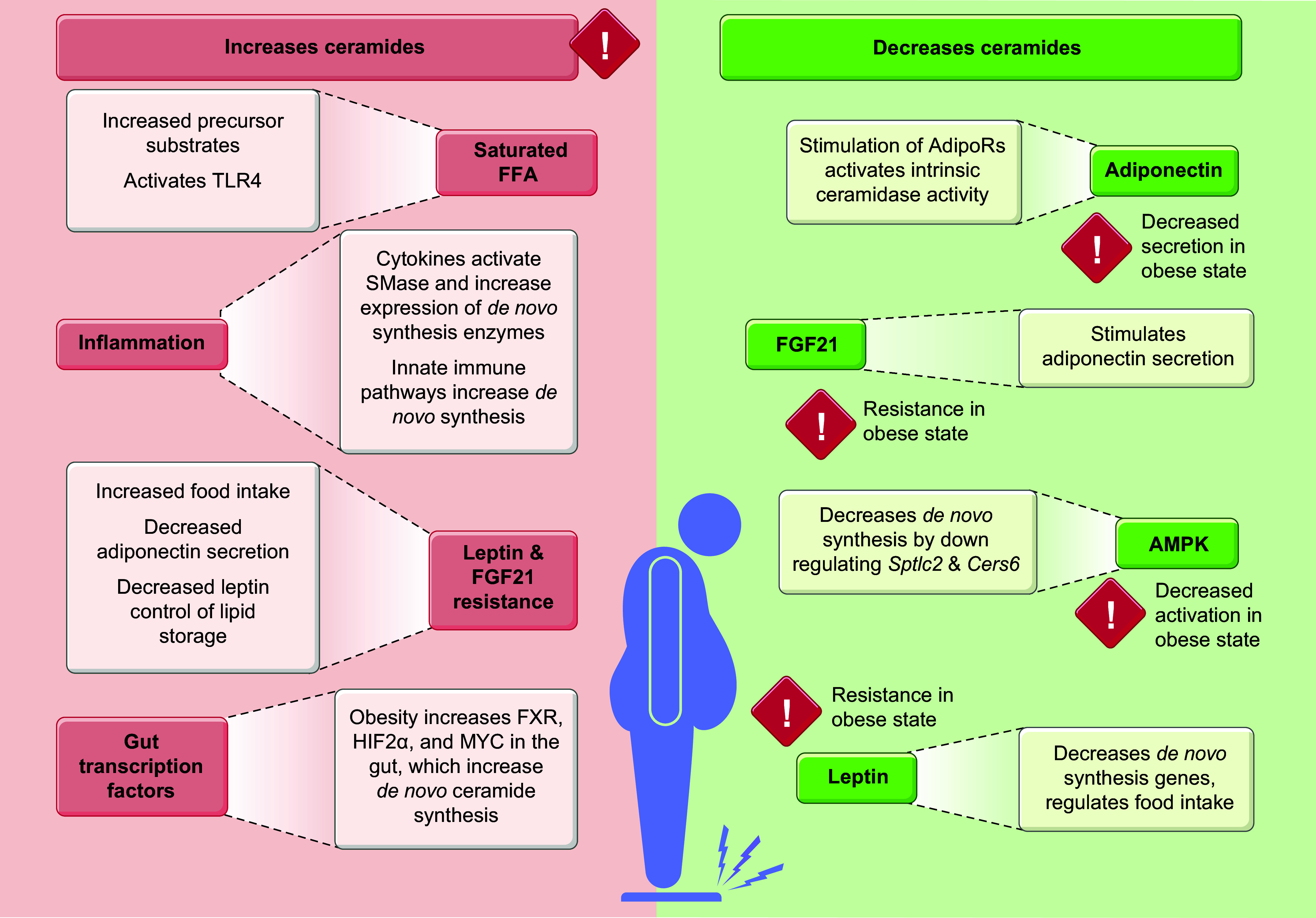
Ceramides are signals of fatty acid excess that accumulate when a cell's energetic needs have been met and its nutrient storage has reached capacity. As these sphingolipids accrue, they alter the metabolism and survival of cells throughout the body including in the heart, liver, blood vessels, skeletal muscle, brain, and kidney. These ceramide actions elicit the tissue dysfunction that underlies cardiometabolic diseases such as diabetes, coronary artery disease, metabolic-associated steatohepatitis, and heart failure. Here, we review the biosynthesis and degradation pathways that maintain ceramide levels in normal physiology and discuss how the loss of ceramide homeostasis drives cardiometabolic pathologies. We highlight signaling nodes that sense small changes in ceramides and in turn reprogram cellular metabolism and stimulate apoptosis. Finally, we evaluate the emerging therapeutic utility of these unique lipids as biomarkers that forecast disease risk and as targets of ceramide-lowering interventions that ameliorate disease.

Keywords: ceramides; diabetes; lipotoxicity; metabolic disease; sphingolipids.


Conflict of interest statement

S.A.S. is a consultant, cofounder, and shareholder in Centaurus Therapeutics, LLC. None of the other authors has any conflicts of interest, financial or otherwise, to disclose.

Figures

None
Graphical abstract
FIGURE 1.
FIGURE 1.
De novo synthesis of ceramides. The synthesis of new ceramides from palmitoyl-CoA and the amino acid serine begins with the serine palmitoyltransferase enzymatic complex. Acyl-chains (R) are inserted into the lipids via the CERS1-–6 enzymes, which have specificity for fatty acids with defined carbon chain lengths. Next, a double bond is formed in the sphingoid backbone by DES1 or DES2. Common pharmaceutical inhibitors that inhibit each step are shown in red.
FIGURE 2.
FIGURE 2.
Ceramides are used to synthesize other sphingolipid species. Ceramides are modified in various ways to create higher-order sphingolipids, such as sphingomyelin or glucosylceramides. Ceramides can also be degraded to form sphingosine and its product sphingosine-1-phosphate (S1P). The various complex sphingolipids and sphingosine can also follow reverse reactions allowing them to be converted back into ceramides. Sugar moieties in the glycosphingolipid pathway: blue circle, glucose; yellow circle, galactose; pink square, sialic acid; yellow square, N-acetyl-d-galactosamine. C1P, ceramide-1-phosphate.
FIGURE 3.
FIGURE 3.
Obesity influences ceramide metabolism. In obesity, factors such as increased free fatty acids (FFA) and systemic inflammation contribute to the increased synthesis of ceramides. In parallel, obesity constrains factors that decrease ceramide concentrations. AdipoR, adiponectin receptor; AMPK, 5′ adenosine monophosphate-activated protein kinase; FXR, farnesoid X receptor; HIF2α, hypoxia-inducible factor 2α; TLR4, Toll-like receptor-4.
FIGURE 4.
FIGURE 4.
Progressive increases in ceramides explain the metabolic reprogramming and subsequent cell death that drives cardiometabolic disorders. As ceramides accrue in aging or disease, they initiate a reprogram metabolism by inhibiting glucose utilization, increasing triglyceride deposition, and inducing oxidative stress. These are characteristic features of the metabolic syndrome. As ceramides continue to accrue, they drive the cell death and fibrosis that defines irreversible features of cardiometabolic diseases. MAFLD, metabolic dysfunction-associated fatty liver disease; MASH, metabolic dysfunction-associated steatohepatitis.
FIGURE 5.
FIGURE 5.
Ceramides block insulin signaling. Insulin signaling induces phosphorylation of AKT, a key anabolic enzyme. One of AKT’s critical functions is to stimulate the translocation of the GLUT4 glucose transporter to the plasma membrane. Ceramides stabilize protein phosphatase 2A (PP2A) heterotrimers and displace the PP2A inhibitor inhibitor 2 of protein phosphatase 2A (I2PP2A), allowing PP2A to dephosphorylate AKT and sequester GLUT4. Ceramides also activate PKCζ, which phosphorylates AKT, creating a strong interaction between AKT and PKCζ and blocking AKT’s ability to interact with phosphatidylinositol 3,4,5-trisphosphate (PIP3). These dual mechanisms block the phosphorylation and activation of this important enzyme.
FIGURE 6.
FIGURE 6.
Hepatic ceramides increase fatty acid uptake and triglyceride storage, enhance gluconeogenesis, and induce mitochondrial dysfunction. Ceramides induce translocation of CD36 from the endoplasmic reticulum (ER) to the plasma membrane to increase fatty acid import into the cell. Ceramides also induce Srebp1f expression to increase glycerolipid synthesis. At the same time, ceramide de novo synthesis gene expression rises, and the increased intracellular free fatty acids (FFA) can then be used as substrate to generate more ceramides. As CERS6 genes become more heavily expressed, the expression of lipase genes is diminished. This creates an environment in the cell where the concentration of both glycerolipids and sphingolipids is rising. Without the means to liberate FFA from the more complex lipid molecules via lipases, these lipids then must be stored in lipid droplets. Ceramides, as in other cells, also activate protein phosphatase 2A (PP2A) and PKCζ, which inhibit the actions of AKT, effectively inhibiting gluconeogenesis. They also diminish mitochondrial efficiency, induce mitochondrial fission, increase reactive oxygen species (ROS), and stimulate the leakage of cytochrome c to initiate apoptosis. TCA, tricarboxylic acid.
FIGURE 7.
FIGURE 7.
Ceramides in beta cells. The increase in beta cell ceramides can lead to increased apoptosis. Ceramides also influence insulin production and secretion by inhibiting gene transcription, increasing endoplasmic reticulum (ER) stress, impairing electron transport chain (ETC) complex activity, and diminishing processing of proinsulin in the Golgi. ROS, reactive oxygen species.
FIGURE 8.
FIGURE 8.
Ceramides influence many major factors that lead to heart disease. Increased ceramides have been observed in atherosclerosis and ischemic injury, which leads to myocardial infarction. Ceramides are also implicated in increased apoptosis resulting in heart failure with reduced ejection fraction (HFrEF) and likely alter fuel choice to drive cardiac hypertrophy. Ceramides also increase the chance of developing multiple disease states that are implicated in the development of both heart failure with preserved ejection fraction (HFpEF) and HFrEF.
FIGURE 9.
FIGURE 9.
Ceramides in cardiomyocytes. Increased influx of free fatty acids (FFA) into cardiomyocytes and β-adrenergic stimulation increase de novo ceramide synthesis. Concurrently, cytokine signaling increases the catabolism of sphingomyelin into ceramides. The increase of ceramides leads to a loss of mitochondrial efficiency and eventually apoptosis. Ceramides are also linked to an increase in fibrosis, hypertrophy, and disrupted autophagy. aSMase, acid sphingomyelinase; ETC, electron transport chain; ROS, reactive oxygen species; TCA, tricarboxylic acid.
FIGURE 10.
FIGURE 10.
Elevated ceramides lead to endothelial cell dysfunction. Impairment of the vascular endothelium is largely due to a loss of nitric oxide (NO) production and availability. Ceramides can reduce NO by inhibiting the activity of endothelial nitric oxide synthase (eNOS). In the mitochondria, ceramides increase reactive oxygen species (ROS), which decrease eNOS activity. Ceramides also stabilize protein phosphatase 2A (PP2A) and block the inhibitory action of inhibitor 2 of protein phosphatase 2A (I2PP2A). This results in PP2A directly interacting with eNOS and AKT to block their interaction, resulting in diminished NO and impaired vasodilation.
FIGURE 11.
FIGURE 11.
Ceramides contribute to metabolic dysfunction-associated fatty liver disease (MAFLD)/metabolic dysfunction-associated steatohepatitis (MASH). Ceramides have been implicated in various features of MAFLD and MASH. Decreasing ceramides through pharmacological inhibition or genetic ablation of de novo ceramide synthesis genes improves outcomes related to these steatotic liver diseases. TAG, triacylglycerol.

References

    1. Llorca-Colomer F, Murillo-Llorente MT, Legidos-Garcia ME, Palau-Ferre A, Pérez-Bermejo M. Differences in classification standards for the prevalence of overweight and obesity in children. a systematic review and meta-analysis. Clin Epidemiol 14: 1031–1052, 2022. doi: 10.2147/CLEP.S375981. - DOI - PMC - PubMed
    1. Holland WL, Miller RA, Wang ZV, Sun K, Barth BM, Bui HH, Davis KE, Bikman BT, Halberg N, Rutkowski JM, Wade MR, Tenorio VM, Kuo MS, Brozinick JT, Zhang BB, Birnbaum MJ, Summers SA, Scherer PE. Receptor-mediated activation of ceramidase activity initiates the pleiotropic actions of adiponectin. Nat Med 17: 55–63, 2011. doi: 10.1038/nm.2277. - DOI - PMC - PubMed
    1. Holland WL, Scherer PE. PAQRs: a counteracting force to ceramides? Mol Pharmacol 75: 740–743, 2009. doi: 10.1124/mol.109.054817. - DOI - PMC - PubMed
    1. Holland WL, Bikman BT, Wang LP, Yuguang G, Sargent KM, Bulchand S, Knotts TA, Shui G, Clegg DJ, Wenk MR, Pagliassotti MJ, Scherer PE, Summers SA. Lipid-induced insulin resistance mediated by the proinflammatory receptor TLR4 requires saturated fatty acid-induced ceramide biosynthesis in mice. J Clin Invest 121: 1858–1870, 2011. doi: 10.1172/JCI43378. - DOI - PMC - PubMed
    1. Gonzalez FJ, Jiang C, Patterson AD. An intestinal microbiota-farnesoid X receptor axis modulates metabolic disease. Gastroenterology 151: 845–859, 2016. doi: 10.1053/j.gastro.2016.08.057. - DOI - PMC - PubMed

Publication types

LinkOut - more resources