Review
. 2024 Feb 2;134(3):328-342.
doi: 10.1161/CIRCRESAHA.123.321553. Epub 2024 Feb 1.

Mechanism of Ventricular Tachycardia Occurring in Chronic Myocardial Infarction Scar

Affiliations
Review

Mechanism of Ventricular Tachycardia Occurring in Chronic Myocardial Infarction Scar

J Kevin Donahue et al. Circ Res. .

Abstract

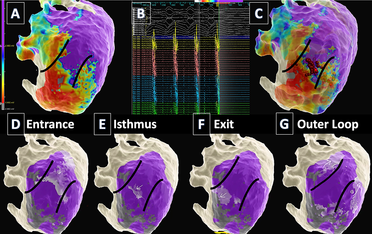
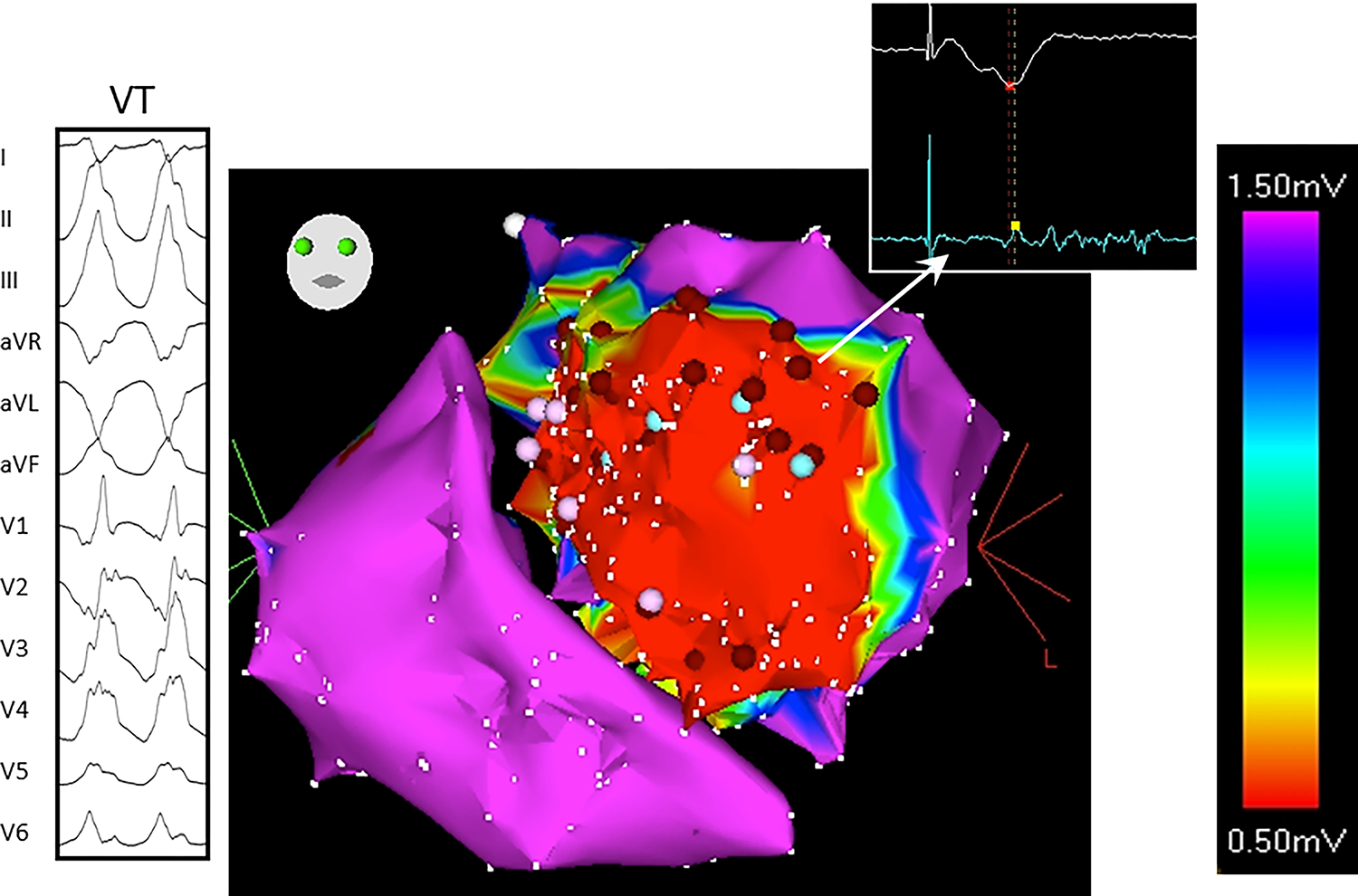
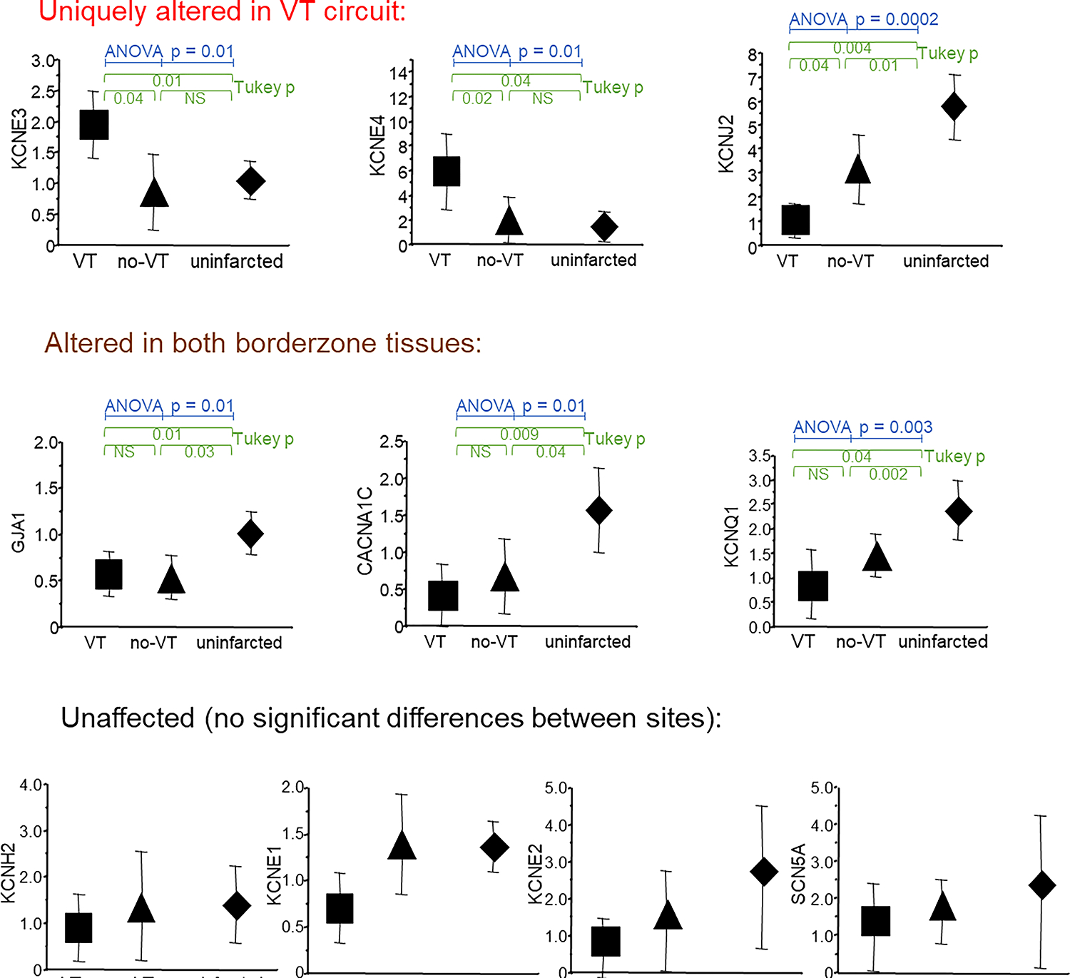
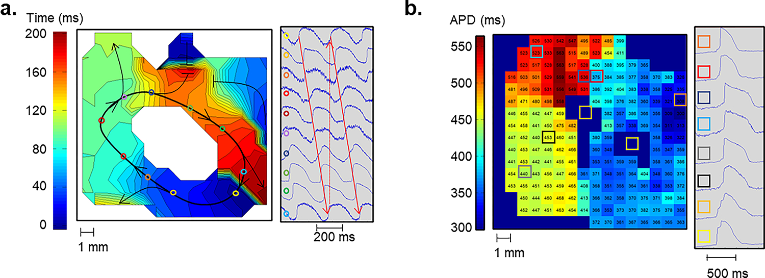
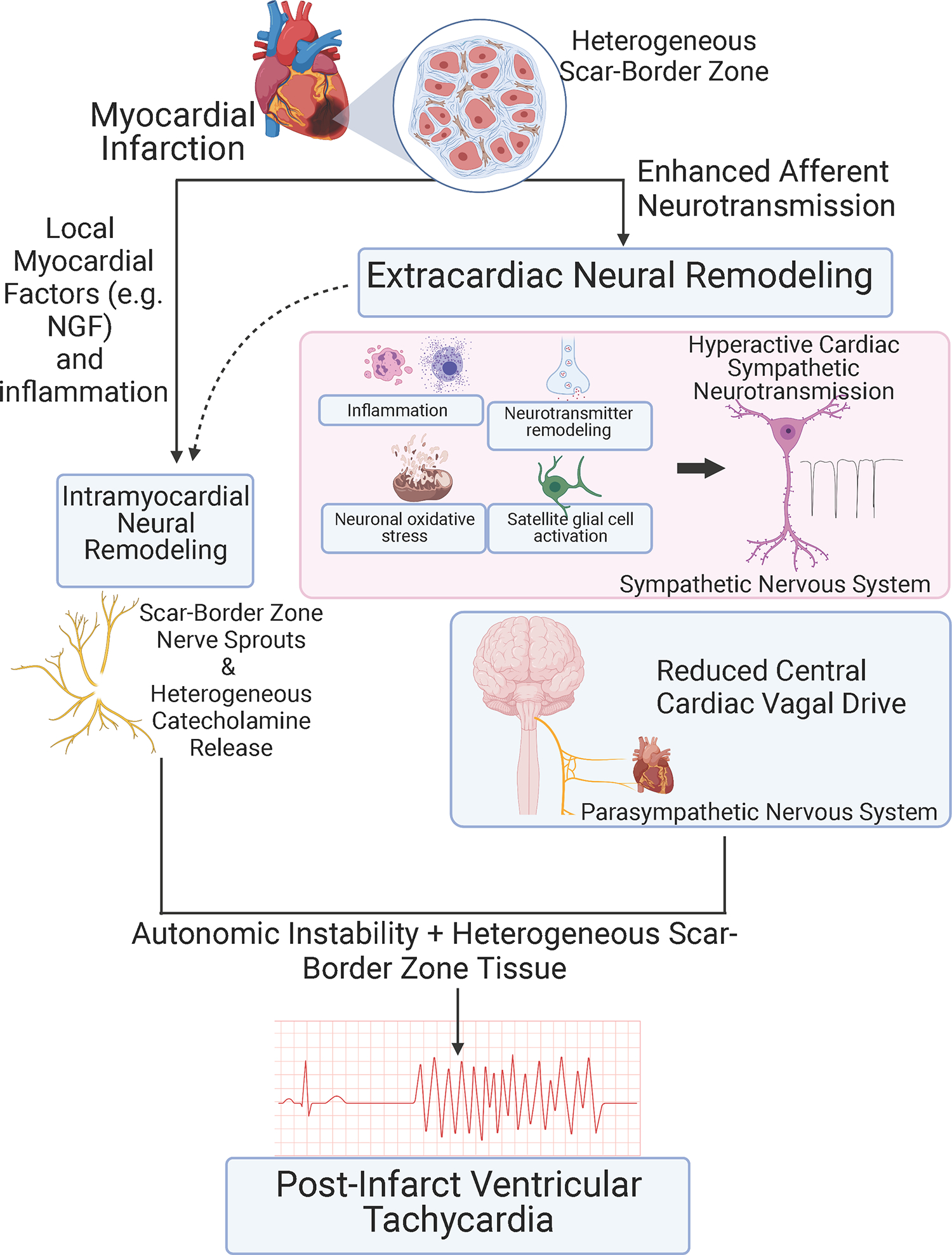
Cardiac arrest is the leading cause of death in the more economically developed countries. Ventricular tachycardia associated with myocardial infarct is a prominent cause of cardiac arrest. Ventricular arrhythmias occur in 3 phases of infarction: during the ischemic event, during the healing phase, and after the scar matures. Mechanisms of arrhythmias in these phases are distinct. This review focuses on arrhythmia mechanisms for ventricular tachycardia in mature myocardial scar. Available data have shown that postinfarct ventricular tachycardia is a reentrant arrhythmia occurring in circuits found in the surviving myocardial strands that traverse the scar. Electrical conduction follows a zigzag course through that area. Conduction velocity is impaired by decreased gap junction density and impaired myocyte excitability. Enhanced sympathetic tone decreases action potential duration and increases sarcoplasmic reticular calcium leak and triggered activity. These elements of the ventricular tachycardia mechanism are found diffusely throughout scar. A distinct myocyte repolarization pattern is unique to the ventricular tachycardia circuit, setting up conditions for classical reentry. Our understanding of ventricular tachycardia mechanisms continues to evolve as new data become available. The ultimate use of this information would be the development of novel diagnostics and therapeutics to reliably identify at-risk patients and prevent their ventricular arrhythmias.

Keywords: cause of death; chronic total occlusion; complications; death; mortality.


Conflict of interest statement

Disclosures J.K. Donahue has filed a patent for intellectual property owned by the UMass Chan Medical School in the area of repolarization heterogeneity in VT. J. Chrispin reports honoraria from Abbott. O.A. Ajijola has filed a patent for intellectual property owned by University of California Regents in the areas of catheter ablation and neuromodulation. O.A. Ajijola reports honoraria from Biosense Webster.

Figures

Figure 1:
Figure 1:
VT mapping and ablation in a patient with prior anterior MI. (TOP) A. Epicardial voltage mapping identified a large anteroseptal scar extending from the base to apex. Myocardium with normal bipolar voltage is purple. Dense scar is red and borderzone is blue-yellow-orange. B. VT critical isthmus was identified when contact of the catheter with myocardium “bump” terminated the VT. C. Ablation (red dots) in the isthmus rendered the patient non-inducible. (BOTTOM) Activation mapping during VT identified a “figure of 8” circuit with entrance (D), isthmus (E), exit (F), and outer loop (G) on the epicardial surface. The lighter purple region marked with the arrows is the activation wavefront. Darker purple is live myocardium, and gray is dense scar. Arrows denote the circuit.
Figure 2:
Figure 2:
Typical low voltage, fractionated electrogram recorded from scarred myocardium during VT. The twelve-lead ECG of the VT and bipolar electrogram from the ablation catheter are shown in the left and right insets respectively. The catheter is placed at the site within scarred myocardium where the VT was ultimately ablated. The ablation electrogram shows a low amplitude, split and fractionated electrogram often seen in scar, both at successful and unsuccessful ablation sites. Complexity of this signal illustrates the difficulty in assigning a local activation time for either activation mapping or conduction velocity calculation.
Figure 3:
Figure 3:
Comparison of mRNA expression of ion channels and connexin43 at the mapped VT site (square), scarred myocardium unassociated with any VT (triangle) and uninfarcted basal lateral myocardium (diamond). (Adapted with permission from Kelemen et al.)
Figure 4:
Figure 4:
Complete VT circuit observed during optical mapping with a voltage sensitive dye in a perfused myocardial scar tissue wedge. (A) An isochronal map showing activation of a complete VT circuit is shown. The colored circles indicate locations on the activation map where example pixels at right were located. The white central region had 2:1 activation and did not participate in the VT. (B) An APD map during 1000 ms fixed rate pacing of the full VT circuit tissue from panel A. The colored boxes show locations on the APD map, for example electrograms at right. All observed VT circuits had a tract of tissue with short APDs in contact with a tract of tissue with long APDs. This APD pattern was only seen in optical maps of complete VT circuits. (Adapted with permission from Kelemen et al.)
Figure 5:
Figure 5:
Schematic showing identified autonomic mechanisms in post-infarct VT. Autonomic instability is the imbalance between sympathetic (proarrhythmic) and parasympathetic (antiarrhythmic) signaling.

References

    1. Bridges C, Burkman J, Malekan R, Konig S, Chen H, Yarnall C, Gardner T, Stewart A, Stecker M, Patterson T and Stedman H. Global cardiac-specific transgene expression using cardiopulmonary bypass with cardiac isolation. Ann Thorac Surg. 2002;73:1939–1946. - PubMed
    1. Berdowski J, Berg RA, Tijssen JG and Koster RW. Global incidences of out-of-hospital cardiac arrest and survival rates: Systematic review of 67 prospective studies. Resuscitation. 2010;81:1479–87. - PubMed
    1. Pouleur AC, Barkoudah E, Uno H, Skali H, Finn PV, Zelenkofske SL, Belenkov YN, Mareev V, Velazquez EJ, Rouleau JL, Maggioni AP, Kober L, Califf RM, McMurray JJ, Pfeffer MA and Solomon SD. Pathogenesis of sudden unexpected death in a clinical trial of patients with myocardial infarction and left ventricular dysfunction, heart failure, or both. Circulation. 2010;122:597–602. - PubMed
    1. Steinhaus DA, Vittinghoff E, Moffatt E, Hart AP, Ursell P and Tseng ZH. Characteristics of sudden arrhythmic death in a diverse, urban community. Am Heart J. 2012;163:125–31. - PMC - PubMed
    1. Janse MJ and Wit AL. Electrophysiological mechanisms of ventricular arrhythmias resulting from myocardial ischemia and infarction. Physiol Rev. 1989;69:1049–1169. - PubMed

Publication types

MeSH terms