. 2024 Jan 16;165(3):bqae014.
doi: 10.1210/endocr/bqae014.

Adrenal Abcg1 Controls Cholesterol Flux and Steroidogenesis

Affiliations

Adrenal Abcg1 Controls Cholesterol Flux and Steroidogenesis

Jani Liimatta et al. Endocrinology. .

Abstract

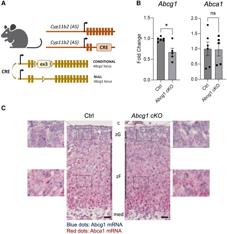
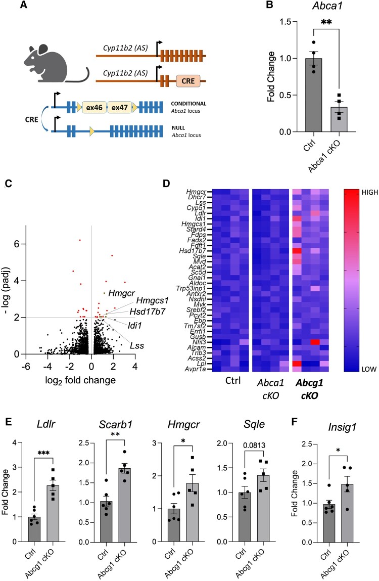
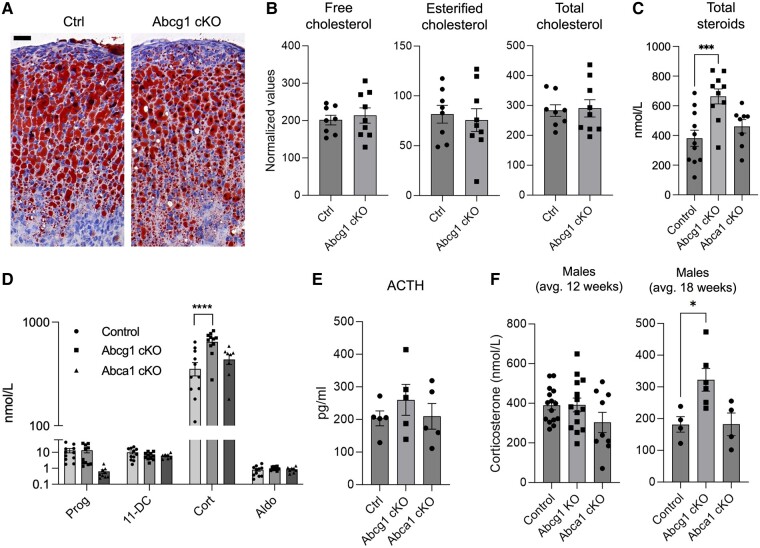
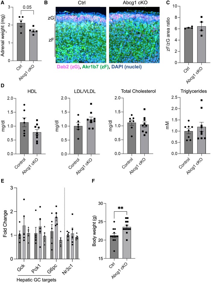
Cholesterol is the precursor of all steroids, but how cholesterol flux is controlled in steroidogenic tissues is poorly understood. The cholesterol exporter ABCG1 is an essential component of the reverse cholesterol pathway and its global inactivation results in neutral lipid redistribution to tissue macrophages. The function of ABCG1 in steroidogenic tissues, however, has not been explored. To model this, we inactivated Abcg1 in the mouse adrenal cortex, which led to an adrenal-specific increase in transcripts involved in cholesterol uptake and de novo synthesis. Abcg1 inactivation did not affect adrenal cholesterol content, zonation, or serum lipid profile. Instead, we observed a moderate increase in corticosterone production that was not recapitulated by the inactivation of the functionally similar cholesterol exporter Abca1. Altogether, our data imply that Abcg1 controls cholesterol uptake and biosynthesis and regulates glucocorticoid production in the adrenal cortex, introducing the possibility that ABCG1 variants may account for physiological or subclinical variation in stress response.

Keywords: Abcg1; adrenal cortex; cholesterol; glucocorticoids; steroids.


Figures

Figure 1.
Figure 1.
Effective gene recombination in Abcg1 cKO mice. (A) Schematic representation of the mouse model used to inactivate Abcg1 in adrenocortical cells using Cre-mediated recombination of Abcg1's third exon. (B) Quantitation of transcripts encoding Abcg1 and Abca1 in control and Abcg1 cKO adrenal glands. (C) In situ depiction of Abcg1 (blue dots) and Abca1 (red dots) transcripts in control (left) and Abcg1 cKO adrenal sections (right), including insets’ virtual magnifications on each side. All mice used for this figure were 2-month-old females. Scale bar = 25μm. *P ≤ .05.
Figure 2.
Figure 2.
Inactivation of adrenocortical Abcg1 results in increased transcripts for cholesterol uptake and synthesis. (A) Schematic representation of the mouse model used to inactivate Abca1 in adrenocortical cells using Cre-mediated recombination of exons 46 and 47. (B) Quantification of Abca1 transcripts in control and Abca1 cKO adrenal glands. (C) Volcano plot depicting 12 downregulated and 19 upregulated genes (red or beige dots) in Abcg1 cKO adrenals compared to the combined (summed) datasets of controls and Abca1 cKO counterparts, using cutoffs of 1.2 for fold change and 0.01 for adjusted P-value. Each beige dot is associated with a gene name as indicated in the plot. (D) Heat map depicting color-coded expression levels of 34 transcripts responsible for the enrichment of the HALLMARK_CHOLESTEROL_HOMEOSTASIS dataset in Gene Set Enrichment Analysis. (E and F) Quantitation of transcripts involved in cholesterol regulation, uptake, and de novo synthesis in control and Abcg1 cKO adrenal glands. All mice used for this figure were 2-month-old females. *P ≤ .05; **P ≤ .01; ***P ≤ .001.
Figure 3.
Figure 3.
Inactivation of adrenocortical Abcg1 results in increased corticosterone synthesis. (A) Oil Red O staining (red) of control and Abcg1 cKO adrenocortical sections. Mayer's hemalum was used to counterstain nuclei (blue). Images are representative of 4 animals per genotype. Scale bar = 25 µm. (B) Free, esterified, and total cholesterol in whole adrenal glands from control and Abcg1 cKO animals. (C) Aggregated quantification of adrenal steroids detected in mouse sera using liquid chromatography-mass spectrometry, ie, pregnenolone, Prog, 11-DC, Cort, and Aldo. (D) Steroid concentrations in sera of control and Abcg1 cKO mice. Most pregnenolone values were below the threshold of accurate quantification, likely because of intense processivity into downstream products, and are not reported in this graph. (E) Levels in sera from control and Abcg1 cKO mice. (F) Levels of corticosterone in male control and Abcg1 cKO mice at different ages. Except for panel F, all mice used for this figure were 2-month-old females. *P ≤ .05, ***P ≤ .001, ****P ≤ .0001.
Figure 4.
Figure 4.
Abcg1 cKO mice display increased body weight but unaltered adrenal mass, zonation, and serum lipid profile. (A) Adrenal weight measured on freshly dissected whole adrenals in control and Abcg1 cKO mice. (B) Representative depiction of immunofluorescence assay on adrenocortical sections from control and Abcg1 cKO mice. Images are representative of 4 animals per genotype. Scale bar = 50 µm. (C) Ratio of the zF area (measured as the area stained by Akr1b7) and the zG area (measured as the area stained by Dab2). (D) Lipid profile in control and Abcg1 cKO mouse sera. (E). Quantification of glucocorticoid-sensitive (ie, Gck, Pck1, G6pc) and insensitive (Nr3c1) genes in livers from control, Abcg1 cKO, and Abca1 cKO animals. (F) Quantification of live animal weight. All mice used for this figure were 2-month-old females. **P ≤ .01.

Comment in

References

    1. Miller WL, Auchus RJ. The molecular biology, biochemistry, and physiology of human steroidogenesis and its disorders. Endocr Rev. 2011;32(1):81‐151. - PMC - PubMed
    1. Vaduva P, Bonnet F, Bertherat J. Molecular basis of primary aldosteronism and adrenal cushing syndrome. J Endocr Soc. 2020;4(9):bvaa075. - PMC - PubMed
    1. Pignatti E, Flück CE. Adrenal cortex development and related disorders leading to adrenal insufficiency. Mol Cell Endocrinol. 2021;527:111206. - PubMed
    1. Donoghue SE, Pitt JJ, Boneh A, White SM. Smith-Lemli-Opitz syndrome: clinical and biochemical correlates. J Pediatr Endocrinol Metab. 2018;31(4):451‐459. - PubMed
    1. Bose HS, Sugawara T, Strauss JF III, Miller WL; International Congenital Lipoid Adrenal Hyperplasia Consortium . The pathophysiology and genetics of congenital lipoid adrenal hyperplasia. N Engl J Med. 1996;335(25):1870‐1878. - PubMed