. 2024 Apr:374:114702.
doi: 10.1016/j.expneurol.2024.114702. Epub 2024 Jan 30.

Repetitive head trauma and apoE4 induce chronic cerebrovascular alterations that impair tau elimination from the brain

Affiliations

Repetitive head trauma and apoE4 induce chronic cerebrovascular alterations that impair tau elimination from the brain

Maxwell Eisenbaum et al. Exp Neurol. 2024 Apr.

Abstract

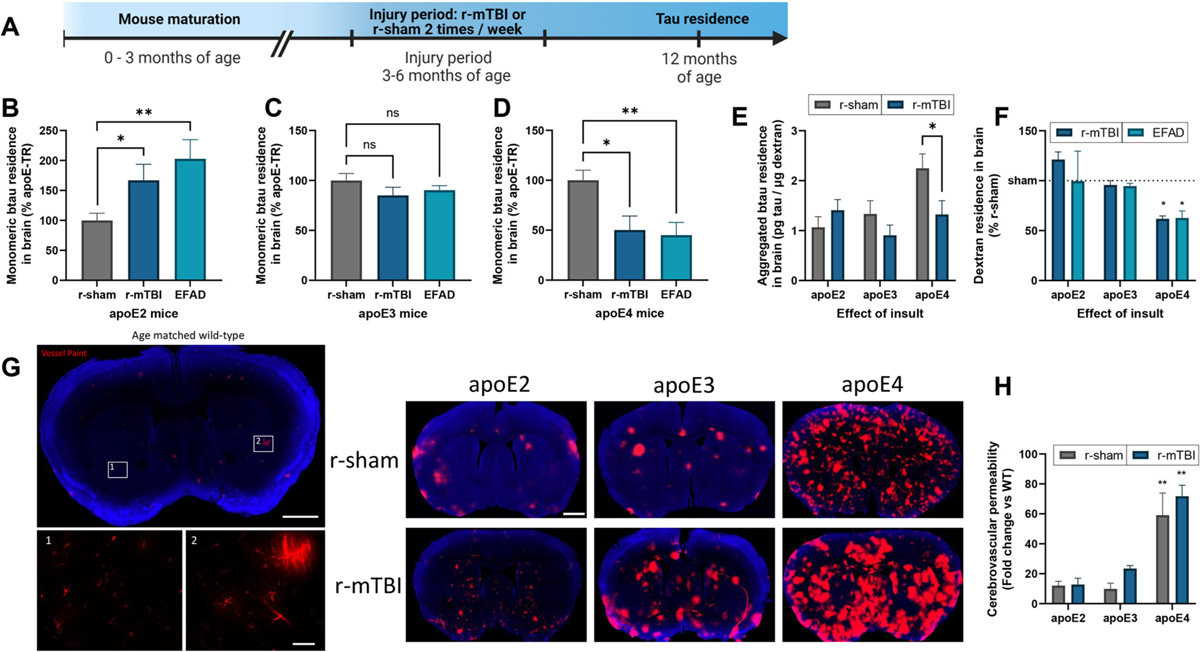
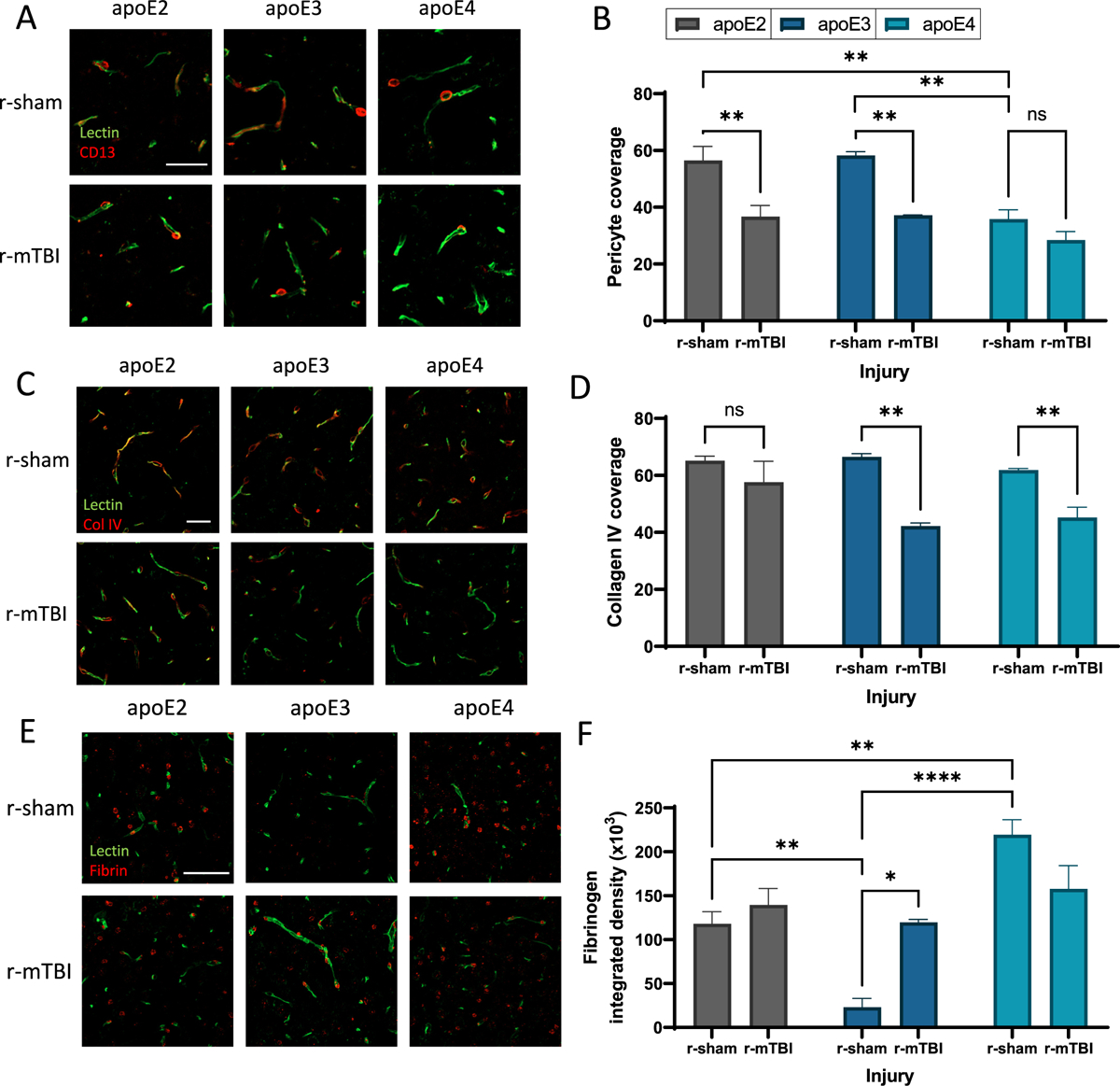
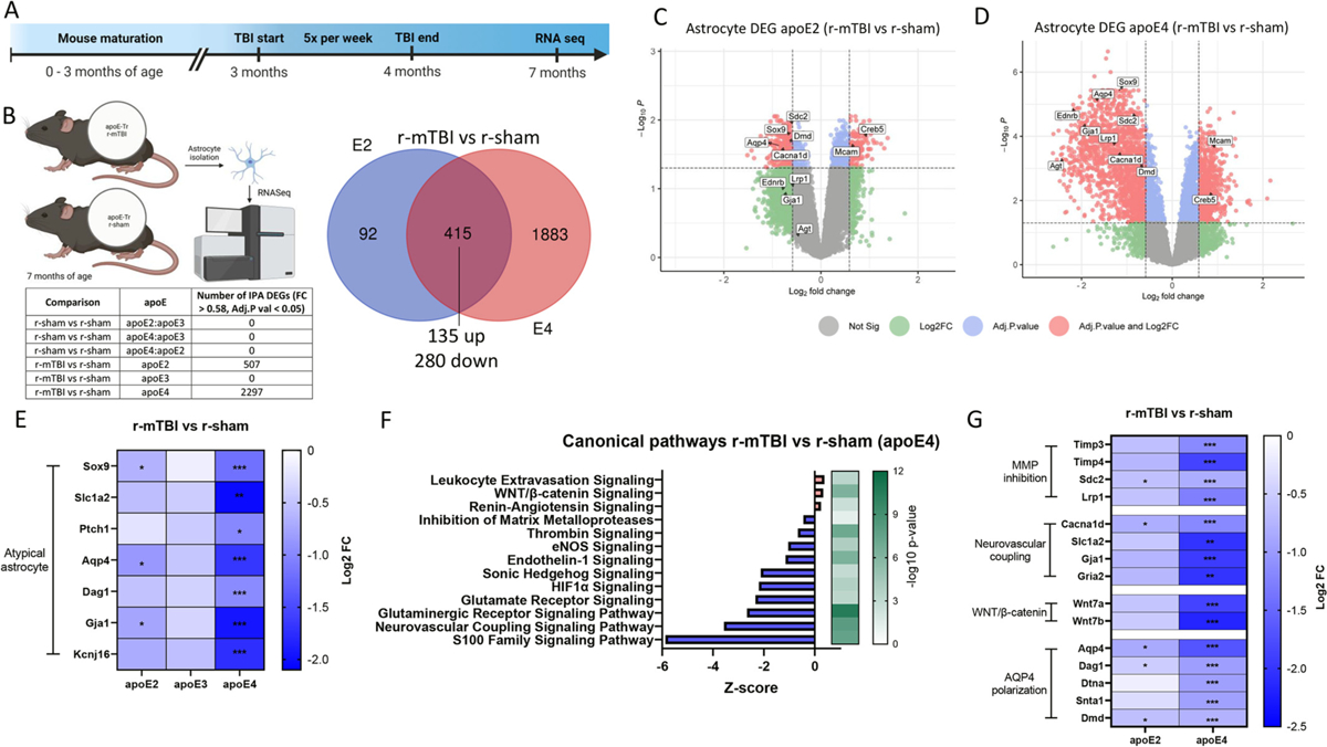
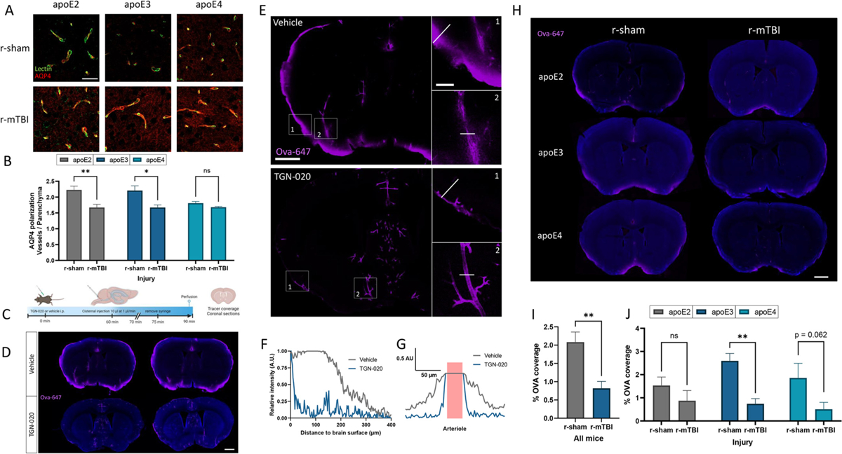
Repetitive mild traumatic brain injuries (r-mTBI) sustained in the military or contact sports have been associated with the accumulation of extracellular tau in the brain, which may contribute to the pathogenesis of neurodegenerative tauopathies. The expression of the apolipoprotein E4 (apoE4) isoform has been associated with higher levels of tau in the brain, and worse clinical outcomes after r-mTBI, though the influence of apoE genotype on extracellular tau dynamics in the brain is poorly understood. We recently demonstrated that extracellular tau can be eliminated across blood-brain barrier (BBB), which is progressively impaired following r-mTBI. The current studies investigated the influence of repetitive mild TBI (r-mTBI) and apoE genotype on the elimination of extracellular solutes from the brain. Following intracortical injection of biotin-labeled tau into humanized apoE-Tr mice, the levels of exogenous tau residing in the brain of apoE4 mice were elevated compared to other isoforms, indicating reduced tau elimination. Additionally, we found exposure to r-mTBI increased tau residence in apoE2 mice, similar to our observations in E2FAD animals. Each of these findings may be the result of diminished tau efflux via LRP1 at the BBB, as LRP1 inhibition significantly reduced tau uptake in endothelial cells and decreased tau transit across an in vitro model of the BBB (basolateral-to-apical). Notably, we showed that injury and apoE status, (particularly apoE4) resulted in chronic alterations in BBB integrity, pericyte coverage, and AQP4 polarization. These aberrations coincided with an atypical reactive astrocytic gene signature indicative of diminished CSF-ISF exchange. Our work found that CSF movement was reduced in the chronic phase following r-mTBI (>18 months post injury) across all apoE genotypes. In summary, we show that apoE genotype strongly influences cerebrovascular homeostasis, which can lead to age-dependent deficiencies in the elimination of toxic proteins from the brain, like tau, particularly in the aftermath of head trauma.

Keywords: ApoE; Blood brain barrier; Glymphatic system; Tau; Traumatic brain injury.


Conflict of interest statement

Declaration of competing interest The authors declare they have no conflict of interest.

Figures

Fig. 1.
Fig. 1.
Effect of apoE isoform on tau brain residence. (a) The time course of tau elimination from the brain of apoE3 mice (12 months of age) was established by examining monomeric biotin-labeled tau (btau) levels (n = 6) and 10 kDa LyD (n = 5) levels in the brain at various time points following intracortical injection. Btau content was analyzed using ELISA while LyD was analyzed via fluorescence and normalized to total protein using the BCA protein assay. The half-life for both btau and LyD were determined using nonlinear regression and a one phase decay fit. (b-d) Following intracortical injection in apoE2, apoE3, or apoE4 mice (12 months of age), the amount of exogenous btau and co-injected LyD residing in the brain was determined at 2 h post injection. Monomeric (b) or aggregated (c) btau content was analyzed using an ELISA, while LyD was analyzed via fluorescence. Values represent mean + SEM (n = 5) and are expressed as pg of btau per μg of LyD. *P < 0.05 as determined by one-way ANOVA and Bonferroni’s multiple comparisons test. (d) Dextran residence values represent mean + SEM (n = 10) and are expressed as μg of LyD per mg protein. *P < 0.05 as determined by one-way ANOVA and Bonferroni’s multiple comparisons test. (e) Isolated cerebrovascular lysates from apoE2, apoE3 or apoE4 mice (12 months of age) were analyzed for caveolin-1 by ELISA and normalized to total protein using the BCA protein assay. Values represent mean + SEM (n = 5). *P < 0.05 as determined by one-way ANOVA and Bonferroni’s multiple comparisons test. (f) Schematic diagram of in vitro BBB model used in (g, h). (g) Monomeric btau was added alongside the known paracellular marker 10 kDa LyD to the basolateral compartment of the monoculture or coculture in vitro BBB model. (h) Monomeric or aggregate enriched btau was added alongside LyD to the basolateral compartment of the coculture in vitro BBB model in the presence or absence of the LRP1 antagonist RAP (100 nM). (g, h) Samples were collected from the apical compartment at 0, 30, and 60 min to determine the permeability of btau and LyD across the BBB model. Values represent mean ± SEM (n = 3) and are expressed as the apparent permeability coefficient (Papp). *P < 0.05 as determined by one-way ANOVA and Bonferroni’s multiple comparisons test. (i) HBMECs were exposed to 1 μg/mL of monomeric btau in the presence or absence of RAP (100 nM) for 1 h at 37 °C. The cell lysates were analyzed for btau content by ELISA and normalized to total protein using the BCA protein assay. Values represent mean + SEM (n = 3). * P < 0.05 as determined by unpaired two-tailed Student’s t-test.
Fig. 2.
Fig. 2.
Influence of apoE and insult on tau brain residence. (a) Timeline of closed head injury paradigm. (b-f) Following intracortical injection in 12 month old r-sham, r-mTBI and EFAD (apoE2, apoE3, and apoE4) mice, the amount of exogenous btau and co-injected LyD residing in the brain was determined at 2 h post injection. Monomeric (b-d) or aggregated (e) btau content was analyzed using an ELISA, while LyD (f) was analyzed via fluorescence. (b-d) Values represent mean + SEM (n = 5), and are expressed as the percentage tau residence normalized to each respective r-sham. *P < 0.05, **P < 0.01 as determined by one-way ANOVA and Bonferroni’s multiple comparisons test. (e) Values represent mean + SEM (n = 5) and are expressed as pg of aggregated btau per μg of LyD. *P < 0.05 as determined by one-way ANOVA and Bonferroni’s multiple comparisons test. (f) Values represent mean + SEM (n = 10) and are expressed as the percentage dextran residence normalized to each respective r-sham. *P < 0.05 as determined by two-way ANOVA and Bonferroni’s multiple comparisons test. (g, h) Vessel paint was applied to 12 month old r-sham and r-mTBI mice (apoE2, apoE3 and apoE4) via transcardiac injection. (g) Representative images of an age matched wild-type mouse showing vessel paint (red) leakage into the cortex with Dapi staining (blue). Insets 1 and 2 show boxed areas in (g). (h) Cerebrovascular permeability was quantified as the percent area of total cortex vessel paint signal normalized to age matched wild-type mice. Values represent mean + SEM (n = 3) and are expressed as the fold change normalized to age matched wild-type mice. *P < 0.05 as determined by two-way ANOVA and Bonferroni’s multiple comparisons test.
Fig. 3.
Fig. 3.
Influence of apoE and insult on pericyte coverage and BBB integrity. (a) Confocal microscopy of CD13 immunodetection showing pericyte coverage (red) of lectin-positive brain capillaries (green) in the somatosensory cortex of 24 month old r-sham and r-mTBI mice (apoE2, apoE3 and apoE4). Scale bar, 25 μm. (b) Quantification of pericyte coverage on capillaries in r-sham and r-mTBI (apoE2, apoE3 and apoE4) mice. Values represent mean + SEM (n = 4). **P < 0.01 as determined by two-way ANOVA and Bonferroni’s multiple comparisons test. (c) Confocal microscopy of Collagen IV (red) and lectin-positive brain capillaries (green) in the somatosensory cortex of 24 month old r-sham and r-mTBI mice (apoE2, apoE3 and apoE4). Scale bar, 25 μm. (d) Quantification of Collagen IV coverage of lectin-positive capillaries. Values represent mean + SEM (n = 4). **P < 0.01 as determined by two-way ANOVA and Bonferroni’s multiple comparisons test. (e) Confocal microscopy of fibrin (red) and lectin-positive brain capillaries (green) in the somatosensory cortex of 24 month old r-sham and r-mTBI mice (apoE2, apoE3 and apoE4). Scale bar, 50 μm. (f) Quantification of extravascular pericapillary fibrinogen deposits. Values represent mean + SEM (n = 4). **P < 0.01 as determined by two-way ANOVA and Bonferroni’s multiple comparisons test.
Fig. 4.
Fig. 4.
Enriched atypical reactive astrocyte profile by apoE4 in r-mTBI mouse model. (a-f) RNA was extracted from astrocytes isolated from 7 month old r-sham and r-mTBI (apoE2, apoE3 and apoE4) mice, and gene ontology analysis of r-mTBI astrocytes (versus each respective r-sham) was performed to identify enriched canonical pathways. (a) Timeline of accelerated closed head injury paradigm. (b) Venn diagram of the significantly differentially expressed genes (DEGs) affected by injury for apoE2 and apoE4 (adjusted P < 0.05, |fold change| > 0.58). All common DEGs between apoE2 and apoE4 r-mTBI (compared to their respective shams) changed in a consistent direction. (c, d) Volcano plots of DEGs in r-mTBI versus sham astrocytes isolated from (c) apoE2 and (d) apoE4 mice (significant cut off set at adjusted P < 0.05, |fold change| > 0.58, red points indicate DEGs above both the adjusted p value and Log2FC cut off threshold). (e) Heatmap showing fold change (FC) values (compared to respective r-shams) of genes correlating with the atypical reactive astrocyte phenotype signatures across injury groups. (f) Results of enrichment analysis comparing the DEGs in astrocytes isolated from r-mTBI and r-sham apoE4 mice to the ingenuity knowledge base, showing top significantly dysregulated canonical pathways. Orange, upregulated; blue, downregulated genes/functions. (g) Heatmap showing downregulated FC values compared to values in respective r-shams of DEGs involved in Matrix metalloprotease inhibition (Timp3, Timp4, Sdc2, Lrp1), Neurovascular coupling (Cacna1d, Sl1a2, Gja1, Gria2), and WNT/β-catenin signaling (Wnt7a, Wnt7b), and glymphatic system-associated AQP4 polarization (Aqp4, Dag1, Dtna, Snta1, Dmd). Significance reflects adjusted P < 0.05, |FC| > 0.58.
Fig. 5.
Fig. 5.
Influence of apoE and insult on glymphatic influx. (a) Confocal microscopy of AQP4 (red) and lectin-positive brain capillaries (green) in the somatosensory cortex of 24 month old r-sham and r-mTBI mice (apoE2, apoE3 and apoE4). Scale bar, 25 μm. (b) Quantification of AQP4 polarization measured as the ratio of signal around the capillary divided by the signal in the surrounding parenchyma. Values represent mean + SEM (n = 4). *P < 0.05, **P < 0.01 as determined by two-way ANOVA and Bonferroni’s multiple comparisons test. (c-g) The effect of pharmacological inhibition of AQP4 on the CSF-ISF exchange was examined in wild-type mice (6 months) by injecting TGN-020 (250 mg/kg) or vehicle i.p., followed by an intracisternal injection of the CSF tracer ovalbumin-647. 30 min after the intracisternal injection, mice were perfused with 4% PFA and the distribution of the CSF influx tracer was probed in brain slices. (d) Representative confocal microscopic images showing Ova (magenta) influx with Dapi staining (blue). Scale bar, 1.0 mm. (e) Representative images showing Ova (magenta) distribution and influx in coronal sections. Insets 1 and 2 show the boxed areas in (e). Scale bar full images, 1.0 mm. Scale bar insets, 250 μm. (f) Tracer penetration depth profile, normalized to the fluorescence at the pial surface of a coronal section. The line in insert 1 was placed orthogonal to the cortical surface at the most dorsal position in the TGN-020 treated mouse where tracer could be found at the pial surface, and depth was measured at the same position in the untreated mouse. (g) Tracer perivascular influx profile, normalized to the fluorescence at the pial surface of a cortical section. The line in insert 2 was placed across a penetrating artery, and distribution of Ova was assessed from the arterial lumen. Vessel lumen in pink. (h-j) The effect of r-mTBI on CSF-ISF exchange was examined in 24 month old r-sham and r-mTBI (apoE2, apoE3, and apoE4) mice by injecting the CSF tracer Ova into the cisterna magna. Brains were harvested 30 min following the injection, and the Ova influx was quantified. (h) Representative confocal microscopic images showing Ova (magenta) coverage with Dapi staining (blue). Scale bar, 1.0 mm. (i, j) Quantification of Ova coverage on brain coronal sections. (i) Values represent mean + SEM (n = 12). (j) Values represent mean + SEM (n = 4). **P < 0.01 as determined by two-way ANOVA and Bonferroni’s multiple comparisons test.

Similar articles

Cited by

References

    1. Afgan E, Baker D, Batut B, van den Beek M, Bouvier D, Ech M, Chilton J, Clements D, Coraor N, Grüning BA, Guerler A, Hillman-Jackson J, Hiltemann S, Jalili V, Rasche H, Soranzo N, Goecks J, Taylor J, Nekrutenko A, Blankenberg D, 2018. The galaxy platform for accessible, reproducible and collaborative biomedical analyses: 2018 update. Nucleic Acids Res 46 (W1), W537–W544. 10.1093/nar/gky379. - DOI - PMC - PubMed
    1. Aldea R, Weller RO, Wilcock DM, Carare RO, Richardson G, 2019. Cerebrovascular smooth muscle cells as the drivers of intramural periarterial drainage of the brain. Front. Aging Neurosci 11 (JAN) 10.3389/fnagi.2019.00001. - DOI - PMC - PubMed
    1. Alsaqati M, Thomas RS, Kidd EJ, 2023. Upregulation of endocytic protein expression in the Alzheimer’s disease male human brain. Aging Brain 4, 100084. 10.1016/j.nbas.2023.100084. - DOI - PMC - PubMed
    1. Andreone BJ, Chow BW, Tata A, Lacoste B, Ben-Zvi A, Bullock K, Deik AA, Ginty DD, Clish CB, Gu C, 2017. Blood-brain barrier permeability is regulated by lipid transport-dependent suppression of caveolae-mediated transcytosis. Neuron 94 (3), 581–594.e5. 10.1016/j.neuron.2017.03.043. - DOI - PMC - PubMed
    1. Andrews S, 2010. FastQC: A Quality Control Tool for High Throughput Sequence Data [Online]. Available Online at. Http://Www.Bioinformatics.Babraham.Ac.Uk/Projects/Fastqc/.