Review
. 2024 Mar;300(3):105692.
doi: 10.1016/j.jbc.2024.105692. Epub 2024 Jan 30.

Protein kinase C signaling "in" and "to" the nucleus: Master kinases in transcriptional regulation

Affiliations
Review

Protein kinase C signaling "in" and "to" the nucleus: Master kinases in transcriptional regulation

Marcelo G Kazanietz et al. J Biol Chem. 2024 Mar.

Abstract

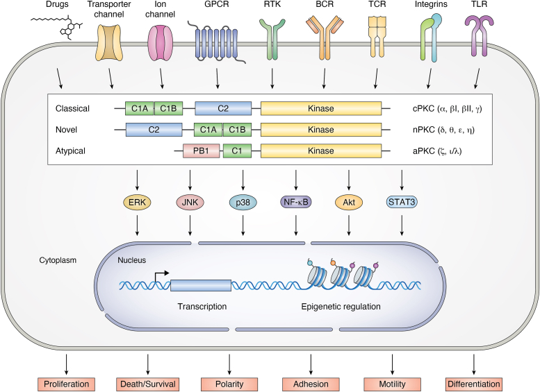
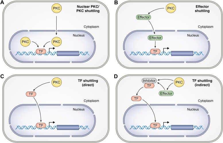
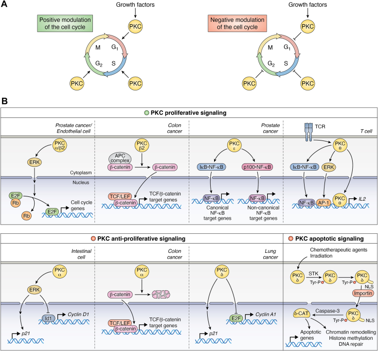
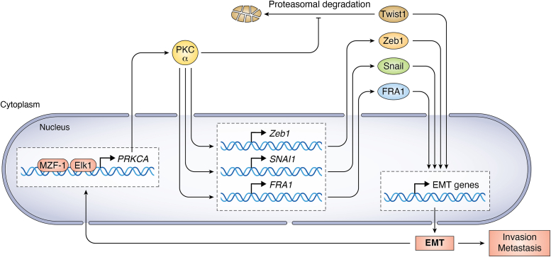
PKC is a multifunctional family of Ser-Thr kinases widely implicated in the regulation of fundamental cellular functions, including proliferation, polarity, motility, and differentiation. Notwithstanding their primary cytoplasmic localization and stringent activation by cell surface receptors, PKC isozymes impel prominent nuclear signaling ultimately impacting gene expression. While transcriptional regulation may be wielded by nuclear PKCs, it most often relies on cytoplasmic phosphorylation events that result in nuclear shuttling of PKC downstream effectors, including transcription factors. As expected from the unique coupling of PKC isozymes to signaling effector pathways, glaring disparities in gene activation/repression are observed upon targeting individual PKC family members. Notably, specific PKCs control the expression and activation of transcription factors implicated in cell cycle/mitogenesis, epithelial-to-mesenchymal transition and immune function. Additionally, PKCs isozymes tightly regulate transcription factors involved in stepwise differentiation of pluripotent stem cells toward specific epithelial, mesenchymal, and hematopoietic cell lineages. Aberrant PKC expression and/or activation in pathological conditions, such as in cancer, leads to profound alterations in gene expression, leading to an extensive rewiring of transcriptional networks associated with mitogenesis, invasiveness, stemness, and tumor microenvironment dysregulation. In this review, we outline the current understanding of PKC signaling "in" and "to" the nucleus, with significant focus on established paradigms of PKC-mediated transcriptional control. Dissecting these complexities would allow the identification of relevant molecular targets implicated in a wide spectrum of diseases.

Keywords: diacylglycerol; gene expression; nucleus; protein kinase C; signal transduction; transcription factor.


Conflict of interest statement

Conflict of interest The authors declare that they have no conflicts of interest with the contents of this article.

Figures

Figure 1
Figure 1
PKC isozymes and signal transduction. PKCs are classified into classical/conventional, novel, and atypical based on their distinctive biochemical and structural properties. Cell surface receptors or drugs can trigger the activation of discrete PKCs, which phosphorylate many cellular effectors, including signal transduction proteins that impact nuclear function. BCR, B cell receptor; GPCR, G protein–coupled receptor; RTK, receptor tyrosine kinase; TCR, T cell receptor; TLR, toll-like receptor.
Figure 2
Figure 2
Hypothetical models for PKC signaling driving nuclear function. The cartoon depicts different models for transcriptional control by PKC. A, PKC isozymes can be either located in the nucleus or translocated to the nucleus, where they phosphorylate components of the transcriptional complexes, including transcription factors (TFs), and turn on or off the transcriptional activation of selected genes. B, PKCs can regulate transcription via their downstream effectors, which shuttle to the nucleus upon phosphorylation by PKC or PKC effector kinases. C, PKCs localize in the cytoplasmic compartment and upon activation phosphorylate TFs, which in turn shuttle to the nucleus. D, PKCs (or PKC effector kinases) can phosphorylate proteins that bind (and inhibit) transcription factors. Phosphorylation of these inhibitory proteins in the complex leads to the dissociation of the transcription factor and its translocation to the nucleus.
Figure 3
Figure 3
PKC regulation of nuclear function in the control of proliferation and apoptosis. PKC isozymes exert proliferative/prosurvival and antiproliferative/apoptotic roles in distinct cell contexts. A, the dual roles of PKC isozymes in proliferation are indicative of either positive (upper panel) or negative (lower panel) control of the cell cycle, acting in different phases with characteristic isozyme and cell type specificity. B, representative examples for positive (upper panels) and negative (lower panels) controls of nuclear events by PKC isozymes. The lower panel also includes an established paradigm for PKCδ in apoptotic signaling in response to chemotherapeutic drugs and irradiation. These stimuli promote PKCδ phosphorylation by soluble tyrosine kinases (STKs), exposure of a cryptic NLS, binding to importin, cytoplasmatic-nuclear shuttling, and nuclear cleavage by caspase-3, leading to the generation of an active PKCδ catalytic fragment (δ-CAT). NLS, nuclear localization signal.
Figure 4
Figure 4
PKCα and the control of EMT. PKCα is up regulated in mesenchymally transformed cells in TNBC and likely in other cancer types. Transcriptional deregulation of the PKCα gene (PRKCA) in EMT is mediated by MZF-1 and Elk-1 transcription factor complexes. PKCα controls the expression of selected EMT transcription factors by promoting transcriptional activation of their corresponding genes or by enhancing their stability, thus leading to a vicious cycle for cancer cell invasiveness. Elk-1, Ets-like protein-1; EMT, epithelial-to-mesenchymal transition; triple-negative breast cancer; MZF-1, myeloid zinc finger-1.

Similar articles

Cited by

References

    1. Newton A.C. Regulation of protein kinase C. Curr. Opin. Cell Biol. 1997;9:161–167. - PubMed
    1. Mellor H., Parker P.J. The extended protein kinase C superfamily. Biochem. J. 1998;332:281–292. - PMC - PubMed
    1. Toker A. Signaling through protein kinase C. Front. Biosci. 1998;3:D1134–D1147. - PubMed
    1. Griner E.M., Kazanietz M.G. Protein kinase C and other diacylglycerol effectors in cancer. Nat. Rev. Cancer. 2007;7:281–294. - PubMed
    1. Newton A.C. Lipid activation of protein kinases. J. Lipid Res. 2009;50:S266–S271. - PMC - PubMed

Publication types