. 2024 Mar;57(3):155-160.
doi: 10.5483/BMBRep.2023-0228.

Calnexin as a dual-role biomarker: antibody-based diagnosis and therapeutic targeting in lung cancer

Affiliations

Calnexin as a dual-role biomarker: antibody-based diagnosis and therapeutic targeting in lung cancer

Soyeon Lim et al. BMB Rep. 2024 Mar.

Abstract

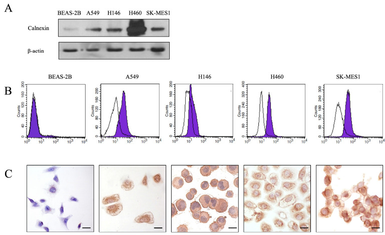
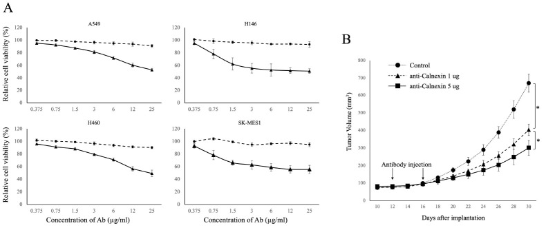
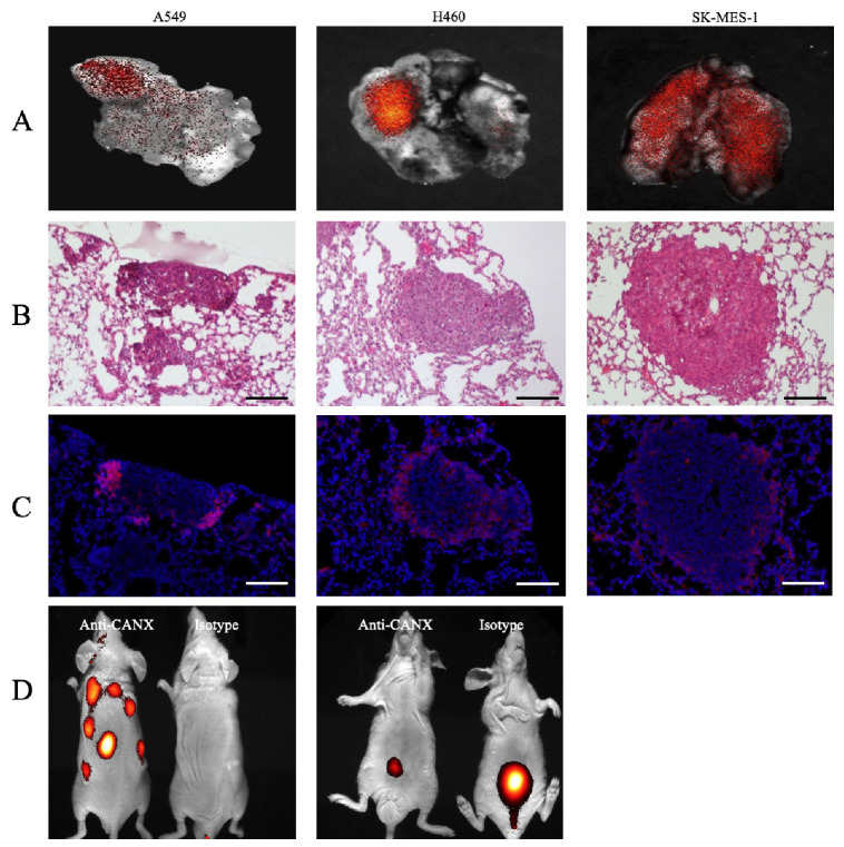
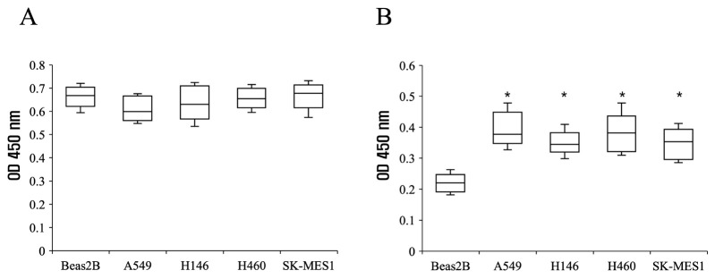
Lung cancer carries one of the highest mortality rates among all cancers. It is often diagnosed at more advanced stages with limited treatment options compared to other malignancies. This study focuses on calnexin as a potential biomarker for diagnosis and treatment of lung cancer. Calnexin, a molecular chaperone integral to N-linked glycoprotein synthesis, has shown some associations with cancer. However, targeted therapeutic or diagnostic methods using calnexin have been proposed. Through 1D-LCMSMS, we identified calnexin as a biomarker for lung cancer and substantiated its expression in human lung cancer cell membranes using Western blotting, flow cytometry, and immunocytochemistry. Anti-calnexin antibodies exhibited complement-dependent cytotoxicity to lung cancer cell lines, resulting in a notable reduction in tumor growth in a subcutaneous xenograft model. Additionally, we verified the feasibility of labeling tumors through in vivo imaging using antibodies against calnexin. Furthermore, exosomal detection of calnexin suggested the potential utility of liquid biopsy for diagnostic purposes. In conclusion, this study establishes calnexin as a promising target for antibody-based lung cancer diagnosis and therapy, unlocking novel avenues for early detection and treatment. [BMB Reports 2024; 57(3): 155-160].


Conflict of interest statement

CONFLICTS OF INTEREST

The authors have no conflicting interests.

Figures

Fig. 1
Fig. 1
Calnexin protein expression in lung cancer cells. (A) Western blot analysis revealing elevated calnexin expression in A549, NCI-H146, NCI-H460, and SK-MES-1 lung cancer cell lines compared to a normal epithelial cell line Beas2B. (B) Flow cytometry histogram showing that the calnexin antibody can strongly bind to the surface of lung cancer cells (purple shaded area) with minimal binding to Beas2B (black curve). The experiment was performed in duplicate at least three times. (C) Lung cancer cells exhibit abundant membrane and ER binding, while Beas2B cells show minimal binding, particularly on the ER (× 400; scale bar, 20 μm).
Fig. 2
Fig. 2
In vitro and in vivo tumor toxicity of anti-calnexin antibody. (A) The dotted line illustrates concentration-dependent cytotoxicity, while the solid line represents complement-dependent cytotoxicity when the complement system is activated by adding goat serum to achieve a final concentration of 5% (vol/vol). Experiments were performed in duplicate at least three times. (B) Antitumor effect of anti-calnexin antibody. A549 cells were subcutaneously injected into backs of nude mice and treated intraperitoneally with 1 μg and 5 μg of anti-calnexin antibody on days 12 and 16 when the tumor size reached 70-150 mm3 after 10 days. *P-value < 0.05 (n = 9).
Fig. 3
Fig. 3
In vivo and ex vivo imaging of a tumor xenograft model. (A) Ex vivo imaging of lungs in a tumor xenograft mouse model. (B) Lung tissues stained with H&E for ex vivo imaging. (C) Serial sections stained with DAPI and observed under a fluorescence microscope (× 100; scale bar, 100 μm). (D) In vivo imaging of a subcutaneous tumor xenografted model. Imaging results were obtained at 3 hours after intravenous injection of labeled antibodies (n = 6).
Fig. 4
Fig. 4
ELISA of exosomes from lung cancer xenograft mouse serum. ELISA utilized lung cancer xenograft mouse serum exosomes with normal and Beas2B-injected mouse sera as controls. Comparable OD values were observed with anti-CD9 (A). However, the anti-calnexin antibody revealed higher OD values in cancer cell-injected mouse serum compared to controls (n = 6, *P < 0.05) (B).

References

    1. Organization WHO. https://www.who.int/news-room/fact-sheets/detail/lung-cancer.
    1. Siegel RL, Miller KD, Fuchs HE, Jemal A. Cancer statistics, 2022. CA Cancer J Clin. 2022;72:7–33. doi: 10.3322/caac.21708. - DOI - PubMed
    1. Oberije C, De Ruysscher D, Houben R, et al. A validated prediction model for overall survival from stage III non-small cell lung cancer: toward survival prediction for individual patients. Int J Radiat Oncol Biol Phys. 2015;92:935–944. doi: 10.1016/j.ijrobp.2015.02.048. - DOI - PMC - PubMed
    1. Bernabe-Caro R, Chen Y, Dowlati A, Eason P. Current and emerging treatment options for patients with relapsed small-cell lung carcinoma: a systematic literature review. Clin Lung Cancer. 2023;24:185–208. doi: 10.1016/j.cllc.2023.01.012. - DOI - PubMed
    1. Mitchell JM, Flight RM, Moseley HNB. Untargeted lipidomics of non-small cell lung carcinoma demonstrates differentially abundant lipid classes in cancer vs. non-cancer tissue. Metabolites. 2021;11:740. doi: 10.3390/metabo11110740.71d7cd8c2dff47edba9bb2a5e5e4c12f - DOI - PMC - PubMed