. 2024 Jan 9;27(2):108838.
doi: 10.1016/j.isci.2024.108838. eCollection 2024 Feb 16.

Extracellular matrix protein composition dynamically changes during murine forelimb development

Affiliations

Extracellular matrix protein composition dynamically changes during murine forelimb development

Kathryn R Jacobson et al. iScience. .

Abstract

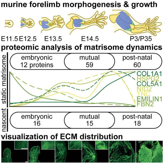
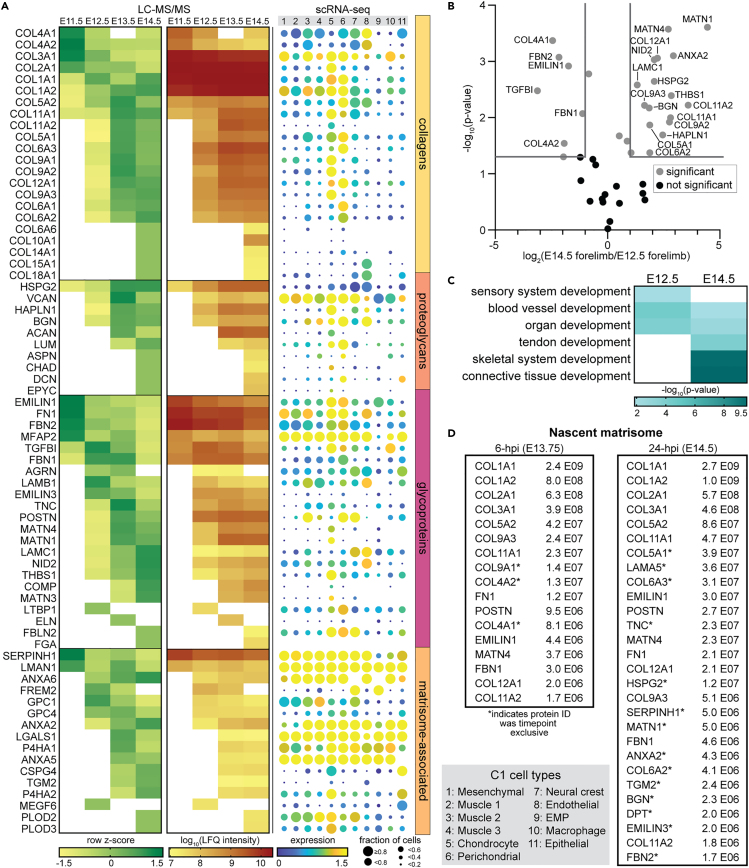
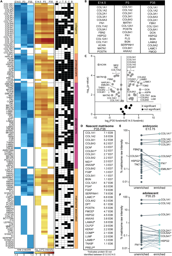
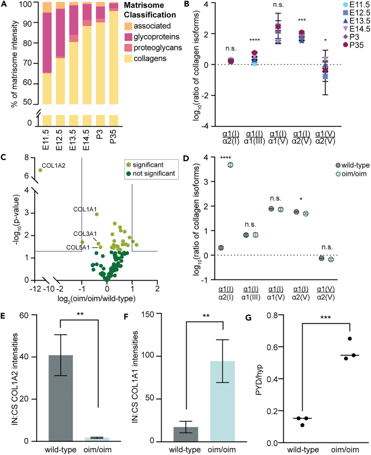
The extracellular matrix (ECM) is an integral part of multicellular organisms, connecting different cell layers and tissue types. During morphogenesis and growth, tissues undergo substantial reorganization. While it is intuitive that the ECM remodels in concert, little is known regarding how matrix composition and organization change during development. Here, we quantified ECM protein dynamics in the murine forelimb during appendicular musculoskeletal morphogenesis (embryonic days 11.5-14.5) using tissue fractionation, bioorthogonal non-canonical amino acid tagging, and mass spectrometry. Our analyses indicated that ECM protein (matrisome) composition in the embryonic forelimb changed as a function of development and growth, was distinct from other developing organs (brain), and was altered in a model of disease (osteogenesis imperfecta murine). Additionally, the tissue distribution for select matrisome was assessed via immunohistochemistry in the wild-type embryonic and postnatal musculoskeletal system. This resource will guide future research investigating the role of the matrisome during complex tissue development.

Keywords: Biological sciences; Developmental biology; Natural sciences.


Conflict of interest statement

The authors declare no competing interests.

Figures

None
Graphical abstract
Figure 1
Figure 1
The matrisome of the forelimb is specialized during murine embryogenesis Tissue fractionation was combined with LC-MS/MS to analyze the matrisome of embryonic day (E)11.5-E14.5 whole murine embryos and forelimbs (n = 3 biological replicates/time point). Cytosolic (C), nuclear (N), and membrane (M) fractions from whole embryos were combined into one CNM fraction (Figure S1A) and analyzed, along with cytoskeletal (CS) and insoluble (IN) fractions of the whole embryos (E) and forelimbs (F), by LC-MS/MS (Table S1). (A) The distribution of cellular compartments, in the IN, CS, and CNM fractions, plotted as average of the biological replicates. For all tissues and time points, there was significantly more matrisome, based on the percentage of total raw intensity attributed to ECM proteins, in the IN fractions compared to CS and CNM (p < 0.001, one-way ANOVA). (B and C) Volcano plots comparing differences in ECM protein composition between (B) E11.5 forelimb and embryo, and (C) E14.5 forelimb and embryo. Gray lines denote >2-fold change and p < 0.05 (two-tailed t-test). (D) E14.5 WT brains (n = 3) were processed as described for the forelimbs, and revealed there were distinct matrisome compositions in functionally different tissues (Table S1). Gray lines denote >2-fold change and p < 0.05 (two-tailed t-test). (E) Development-associated Gene Ontology (GO) terms generated by analyzing ECM proteins that were significantly more abundant or exclusively identified in the forelimb and brain, and the corresponding log10-transformed p values.
Figure 2
Figure 2
ECM protein composition varies as a function of murine musculoskeletal development (A, left) LFQ intensities of the matrisome composition of E11.5-E14.5 WT forelimbs were normalized and combined from both CS and IN fractions for each timepoint (Table S2). Row z-scores were calculated and averaged across biological replicates (n = 3) and proteins were clustered first by matrisome classification, then by relative abundance starting at E11.5. (A, center) Corresponding log10-transformed LFQ intensities for proteins shown in (A, left). White boxes signify proteins not identified in n ≥ 2 biological replicates. (A, right) C1 Fluidigm single-cell RNA-sequencing (scRNA-seq) data of the matrisome identified in E10-E15.5 limbs (Table S3), generated by He et al. Color indicates the log2(average expression +1) and circle size represents the percentage of cells expressing that transcript. Cell types defined by He et al. See Figure S2 for similar dot plot generated with 10X scRNA-seq data. (B) Many ECM proteins were significantly different in abundance between E12.5 and E14.5 forelimbs. Gray lines denote >2-fold change and p < 0.05 (two-tailed t-test). (C) GO analysis of ECM proteins more abundant in, or exclusive to, E12.5 or E14.5 forelimbs generated distinct developmental terms. (D) In vivo Aha-tagging (BONCAT), tissue fractionation, and enrichment of Aha-labeled ECM proteins were combined with LC-MS/MS analysis to identify the nascent matrisome in embryonic forelimbs (Table S2). Newly synthesized proteins (NSPs) that were part of the matrisome, identified in embryonic forelimbs 6- or 24-h post injection (hpi) of Aha at E13.5, were ranked in order of abundance. Proteins exclusively identified at E13.75 (6-hpi) or E14.5 (24-hpi) were denoted (∗) and the values displayed report the average raw protein intensity (n = 3).
Figure 3
Figure 3
The matrisome continued to change during postnatal musculoskeletal growth (A, left) Heatmap of row z-scores and (A, center) corresponding combined LFQ intensities for ECM proteins identified in the E14.5, P3, and P35 forelimbs (Table S2). White boxes signify proteins not identified in n ≥ 2 biological replicates. (A, right) The distribution of individual ECM identified by proteomic studies of isolated tissues, including muscle (M), myotendinous junction (J), tendon (T), enthesis (E), cartilage (C), and bone (B).,,,,, (B) Top 10 abundant ECM proteins in E14.5, P3, and P35 forelimbs show differences in matrisome composition as a function of development. (C) Volcano plot comparing ECM proteins identified at E14.5 and P35. Gray lines denote >2-fold change and p < 0.05 (two-tailed t-test). (D) Nascent matrisome from P35-P36 forelimbs identified by BONCAT. ECM proteins were ranked by abundance at 6-hpi; PRELP was only identified 24-hpi. Values are the average raw intensity for n = 3. Matrisome components that were exclusive to adolescence, compared to embryonic forelimbs (Figure 2D), are marked with #. (E and F) Comparison of the relative percentage of matrisome intensity between unenriched and enriched samples. Points are the average of n = 3 biological replicates. Labels on the left indicate ECM proteins of interest that were only identified in unenriched samples. Lines connect proteins identified in both unenriched and enriched samples (labeled on the right). Darker, bolded lines highlight ECM proteins of interest and ∗ indicates a significant change in intensity percentage between unenriched and enriched sample (two-tailed t-test, p < 0.05).
Figure 4
Figure 4
The ECM is differentially distributed within musculoskeletal tissues during forelimb development (A–R) Cryosections from E11.5-E14.5, P3 and adult forelimbs were stained with antibodies against: (A–F, A′–D′) type I collagen (COLI; green), type V collagen (COLV; red), and myosin heavy chain, a marker for differentiated skeletal muscle (MY32; blue); (G–L, G′–J′) tenascin-C (TNC; green), fibrillin-2 (FBN2; red), and MY32 (blue); (M–R, M′–P′) elastin microfibril interfacer-1 (EMILIN1; green), perlecan (HSPG2; red), and nidogen-2 (NID2; blue). Insets (indicated with ’) are a 3× enlargement of the region containing the nascent elbow (∗) for E11.5-E14.5. Scale bars represent 200 μm. Individual channels and secondary antibody only negative control panels are shown in Figures S3–S5. (S) Graphical summary of protein dynamics.
Figure 5
Figure 5
Altered collagen dynamics in osteogenesis imperfecta murine (oim/oim) forelimbs (A) During WT forelimb development (E11.5-E14.5) and growth (P3, P35), collagen content significantly increased, and (B) the ratios of collagen isoforms associated with type I fibrillogenesis were dependent on age (p < 0.0001; one-way ANOVA). Values shown are average of n=3 replicates and scale bars represent the standard deviation (Table S2). (C) LC-MS/MS analysis of CS and IN fractions of oim/oim and wild-type forelimbs validated a decrease in COL1A2 abundance (Table S5; two-tailed t-test; ∗∗∗∗p < 0.0001). (D) The ratios of collagen chains associated with type I collagen fibrillogenesis in oim/oim and wild-type forelimbs. Although the ratios between α1(I):α2(I) and α1(I):α2(V) were significantly different in oim/oim forelimbs (p < 0.05, two-tailed t-test), the other ratios were not affected by the mutation. (E) In the mutant forelimbs, less COL1A2 was found in the IN fraction, relative to the CS fraction (two-tailed t-test; ∗∗p < 0.01), indicating increased solubility of this collagen chain in oim/oim forelimb, compared to the wild-type. (F) In contrast, significantly more COL1A1 was found in the IN fraction, relative to the CS fraction, compared to controls (two-tailed t-test; ∗∗p < 0.01). (G) Increased pyrrolidine cross-links (PYD), relative to the hydroxyproline content (Hyp), was observed in mutant TA tendons from 10-weeks-old mice (n=3; two-tailed t-test; ∗∗∗p < 0.001).

References

    1. Charvet B., Ruggiero F., Le Guellec D. The development of the myotendinous junction. A review. Muscles Ligaments Tendons J. 2012;2:53–63. - PMC - PubMed
    1. Huang A.H. Coordinated development of the limb musculoskeletal system: Tendon and muscle patterning and integration with the skeleton. Dev. Biol. 2017;429:420–428. - PMC - PubMed
    1. He P., Williams B.A., Trout D., Marinov G.K., Amrhein H., Berghella L., Goh S.-T., Plajzer-Frick I., Afzal V., Pennacchio L.A., et al. The changing mouse embryo transcriptome at whole tissue and single-cell resolution. Nature. 2020;583:760–767. - PMC - PubMed
    1. Kelly N.H., Huynh N.P.T., Guilak F. Single cell RNA-sequencing reveals cellular heterogeneity and trajectories of lineage specification during murine embryonic limb development. Matrix Biol. 2020;89:1–10. - PMC - PubMed
    1. McQueen C., Towers M. Establishing the pattern of the vertebrate limb. Development. 2020;147 - PubMed

LinkOut - more resources