

Utilization of third-trimester fetal transcerebellar diameter measurement for gestational age estimation: a comparative study using Bland-Altman analysis
- PMID: 38304306
- PMCID: PMC10832473
- DOI: 10.1016/j.xagr.2024.100307
Utilization of third-trimester fetal transcerebellar diameter measurement for gestational age estimation: a comparative study using Bland-Altman analysis
Abstract
Background: Several studies show that gestational age estimation during the third trimester of pregnancy using fetal transcerebellar diameter is superior to that measured using fetal biometry (biparietal diameter, head circumference, abdominal circumference, and femur diaphysis length). However, the conclusion of the studies stemmed from findings of correlation and regression statistical tests, which are not the recommended statistical analysis methods for comparing the values of 1 variable as measured by 2 different methods.
Objective: This study aimed to compare the accuracy of gestational age estimation using transcerebellar diameter to that using fetal biometry during the third trimester of pregnancy using Bland-Altman statistical analysis.
Study design: This was a cross-sectional study on pregnant women who presented for routine antenatal care follow-up in the third trimester of pregnancy (28-41 weeks of gestation) at St. Paul's Hospital Millennium Medical College (Ethiopia) between November 1, 2020, and February 28, 2021. Data were collected prospectively using a structured questionnaire on the Open Data Kit. The primary outcome of our study was the mean bias of gestational age estimation (error in estimating gestational age) using transcerebellar diameter and composite fetal biometry (composite gestational age). Data were analyzed using Stata (version 15; StataCorp, College Station, TX). Simple descriptive analysis, Bland-Altman analysis, and the Kendall τa discordance measurement were performed as appropriate. The mean bias (error) and limits of agreement were used to present the significance of the finding.
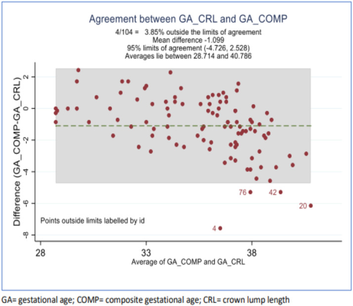
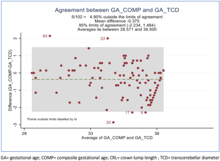
Results: A total of 104 pregnant women in the third trimester were included in the study. The mean error (bias) when transcerebellar diameter was used to estimate the gestational age was 0.65 weeks vs a bias of 1.1 weeks using composite biometry, compared with the gold standard method from crown-lump length (in both cases). The calculated estimated limit of agreement was narrower in the case of transcerebellar diameter than in the case of composite fetal biometry (-3.56 to 2.25 vs -4.73 to 2.53). The Kendall τa discordance measurement revealed that gestational age estimations using composite biometry and crown-lump length were 51% to 70%, respectively, more likely to agree than disagree and that gestational age estimations using transcerebellar diameter and crown-lump length were 62% to 77%, respectively, more likely to agree than to disagree (P≤.001).
Conclusion: Gestational age estimation using transcerebellar diameter is more accurate than gestational age estimation using composite gestational age (biparietal diameter, head circumference, femur diaphysis length, and abdominal circumference). Transcerebellar diameter should be used to date third-trimester pregnancies with unknown gestational age (unknown last normal menstrual period with no early ultrasound milestone).
Keywords: biometry; low-income setting; obstetrical ultrasound; pregnancy dating; transcerebellar diameter; unknown date.
© 2024 The Authors.
Figures

References
-
- Iram S, Gilani SA, Hassan Z, Fatima M, Bacha R, Malik SS. Ultrasonographic evaluation of the fetal transverse cerebellar diameter (TCD) measurement for prediction of gestational age in 2nd and 3rd trimesters of pregnancy. Int J Appl Sci Biotechnol. 2018;6:379–385.
-
- Dashottar S, Senger KPS, Shukla Y, Singh A, Sharma S. Transcerebellar diameter: an effective tool in predicting gestational age in normal and IUGR pregnancy. Int J Reprod Contracept Obstet Gynecol. 2018;7:4190–4197.
-
- Balsane R, Vyas J, Rajoria L, Agarwal P, Gupta S. To study the association between initial fetal crown-rump length and subsequent abortion in a viable first trimester pregnancy. Int J Reprod Contracept Obstet Gynecol. 2016;5:1744–1747.
-
- Salomon LJ, Alfirevic Z, Da Silva Costa F, et al. ISUOG Practice Guidelines: ultrasound assessment of fetal biometry and growth. Ultrasound Obstet Gynecol. 2019;53:715–723. - PubMed
-
- Nagesh R, Seetha PVV, Anil KS. Transverse cerebellar diameter–an ultrasonographic parameter for estimation of fetal gestational age. Int J Contemp Med Res. 2016;3:1029–1031.
LinkOut - more resources
Full Text Sources
