Gynecomastia on Thoracic Computed Tomography
- PMID: 38304650
- PMCID: PMC10832307
- DOI: 10.7759/cureus.51509
Gynecomastia on Thoracic Computed Tomography
Abstract
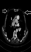
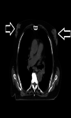
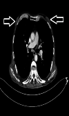
Background and objective Gynecomastia is a benign proliferation of ductal epithelium in the retroareolar region in male patients. The aim of this study was to investigate the frequency of gynecomastia in male patients who underwent thoracic computed tomography (CT) imaging at our clinic, assess possible causes, highlight the imaging characteristics of gynecomastia, and compare our findings with the literature. Materials and methods Male patients over 18 years of age who underwent thoracic CT imaging in our clinic were included in the study. Patients were initially assessed based on age and the presence of gynecomastia. The patients with gynecomastia were evaluated in terms of age, gynecomastia localization (right, left, and bilateral), gynecomastia type (nodular, dentritic, and diffuse), and possible etiology. Results The study included 1500 patients with a mean age of 45.6±21.7 years, and 470 (31.3%) patients had gynecomastia. Gynecomastia was on the right side in 11.3%, on the left side in 11.1%, and bilateral in 77.7% of the patients. Gynecomastia was nodular in 52.1%, dendritic in 35.3%, and diffuse in 17.2% of the patients. The causative factor could not be identified in 44.3% of the patients with gynecomastia. Among cases where the etiology was identified (56.7%), the most common factors were cancer (23.4%), chronic kidney disease (CKD) (13.2%), and chronic hepatitis B (10.7%). Conclusion When evaluating thoracic CT, the breast area, in addition to the lungs, chest wall, and bone structures, should also be evaluated carefully. With the increased use of thoracic CT scans, incidentally detected gynecomastia in patients is also on the rise. Knowing the presence of gynecomastia is very important for the clinician to determine the etiology and treat the underlying disease. Therefore, detecting and reporting gynecomastia on thoracic CT can prevent unnecessary advanced breast imaging methods and play a very important role in treating the underlying etiology.
Keywords: cancer; computed tomography; gynecomastia; male breast imaging; thorax.
Copyright © 2024, Deniz et al.
Conflict of interest statement
The authors have declared that no competing interests exist.
Figures


References
-
- Clinical practice. Gynecomastia. Braunstein GD. N Engl J Med. 2007;357:1229–1237. - PubMed
-
- Imaging in gynecomastia. Billa E, Kanakis GA, Goulis DG. Andrology. 2021;9:1444–1456. - PubMed
-
- EAA clinical practice guidelines-gynecomastia evaluation and management. Kanakis GA, Nordkap L, Bang AK, et al. Andrology. 2019;7:778–793. - PubMed
LinkOut - more resources
Full Text Sources
