. 2024 Mar 19;98(3):e0000724.
doi: 10.1128/jvi.00007-24. Epub 2024 Feb 2.

The 25-kDa linear polyethylenimine exerts specific antiviral activity against pseudorabies virus through interferencing its adsorption via electrostatic interaction

Affiliations

The 25-kDa linear polyethylenimine exerts specific antiviral activity against pseudorabies virus through interferencing its adsorption via electrostatic interaction

Changchao Huan et al. J Virol. .

Abstract

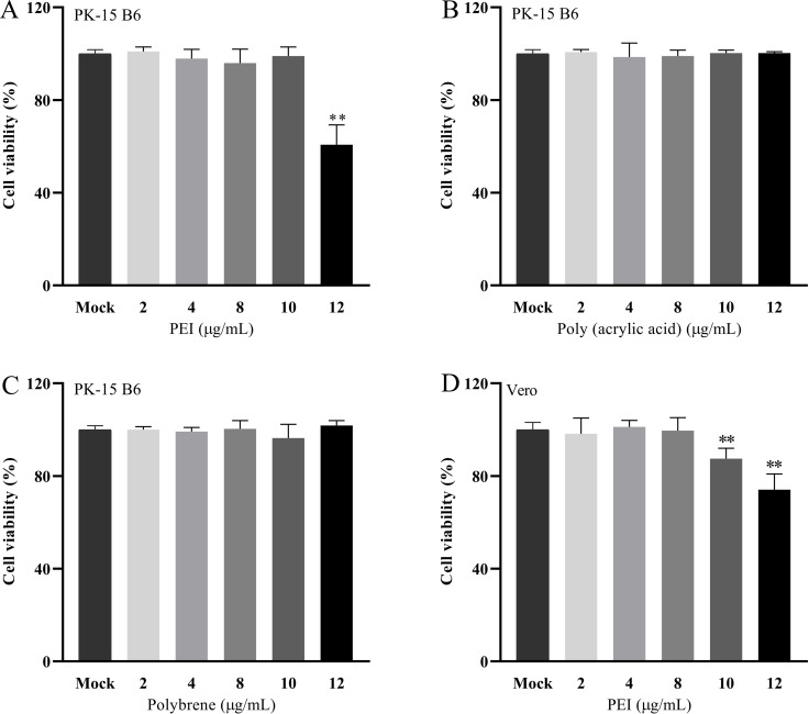
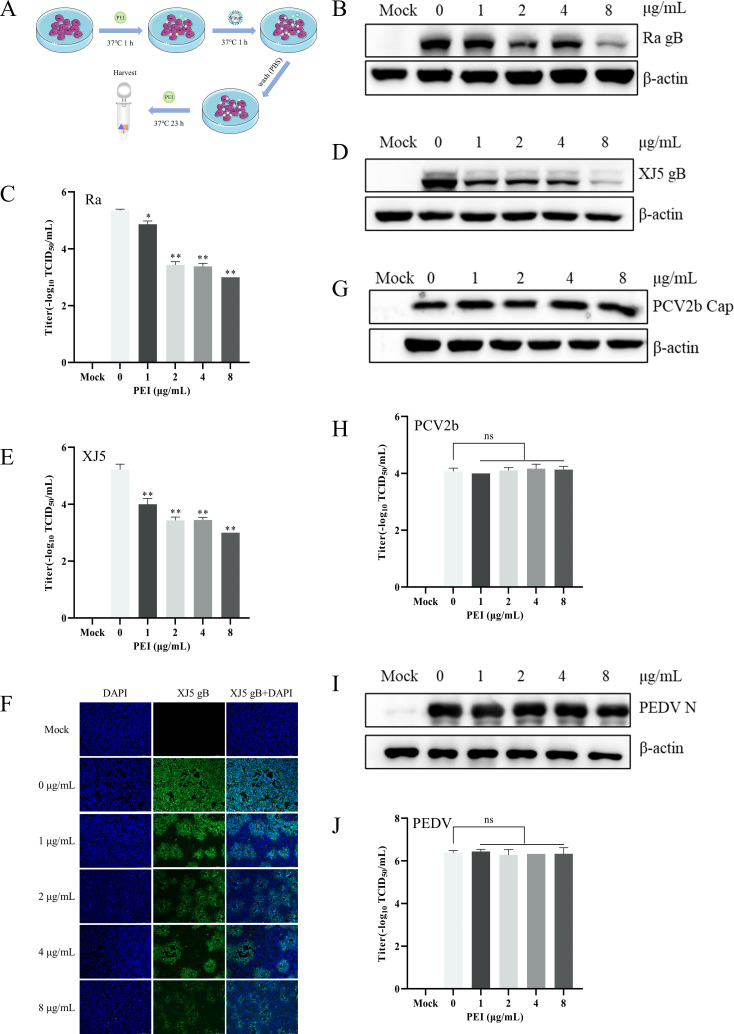
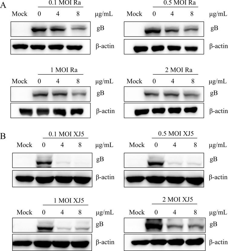
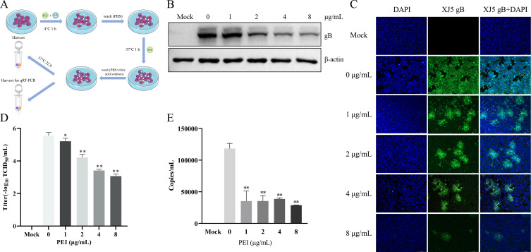
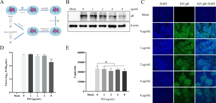
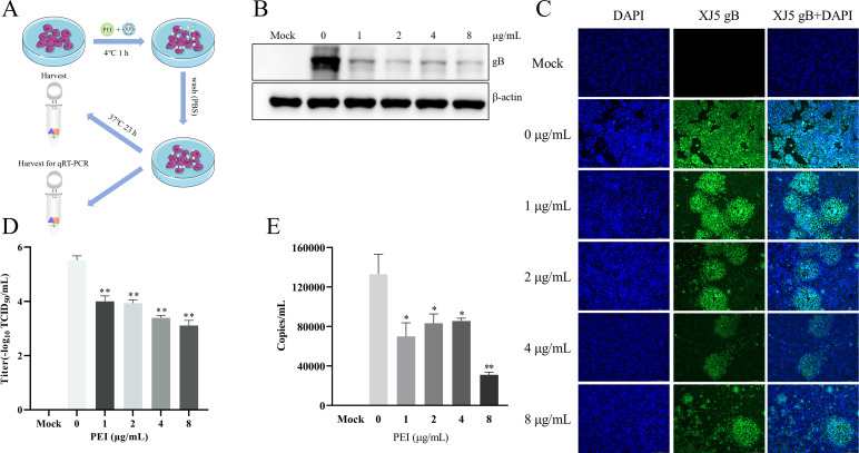
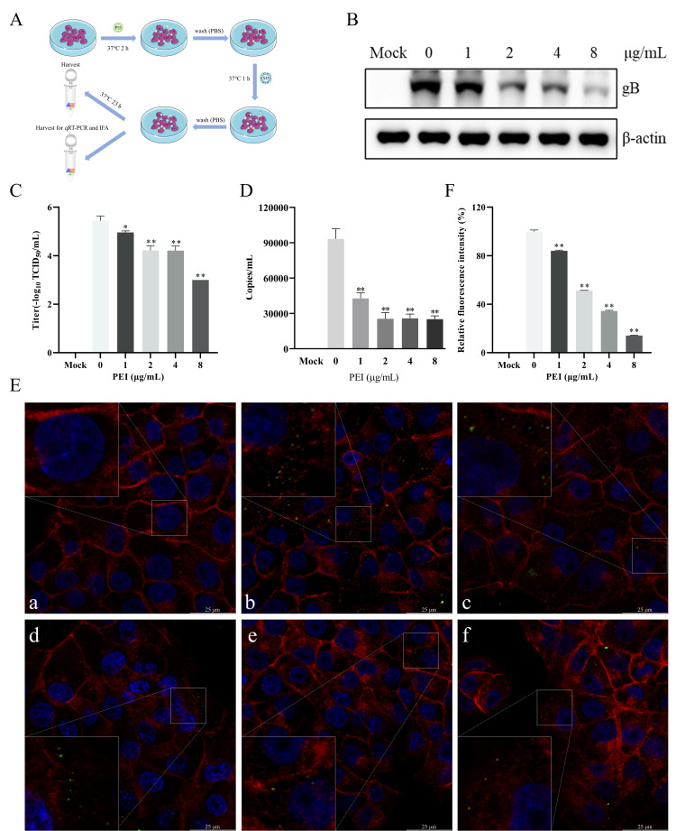
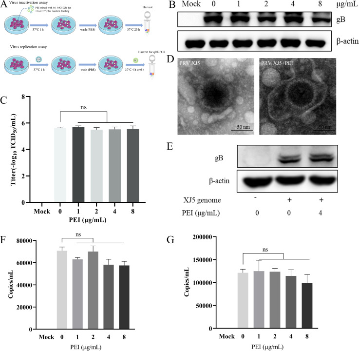
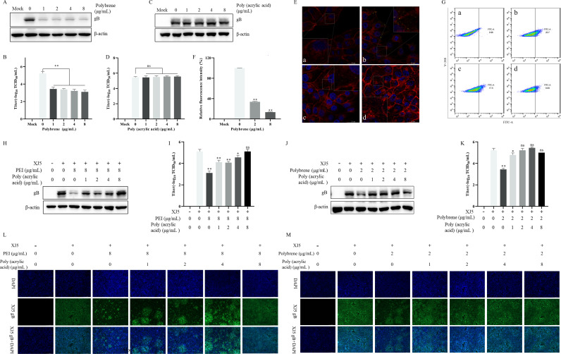
Pseudorabies virus (PRV) is the causative agent of Aujeszky's disease, which is responsible for enormous economic losses to the global pig industry. Although vaccination has been used to prevent PRV infection, the effectiveness of vaccines has been greatly diminished with the emergence of PRV variants. Therefore, there is an urgent need to develop anti-PRV drugs. Polyethylenimine (PEI) is a cationic polymer and has a wide range of antibacterial and antiviral activities. This study found that a low dose of 1 µg/mL of the 25-kDa linear PEI had significantly specific anti-PRV activity, which became more intense with increasing concentrations. Mechanistic studies revealed that the viral adsorption stage was the major target of PEI without affecting viral entry, replication stages, and direct inactivation effects. Subsequently, we found that cationic polymers PEI and Polybrene interfered with the interaction between viral proteins and cell surface receptors through electrostatic interaction to exert the antiviral function. In conclusion, cationic polymers such as PEI can be a category of options for defense against PRV. Understanding the anti-PRV mechanism also deepens host-virus interactions and reveals new drug targets for anti-PRV.IMPORTANCEPolyethylenimine (PEI) is a cationic polymer that plays an essential role in the host immune response against microbial infections. However, the specific mechanisms of PEI in interfering with pseudorabies virus (PRV) infection remain unclear. Here, we found that 25-kDa linear PEI exerted mechanisms of antiviral activity and the target of its antiviral activity was mainly in the viral adsorption stage. Correspondingly, the study demonstrated that PEI interfered with the virus adsorption stage by electrostatic adsorption. In addition, we found that cationic polymers are a promising novel agent for controlling PRV, and its antiviral mechanism may provide a strategy for the development of antiviral drugs.

Keywords: electrostatic interaction; polyethylenimine; pseudorabies virus; viral adsorption.


Conflict of interest statement

The authors declare no conflict of interest.

Figures

Fig 1
Fig 1
The cell viability of the 25-kDa linear PEI, Poly (acrylic acid), and Polybrene was determined by the CCK-8 assay. A, B, C, and D correspond to cell viability of PEI, Poly (acrylic acid), and Polybrene. The results are the averages ± SD of experiments performed six times. Statistical significance is as follows: **P < 0.01.
Fig 2
Fig 2
PEI specifically inhibited PRV infection. PK-15 B6 or Vero cells were pretreated with different concentrations of PEI for 1 h before being infected with PRV/Ra, PRV/XJ5, PCV2b, and PEDV strains (0.1 MOI) in the presence of the PEI, and then, the infected cells were incubated with the corresponding concentration of PEI for 24 hpi. (A) Schematic diagram of the infection experimental setup. B, D, G, and I correspond to the expression levels of viral proteins of the PRV/Ra, PRV/XJ5, PCV2b, and PEDV strains, respectively. (F) The detection of internalized viruses by immunofluorescent assay (IFA). C, E, H, and J correspond to virus titers in the supernatant after infection with the PRV/Ra, PRV/XJ5, PCV2b, and PEDV strains by TCID50 assay, respectively. Statistical significance is as follows: *P < 0.05 and **P < 0.01; ns, not significant.
Fig 3
Fig 3
PEI inhibited PRV infection in PK-15 B6 cells at different MOIs. A and B correspond to the expression levels of gB protein of PRV/XJ5 and PRV/Ra by western blotting, respectively.
Fig 4
Fig 4
PEI suppressed adsorption and entry stages of PRV infection in PK-15 B6 cells. PK-15 B6 cells were infected with PRV/XJ5 at 4°C for 1 h in the presence of different concentrations of PEI, incubated with corresponding concentrations of PEI at 37°C for 1 h, and then washed off the extracellular virus, followed by incubation without PEI to 24 hpi. (A) Schematic diagram of the virus adsorption and entry experimental setup. (B) PRV/XJ5 gB protein levels were detected by western blotting. (C) The detection of internalized viruses by IFA. (D) Virus titers were determined by TCID50 assay. (E) The virus DNA was quantified by quantitative real-time PCR (qRT-PCR). Statistical significance is shown as follows: *P < 0.05 and **P < 0.01.
Fig 5
Fig 5
PEI effects on PRV entry in PK-15 B6 cells. PK-15 B6 cells were infected with PRV/XJ5 at 4°C for 1 h and then incubated with different concentrations of PEI for 1 h at 37°C, followed by washing off the extracellular virus and incubated without PEI for 24 hpi. (A) Schematic diagram of the virus entry experimental setup. (B) PRV/XJ5 gB protein levels were detected by western blotting. (C) The detection of internalized viruses by IFA. (D) Virus titers were determined by TCID50 assay. (E) The intracellular virus DNA was quantified by qRT-PCR. Statistical significance is as follows: **P < 0.01 and ns, not significant.
Fig 6
Fig 6
PEI blocked PRV adsorption in PK-15 B6 cells. PK-15 B6 cells were infected with PRV/XJ5 at 4°C for 1 h in the presence of different concentrations of PEI and then incubated without PEI for 24 hpi. (A) Schematic diagram of the virus adsorption experimental setup. (B) PRV/XJ5 gB protein levels were detected by western blotting. (C) The detection of internalized viruses by IFA. (D) Virus titers were determined by TCID50 assay. (E) The cell surface virus DNA was quantified by qRT-PCR. Statistical significance is as follows: *P < 0.05 and **P < 0.01.
Fig 7
Fig 7
PEI pretreatment inhibited PRV infection. PK-15 B6 cells were pretreated with different concentrations of PEI at 37°C for 2 h and then infected with PRV/XJ5. (A) Schematic diagram of the PEI pretreatment experimental setup. (B) PRV/XJ5 gB protein levels were detected by western blotting. (C) Virus titers were determined by TCID50 assay. (D) The cell surface virus DNA was quantified by qRT-PCR. (E) The adsorbed PRV virions were observed through LSCM, and a, b, c, d, e, and f correspond to the negative control group, PRV-only treated group, and 1, 2, 4, and 8 µg/mL PEI-treated groups, respectively. The green points indicate PRV virions. (F) Relative fluorescence intensity analysis of PRV virions adsorbed to the cell surface. Statistical significance is as follows: *P < 0.05 and **P < 0.01.
Fig 8
Fig 8
PEI did not act on the viral replication stage and disrupt the viral virions. PK-15 B6 cells were infected with the mixture of PEI co-incubated with PRV/XJ5 or added with different concentrations of PEI within the post-infection replication stage to observe the effect on PRV replication. (A) Schematic diagram of the virus replication and inactivation experimental setup. (B) PRV/XJ5 gB protein levels were detected by western blotting. (C) Virus titers were determined by TCID50 assay. (D) The purified PRV/XJ5 virions were incubated with 4 µg/mL PEI for 1 h at 37°C. Then, virus suspensions were prepared for electron microscopy observation. (E) Vero cells were transfected with the genome of PRV/XJ5 for 36 h, and then, the cells were treated with 4 µg/mL PEI for 6 h. After treatment, the cells were collected to detect gB protein levels by western blotting. (F and G) The intracellular virus DNA were quantified by qRT-PCR at 4 and 6 hpi, respectively. Statistical significance is as follows: ns, not significant.
Fig 9
Fig 9
Electrostatic interaction was responsible for the anti-PRV activity of PEI by interfering with virus attachment. PK-15 B6 cells were pretreated with different concentrations of Polybrene and Poly (acrylic acid), the mixtures of 8 µg/mL PEI with different concentrations of Poly (acrylic acid), or the mixtures of 2 µg/mL Polybrene with different concentrations of Poly (acrylic acid) at 37℃ for 2 h and then infected with PRV/XJ5. A, C, H, and J correspond to the gB protein levels detected by western blotting after pretreatment of cells with different concentrations of Polybrene and Poly (acrylic acid), the mixtures of 8 µg/mL PEI with different concentrations of Poly (acrylic acid), or the mixtures of 2 µg/mL Polybrene with different concentrations of Poly (acrylic acid), respectively. B, D, I, and K correspond to virus titers determined by TCID50 assay in the supernatant after pretreatment of cells with different concentrations of Polybrene and Poly (acrylic acid), the mixtures of 8 µg/mL PEI with different concentrations of Poly (acrylic acid), or the mixtures of 2 µg/mL Polybrene with different concentrations of Poly (acrylic acid), respectively. (E) The adsorbed virus particle was observed through LSCM, and a, b, c, and d correspond to the negative control group, PRV-only treated group, 2 and 8 µg/mL Polybrene-treated groups, respectively. The green points indicate PRV virions. (F) Relative fluorescence intensity analysis of PRV virions adsorbed to the cell surface. (G) The detection of cell membrane potential in PEI-pretreated cells by flow cytometry; a, b, c, and d correspond to the blank control group, DiBAC4(3)-only-treated group, and 2 and 8 µg/mL PEI-treated groups, respectively. L and M correspond to the detection of internalized viruses by IFA after pretreatment of cells with the mixtures of 8 µg/mL PEI with different concentrations of Poly (acrylic acid) and the mixtures of 2 µg/mL Polybrene with different concentrations of Poly (acrylic acid), respectively. Statistical significance is as follows: *P < 0.05 and **P < 0.01; ns, not significant.

Similar articles

Cited by

  • Alphaherpesvirus in Pets and Livestock.
    Duan SH, Li ZM, Yu XJ, Li D. Duan SH, et al. Microorganisms. 2025 Jan 4;13(1):82. doi: 10.3390/microorganisms13010082. Microorganisms. 2025. PMID: 39858850 Free PMC article. Review.

References

    1. Pomeranz LE, Reynolds AE, Hengartner CJ. 2005. Molecular biology of pseudorabies virus: impact on neurovirology and veterinary medicine. Microbiol Mol Biol Rev 69:462–500. doi:10.1128/MMBR.69.3.462-500.2005 - DOI - PMC - PubMed
    1. Ferrara G, Longobardi C, D’Ambrosi F, Amoroso MG, D’Alessio N, Damiano S, Ciarcia R, Iovane V, Iovane G, Pagnini U, Montagnaro S. 2021. Aujeszky's disease in South-Italian wild boars (Sus scrofa): a serological survey. Animals (Basel) 11:11. doi:10.3390/ani11113298 - DOI - PMC - PubMed
    1. He W, Auclert LZ, Zhai X, Wong G, Zhang C, Zhu H, Xing G, Wang S, He W, Li K, Wang L, Han GZ, Veit M, Zhou J, Su S. 2019. Interspecies transmission, genetic diversity, and evolutionary dynamics of pseudorabies virus. J Infect Dis 219:1705–1715. doi:10.1093/infdis/jiy731 - DOI - PubMed
    1. Wang G, Zha Z, Huang P, Sun H, Huang Y, He M, Chen T, Lin L, Chen Z, Kong Z, Que Y, Li T, Gu Y, Yu H, Zhang J, Zheng Q, Chen Y, Li S, Xia N. 2022. Structures of pseudorabies virus capsids. Nat Commun 13:1533. doi:10.1038/s41467-022-29250-3 - DOI - PMC - PubMed
    1. Zheng HH, Jin Y, Hou CY, Li XS, Zhao L, Wang ZY, Chen HY. 2021. Seroprevalence investigation and genetic analysis of pseudorabies virus within pig populations in Henan province of China during 2018-2019. Infect Genet Evol 92:104835. doi:10.1016/j.meegid.2021.104835 - DOI - PubMed

LinkOut - more resources