. 2024 Mar 19;98(3):e0182723.
doi: 10.1128/jvi.01827-23. Epub 2024 Feb 2.

Disenfranchised DNA: biochemical analysis of mutant øX174 DNA-binding proteins may further elucidate the evolutionary significance of the unessential packaging protein A

Affiliations

Disenfranchised DNA: biochemical analysis of mutant øX174 DNA-binding proteins may further elucidate the evolutionary significance of the unessential packaging protein A

Samuel D Love et al. J Virol. .

Abstract

Most icosahedral DNA viruses package and condense their genomes into pre-formed, volumetrically constrained capsids. However, concurrent genome biosynthesis and packaging are specific to single-stranded (ss) DNA micro- and parvoviruses. Before packaging, ~120 copies of the øX174 DNA-binding protein J interact with double-stranded DNA. 60 J proteins enter the procapsid with the ssDNA genome, guiding it between 60 icosahedrally ordered DNA-binding pockets formed by the capsid proteins. Although J proteins are small, 28-37 residues in length, they have two domains. The basic, positively charged N-terminus guides the genome between binding pockets, whereas the C-terminus acts as an anchor to the capsid's inner surface. Three C-terminal aromatic residues, W30, Y31, and F37, interact most extensively with the coat protein. Their corresponding codons were mutated, and the resulting strains were biochemically and genetically characterized. Depending on the mutation, the substitutions produced unstable packaging complexes, unstable virions, infectious progeny, or particles packaged with smaller genomes, the latter being a novel phenomenon. The smaller genomes contained internal deletions. The juncture sequences suggest that the unessential A* (A star) protein mediates deletion formation.IMPORTANCEUnessential but strongly conserved gene products are understudied, especially when mutations do not confer discernable phenotypes or the protein's contribution to fitness is too small to reliably determine in laboratory-based assays. Consequently, their functions and evolutionary impact remain obscure. The data presented herein suggest that microvirus A* proteins, discovered over 40 years ago, may hasten the termination of non-productive packaging events. Thus, performing a salvage function by liberating the reusable components of the failed packaging complexes, such as DNA templates and replication enzymes.

Keywords: microviridae; phiX174; single-stranded DNA packaging; single-stranded DNA replication.


Conflict of interest statement

The authors declare no conflict of interest.

Figures

Fig 1
Fig 1
øX174 genome packaging and the DNA-binding protein J. (A) DNA packaging: the parental + strand is displaced into the procapsid as the daughter + strand is synthesized. (B) The amino acid sequence of the øX174, G4, and α3 J proteins. (C) Interior structure of a virion pentamer (PDB 2BPA). The coat proteins are depicted in gray. One J protein is highlighted in purple; the rest are depicted in magenta. Light blue represents the ordered DNA backbone within the DNA-binding pocket.
Fig 2
Fig 2
Phenotypes associated with amino acid substitutions for W30, Y31, and F37. (A) The amino acids found at the W30, Y31, and F37 sites are listed at the top of the grid. Color code: black, lethal; green, viable between 24–42°C; blue, cold sensitive; red, temperature sensitive. The split green/black squares indicate small plaque formation. The red/black color for H at the W30 site indicates a slightly less lethal phenotype at 24°C. See text for details. Symbols: WT: wild type, ND: not determined. (B) The atomic structure of protein J (PDB 2BPA). Basic amino acid side chains (R and K) are highlighted in dark blue. The conserved C-terminus, which contains the aromatic side chains, is depicted in gray.
Fig 3
Fig 3
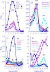
Assembled particle formation in wild-type and øX174 mutant infected cells. Extracts made from infected cells were analyzed by the rate of zonal sedimentation. Gradients were fractionated from the bottom of the tube. Thus, lower fractions contain particles with higher S values. Mutant names and infection temperatures are given in the figure. (A and B). Mutants that produce virions but at reduced yields compared to wild type. (C) Mutants that do not produce soluble assembled particles. The sedimentation profiles of mutants l(J)Y31E, l(J)W30R, l(J)Y31M, and L(J)Y31L are not depicted but yielded similar results. (D). Mutants that produced particles that sediment at ~70S.
Fig 4
Fig 4
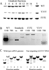
Composition of assembled particles generated by the l(J)F37E and l(J)Y31V packaging mutants. (A) SDS-PAGE of gradient fractions from Fig. 3D. Only the region of the gel with the viral coat protein is depicted. Band intensity corresponds to the A260 readings reported in Fig. 3D. V indicates purified viral coat protein. (B) DNA isolated from 114S and 70S gradient fractions. Symbols: ss, purified single-stranded DNA genomes; RF, replicative form dsDNA. The bracketed band identifies the small aberrant ssDNA associated with the l(J)Y31V 70S material. (C) PCRs were conducted with wild-type and l(J)Y31V 70S DNA. The six PCRs span the entire genome. Reaction numbers utilized the same primers for each template. Bracketed bands identify PCR fragments unique to the l(J)Y31V 70S DNA template.
Fig 5
Fig 5
Internal deletions found in small viral DNAs. (A) Sequencing chromatogram of a PCR product containing a single homogeneous deletion. Numbers indicate the location of the nucleotides within the øX174 sequence, which is 5386 nucleotides in length and circular. The chromatogram corresponds to the top sequence given in part D. (B) Sequencing chromatogram of a PCR product containing heterogeneous deletions. (C) Schematic for the generation of a mottled chromatogram as illustrated in part B. (D) Sequences of internal homogeneous deletions. The 5′ nucleotide sequence before the deletion is depicted in blue. The 3′ sequence is depicted in red. Numbers indicate the location of the nucleotides within the øX174 sequence. Underlined nucleotides could have been donated by either the 5′ or 3′ juncture sequences. Nucleotides in black text represent those found in the juncture that are not present in either the 5′ or 3′ sequences surrounding it. “A* target” indicates a juncture derived from a known target site for the A* protein. “Ori/A*target” indicates a juncture derived from the origin of replication, which is also an A* target site.
Fig 6
Fig 6
The location of the aromatic J residues and the suppressors that restore viability to missense mutations at those sites (PDB 2BPA). The J protein is depicted in dark blue with the W30, Y31, and F37 residues highlighted with space-filled rendering. The body of the coat protein is depicted in gray. The coat protein residues that contact J protein residues W30, Y31, and F37 are depicted with space-filled rendering. The location of the second-site suppressors is highlighted in light blue, space-filled rendering. They are labeled to reflect the wild-type residue, before the number, and the change, after the number. The residues constituting the DNA binding pocket are depicted in magenta, space-filled rendering. Icosahedrally ordered DNA is depicted in cyan ball-and-stick rendering.
Fig 7
Fig 7
A speculative model of protein A* functions during concurrent ssDNA synthesis and packaging. Protein A* binds to target sites on dsDNA, which inhibits helicase function. Slowing genome biosynthesis ensures that it does not outpace packaging. (Bottom line) Packaged ssDNA assumes a confirmation that supports full genome packaging. After one procapsid is filled, the RF DNA template and associated enzymes can be used to fill another procapsid. (Top line) Protein A* inhibition is not sufficient to ensure synthesis-packaging synchrony. ssDNA accumulates outside the capsid. Protein A* binds ssDNA and generates an internal deletion. Rolling circle replication continues until the origin is regenerated. Although this results in a particle without a full genome, the RF DNA template and associated enzymes are salvaged.

Similar articles

References

    1. Gopal A, Egecioglu DE, Yoffe AM, Ben-Shaul A, Rao ALN, Knobler CM, Gelbart WM. 2014. Viral RNAs are unusually compact. PLoS One 9:e105875. doi:10.1371/journal.pone.0105875 - DOI - PMC - PubMed
    1. Speir JA, Johnson JE. 2012. Nucleic acid packaging in viruses. Curr Opin Struct Biol 22:65–71. doi:10.1016/j.sbi.2011.11.002 - DOI - PMC - PubMed
    1. Cerritelli ME, Studier FW. 1996. Assembly of T7 capsids from independently expressed and purified head protein and scaffolding protein. J Mol Biol 258:286–298. doi:10.1006/jmbi.1996.0250 - DOI - PubMed
    1. Aoyama A, Hamatake RK, Hayashi M. 1981. Morphogenesis of phi X174: in vitro synthesis of infectious phage from purified viral components. Proc Natl Acad Sci U S A 78:7285–7289. doi:10.1073/pnas.78.12.7285 - DOI - PMC - PubMed
    1. Mukai R, Hamatake RK, Hayashi M. 1979. Isolation and identification of bacteriophage phi X174 prohead. Proc Natl Acad Sci U S A 76:4877–4881. doi:10.1073/pnas.76.10.4877 - DOI - PMC - PubMed

MeSH terms

LinkOut - more resources