. 2024 Feb 14;32(2):261-275.e4.
doi: 10.1016/j.chom.2024.01.003. Epub 2024 Feb 1.

Evolution of human H3N2 influenza virus receptor specificity has substantially expanded the receptor-binding domain site

Affiliations

Evolution of human H3N2 influenza virus receptor specificity has substantially expanded the receptor-binding domain site

Andrew J Thompson et al. Cell Host Microbe. .

Abstract

Hemagglutinins (HAs) from human influenza viruses descend from avian progenitors that bind α2-3-linked sialosides and must adapt to glycans with α2-6-linked sialic acids on human airway cells to transmit within the human population. Since their introduction during the 1968 pandemic, H3N2 viruses have evolved over the past five decades to preferentially recognize human α2-6-sialoside receptors that are elongated through addition of poly-LacNAc. We show that more recent H3N2 viruses now make increasingly complex interactions with elongated receptors while continuously selecting for strains maintaining this phenotype. This change in receptor engagement is accompanied by an extension of the traditional receptor-binding site to include residues in key antigenic sites on the surface of HA trimers. These results help explain the propensity for selection of antigenic variants, leading to vaccine mismatching, when H3N2 viruses are propagated in chicken eggs or cells that do not contain such receptors.

Keywords: H3N2; epistasis; glycan; hemagglutinin; host adaptation; human; influenza; receptor; receptor-binding site; sialic acid.


Conflict of interest statement

Declaration of interests The authors declare no competing interests.

Figures
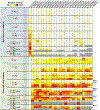
Figure 1.
Figure 1.. Glycan microarray analysis of WT H3 receptor specificity from pre-1968 pandemic to 2018.
Heatmap representation of glycan microarray data comparing receptor binding specificity of major H3(N2) strains, including vaccine strains, over time. Viral strains are listed in abbreviated form across the top row and denoted as either recombinant hemagglutinin (rHA) or whole-virus (WV) samples. Over time, binding of human strains to shorter glycans containing only one or two LacNAc repeats beneath terminal sialic acid (highlighted via blue arrows to the right of the panel) are reduced and is eventually eliminated. The color scale within individual columns (array datasets) are independently scaled to the most intense RFU within that group. Columns depict data collected from various different Sialoside array versions, including V1, V3, V4, & V5. Receptor structures corresponding to glycan numbers for all array versions can be found in Supplementary Data S1, glycan diagrams shown are according to the Symbol Nomenclature for Glycans recommended by the NLM., Grey bars illustrate that a given receptor structure was absent in the array version on which a particular dataset was collected (see Supplementary Data S1). Individual array plots for all HAs are illustrated in Supplementary Data S2.
Figure 2.
Figure 2.. Clade distribution and receptor specificity of clades 3C.2a and 3C.3a
(A) Frequency distribution of human H3N2 clades over the past decade (adapted from Nextstrain data) shows that individual clades arise and recede regularly. However, a major divergence in H3N2 phylogeny in approximately 2012 (see Fig. S2) subsequently led to clades descended from 3C.2a becoming dominant in recent years. (B) Heatmap representation of glycan microarray data (α2–6 sialosides only) for clades 3C.2a- and 3C.3a-descended strains. As shown, 3C.2a maintains length selectivity of prior dominant strains and are ultimately selected for, while 3C.3a viruses regain binding to short receptors (highlighted via blue arrows) and become outcompeted. The color scale within individual columns (array datasets) are independently scaled to the most intense RFU within that group. Columns depict data collected from various different Sialoside array versions, including V1, V3, V4, & V5. Receptor structures corresponding to glycan numbers for all array versions can be found in Supplementary Data S1. Grey bars illustrate that a given receptor structure was absent in the array version on which a particular dataset was collected (see Supplementary Data S1). Individual array plots for all HAs are illustrated in Supplementary Data S2.
Figure 3.
Figure 3.. Correlation of length-dependent receptor binding and HA residue 159
(A) Summary graphic highlighting clades/strains utilized within this study, their receptor-binding specificity with regard to short vs extended glycans, and the identity of the amino acid at position 159 of HA. Interestingly, both within the clade 3C.2a/3C.3a split and historically, a bulky aryl-containing side chain (either tyrosine (Y) or phenylalanine (F) vs a small polar side chain (typically serine (S)) correlates strongly with length-selectivity. Panels (B – D) show engineering of a recombinant contemporary clade 3C.3a strain H3, A/North Dakota/26/2016, to alter its native receptor specificity. (B) WT NDk/16 shows equally strong binding to both long and short receptors, indeed, the most intense individual signal comes from receptor #84, the terminal trisaccharide fragment α2–6-sialyl-LacNAc (6SLN; see array V3 in Supplementary Data S1). Engineering of prior F159 (B) or contemporary (in clade 3C.2a) Y159 (C) either eliminates or strongly reduces binding to nearly all short receptors (see bars highlighted in red). Panels (B – D) depict data collected from Sialoside array versions, V3, V3, & V4, respectively; Receptor structures corresponding to glycan numbers for all array versions can be found in Supplementary Data S1. Full array plots for all HAs/variants shown are illustrated in Supplementary Data S2.
Figure 4.
Figure 4.. STD NMR analysis of early and late H3 receptor binding
STD spectrum of a symmetrical, biantennary α2–6-sialylated N-glycan featuring three LacNAc repeats on each arm (6SLN3-N; panel A) interacting with either post-pandemic HK/68 recombinant H3 HA (B) or contemporary Vic/11 HA (C). Panel A shows the chemical structure of the synthesized glycan ligand, with individual sugar residues number from the reducing end through to Neu5Ac, duplicated residues on the 3’ or 6’ mannose branches are numbered X and X’, respectively. Panels (B & C) show a linear glycan cartoon of the terminal sugar moieties with interacting sugars highlighted in green. For HK/68 (B), binding is dominated by the strong and almost exclusive interactions of terminal Neu5Ac. For Vic/11 (C), binding interactions are clearly spread along the length of the receptor arm. A peak observed at approximately 3.5 ppm in both spectra (B & C) is residual solvent background from TRIS buffer and does not represent HA-glycan STDs.
Figure 5.
Figure 5.. Crystal structure analysis of H3 receptor binding over time
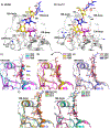
(A & B) Detailed structural views of α2–6-sialyl-(LacNAc)2 (6SLN2) bound to HK68 (early) and A/Texas/73/2017 (late; Tex/17) H3s HAs. Key secondary structural elements forming the RBS and amino acid side chains are labelled in black text (key evolved receptor-binding residues in panel (B) are highlighted in red), while receptor glycan moieties are labeled and highlighted according to CFG (Consortium for Functional Glycomics) color values. Panels (C – G) show triplet overlays of receptor-bound H3 HA structures progressing at intervals over the last five decades. Within each panel, the (invariant) approximately vertical angle of the C1-C2 bond of sialic acid is highlighted by a dashed black line, relative to the position of the sugar ring in the underlying galactose (highlighted by a second dashed line parallel to the C2-C3 bonds). As shown by an arrow in panel (D), between the late 1980s to late 1990s, the angle of the underlying sugar backbone undergoes substantial rearrangement to lie closer to the HA protein surface, highlighted by the C1-C2 vertical plane approximately bisecting the reducing end sugars in panel (C) compared to almost no receptor atoms lying on or over the same axis in panel (G).
Figure 6.
Figure 6.. Receptor binding variants allow evolution of a novel glycan via K160T
Selection of a novel K160T variant in clade 3C.2a H3N2 viruses from 2014 onwards led to formation of a new glycan (at N158) within the head of H3 HAs (A). Engineering of K160T within strains prior to the emergence of clade 3C.2a shows the novel glycan to be deleterious to receptor binding (C & D), with novel variants only able to bind the most extended receptors featuring 4 or 5 LacNAc repeats (D, shown inset). Sequence analyses reveal two functionally linked receptor-binding variants emerging immediately prior to K160T (B) restore native receptor-binding function when combined with the this new variant (E). Panels (C – E) depict data collected from Sialoside array versions, V1, V3, & V5, respectively; Receptor structures corresponding to glycan numbers for all array versions can be found in Supplementary Data S1. Full array plots for all HAs/variants shown are illustrated in Supplementary Data S2.
Figure 7.
Figure 7.. Evolution of NA activity and a novel 159N HA variant
(A) Absolute N2 activity, as measured against the optimized fluorescent substrate, MUNANA, steadily increases over time in line with evolution in H3 receptor binding. (B) Concurrently, within a subset of the same panel of N2 representatives, specific activity measured against 2–3- or 2–6- linked NeuAcα-Gal-pNP substrates reveals a long-term narrowing trend in substrate preference between avian- and human-type receptors, suggesting slow evolution away from the avian origin of these enzymes. (C & D) Glycan microarrays showing the effects on receptor specificity of a recent Y159N variant emerging in clade 3C.2a viruses during 2021. A prior strain, Camb/20 (A/Cambodia/e0826360/2020) maintaining the canonical Y159 shows selectivity for extended α2–6 receptors with at least 3 LacNAc repeats, while the newly evolved Dar/21 (A/Darwin/6/2021) possessing N159 binds species containing 2 or more LacNAc repeats.

Similar articles

Cited by

References

    1. Krammer F, Smith GJD, Fouchier RAM, Peiris M, Kedzierska K, Doherty PC, Palese P, Shaw ML, Treanor J, Webster RG, et al. (2018). Influenza. Nat Rev Dis Primers 4, 3. - PMC - PubMed
    1. Centers for Disease Control and Prevention (2019). 1918 Pandemic (H1N1 virus) (Centers for Disease Control and Prevention). https://www.cdc.gov/flu/pandemic-resources/1918-pandemic-h1n1.html
    1. Taubenberger JK, Kash JC, and Morens DM (2019). The 1918 influenza pandemic: 100 years of questions answered and unanswered. Sci Transl Med 11, eaau5485. - PMC - PubMed
    1. Taubenberger JK, and Kash JC (2010). Influenza virus evolution, host adaptation, and pandemic formation. Cell Host Microbe 7, 440–451. - PMC - PubMed
    1. Reperant LA, Kuiken T, and Osterhaus AD (2012). Adaptive pathways of zoonotic influenza viruses: from exposure to establishment in humans. Vaccine 30, 4419–4434. - PubMed