. 2024 Feb 13;19(2):163-173.
doi: 10.1016/j.stemcr.2024.01.001. Epub 2024 Feb 1.

The LRRK2 kinase substrates RAB8a and RAB10 contribute complementary but distinct disease-relevant phenotypes in human neurons

Affiliations

The LRRK2 kinase substrates RAB8a and RAB10 contribute complementary but distinct disease-relevant phenotypes in human neurons

Adamantios Mamais et al. Stem Cell Reports. .

Abstract

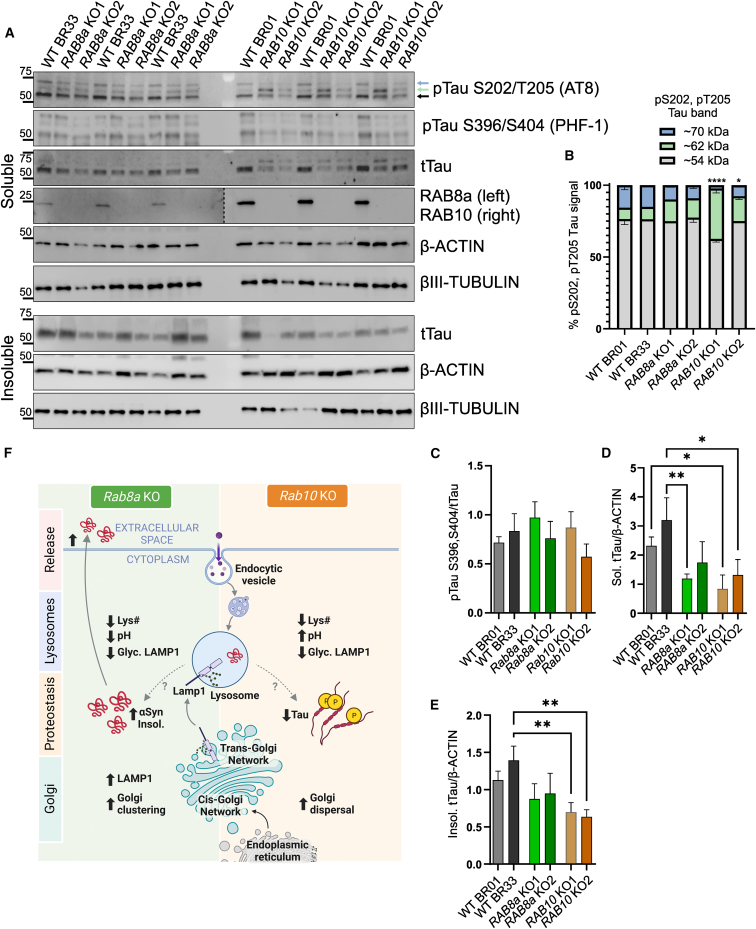
Mutations in the LRRK2 gene cause familial Parkinson's disease presenting with pleomorphic neuropathology that can involve α-synuclein or tau accumulation. LRRK2 mutations are thought to converge upon a pathogenic increase in LRRK2 kinase activity. A subset of small RAB GTPases has been identified as LRRK2 substrates, with LRRK2-dependent phosphorylation resulting in RAB inactivation. We used CRISPR-Cas9 genome editing to generate a novel series of isogenic iPSC lines deficient in the two most well-validated LRRK2 substrates, RAB8a and RAB10, from deeply phenotyped healthy control lines. Thorough characterization of NGN2-induced neurons revealed opposing effects of RAB8a and RAB10 deficiency on lysosomal pH and Golgi organization, with isolated effects of RAB8a and RAB10 ablation on α-synuclein and tau, respectively. Our data demonstrate largely antagonistic effects of genetic RAB8a or RAB10 inactivation, which provide discrete insight into the pathologic features of their biochemical inactivation by pathogenic LRRK2 mutation in human disease.

Keywords: LRRK2; Parkinson Disease; Rab10; Rab8a; Tau; iPSC; synuclein.


Conflict of interest statement

Declaration of interests M.J.L. serves on the scientific advisory board for SPARC, Ltd.

Figures

None
Graphical abstract
Figure 1
Figure 1
RAB8a KO and RAB10 KO human neurons have altered lysosomal morphology and function (A) Representative confocal images of RAB8a KO, RAB10 KO, and isogenic WT control neurons stained with LAMP1, MAP2, and DAPI. (B–I) Lysosomal parameters including lysosomal count, average lysosomal area, and lysosomal pH were assessed by high-content imaging of LysoTracker red or LysoSensor staining. (J and K) Western blot analysis of levels of glycosylated LAMP1. All lysosomal analyses were collated from three to four independent differentiations in separate 96-well plates, with more than eight wells per genotype per plate, on 21-day-old iNs. Replicates in the graphs represent wells across four independent differentiation batches (>32 per line). p < 0.05; ∗∗p < 0.001; ∗∗∗p < 0.0001; ∗∗∗∗p < 0.0001; one-way ANOVA Tukey’s post hoc.
Figure 2
Figure 2
RAB8a KO and RAB10 KO cells exhibit alterations in Golgi distribution RAB8a KO, RAB10 KO, and isogenic WT control iPSCs were fixed and stained for cis-Golgi (GM130), trans-Golgi (TGN46), and LAMP1 (A and D). z stack confocal images were 3D reconstructed in Imaris (Bitplane) and the mean distance between CGN stacks (B), distance of CGN to nucleus (C), TGN volume (E), TGN distance to nucleus (F), colocalization between TGN46 and LAMP1 (G), and number of lysosomes per cell were analyzed (H). (B, C, E, and F represent individual Golgi stacks data points across N > 50 cells over three independent experiments; G and H represent per-cell data). ∗∗p < 0.001; ∗∗∗p < 0.0001; ∗∗∗∗p < 0.0001; one-way ANOVA Tukey’s post hoc.
Figure 3
Figure 3
RAB8a KO human neurons accumulate insoluble α-synuclein and secrete α-synuclein in media RAB8a KO, RAB10 KO, and WT neurons were collected at D21 and extracted in NP-40 containing lysis buffer (soluble fraction), followed by SDS resuspension of the pellet (insoluble). Densitometry analysis represents α-synuclein levels normalized to actin (A–C: p < 0.05; ∗∗p < 0.001; one-way ANOVA Tukey’s post hoc, n = 3 differentiations). (D and E) Medium was collected at D21 and analyzed by dot-blot for total and oligomeric α-synuclein. Protein yields of neuronal lysates were used to normalize for variability in cell density and analyze equivalent amounts of conditioned media (∗∗∗p < 0.0001; ∗∗∗∗p < 0.0001; one-way ANOVA Tukey’s post hoc; n = 4 differentiations).
Figure 4
Figure 4
RAB10 KO increases phospho-Tau species in human neurons Human cortical neurons were collected on D21 and phosphorylated (AT8: pS202/T205 Tau and PHF-1: pS396/S404) and total soluble and insoluble Tau levels were assessed in RAB8a KO and RAB10 KO cells compared with two isogenic control lines (A and B). The ratio of each pTau band (54 kDa, 62 kDa, and 70Da) out of the total pTau signal was plotted (62 kDa groups: one-way ANOVA Tukey post hoc, BR01RAB10KO vs. BR01 or BR33: ∗∗∗∗p < 0.0001, BR33 RAB10 KO vs. BR01 or BR33: p < 0.05, F (5, 12) = 29.83). Densitometry analysis represents PHF-1/tTau (C), soluble tTau normalized to actin (D) and detergent-insoluble tTau over actin (E) (n = 3 differentiations, one-way ANOVA Tukey’s post hoc). Schematic of RAB8a KO and RAB10 KO cellular phenotypes and proposed model (F) (created with BioRender.com).

Update of

References

    1. Andrews S.J., Fulton-Howard B., Goate A. Protective Variants in Alzheimer’s Disease. Curr. Genet. Med. Rep. 2019;7:1–12. - PMC - PubMed
    1. Bailey R.M., Covy J.P., Melrose H.L., Rousseau L., Watkinson R., Knight J., Miles S., Farrer M.J., Dickson D.W., Giasson B.I., Lewis J. LRRK2 phosphorylates novel tau epitopes and promotes tauopathy. Acta Neuropathol. 2013;126:809–827. - PMC - PubMed
    1. Banworth M.J., Li G. Consequences of RAB GTPase dysfunction in genetic or acquired human diseases. Small GTPases. 2018;9:158–181. - PMC - PubMed
    1. Barrett J.C., Hansoul S., Nicolae D.L., Cho J.H., Duerr R.H., Rioux J.D., Brant S.R., Silverberg M.S., Taylor K.D., Barmada M.M., et al. Genome-wide association defines more than 30 distinct susceptibility loci for Crohn’s disease. Nat. Genet. 2008;40:955–962. - PMC - PubMed
    1. Bennett D.A., Buchman A.S., Boyle P.A., Barnes L.L., Wilson R.S., Schneider J.A. Religious Orders Study and Rush Memory and Aging Project. J. Alzheimers Dis. 2018;64:S161–S189. - PMC - PubMed

Publication types

MeSH terms

Substances

LinkOut - more resources