. 2024 Feb 2;14(1):2785.
doi: 10.1038/s41598-024-53270-2.

Comparison of preparation methods of rat kidney single-cell suspensions

Affiliations

Comparison of preparation methods of rat kidney single-cell suspensions

Tiantian Wang et al. Sci Rep. .

Abstract

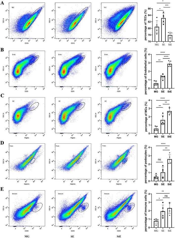
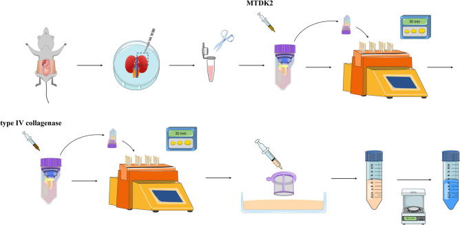
Preparation of kidney tissue single-cell suspensions is the basis of single-cell sequencing, flow cytometry and primary cell culture, but it is difficult to prepare high quality whole kidney single-cell suspensions because of the complex structure of the kidney. We explored a technique called stepwise enzymatic digestion (StE) method for preparing a single-cell suspension of rat whole kidney tissue which contained three main steps. The first step is to cut the kidney into a homogenate. The second step is the digestion of renal tubules using Multi Tissue Dissociation Kit 2 and the last step is the digestion of glomeruli using type IV collagenase. We also compared it with two previous techniques, mechanical grinding method and simple enzymatic digestion method. The StE method had the advantages of high intrinsic glomerular cells and immune cells harvest rate, high singlets rate and high cell viability compared with the other two techniques. In conclusion, the StE method is feasible, highly efficient, and worthy of further research and development.


Conflict of interest statement

The authors declare no competing interests.

Figures

Figure 1
Figure 1
Morphological differences of cell suspensions in the different groups. (A) White flocculent suspended matter was observed in the supernatant of the MG group, while the upper liquid was clarified in the SE group and StE group. (B) The morphology of single-cell suspensions in the MG group, SE group and StE group was observed and photographed under a microscope. The magnification is × 100.
Figure 2
Figure 2
Percentage of singlets in each group. The singlet percentages of the SE and StE groups were significantly higher than that of the MG group, n = 5, *p < 0.05, **p < 0.01.
Figure 3
Figure 3
Proportions of different types of cells in the different groups. (A) The detection rate of renal tubular epithelial cells was highest in the SE group, followed by the MG group and then the StE group. (B,C) The detection rate of endothelial cells or mesangial cells was the highest in the StE group, followed by the SE and MG groups. (D) The detection rate of podocytes was significantly higher in the StE group than in the MG and SE groups, with no difference between the latter two. (E) The detection rates of immune cells in the SE and StE groups were significantly higher than that in the MG group, with no difference between the first two groups, n = 5, *p < 0.05, **p < 0.01, ***p < 0.001, ****p < 0.0001. TECs, renal tubular epithelial cells; MCs, mesangial cells; Podo, podocytes; Endo, endothelial cells.
Figure 4
Figure 4
The expressions of Mme, Pecam1, Pdgfrb, Nphs1 and Ptprc in each type of sorted cells detected by qRT-PCR. (A) The expression of Mme in renal tubular epithelial cells was higher than that of the others. (B) The expression of Pecam1 in endothelial cells was higher than that of the others. (C) The expression of Pdgfrb in mesangial cells was higher than that of the others. (D) The expression of Nphs1 in podocytes was higher than that of the others. (E) The expression of Ptprc in immune cells was higher than that of the others. n = 3, *p < 0.05, **p < 0.01. TECs, renal tubular epithelial cells; MCs, mesangial cells; Podo, podocytes; Endo, endothelial cells.
Figure 5
Figure 5
Schematic description of stepwise enzymatic digestion method.

Similar articles

References

    1. Yamada K, et al. Clusterin is up-regulated in glomerular mesangial cells in complement-mediated injury. Kidney Int. 2001;59:137–146. doi: 10.1046/j.1523-1755.2001.00474.x. - DOI - PubMed
    1. Amon L, et al. Guidelines for DC preparation and flow cytometry analysis of mouse lymphohematopoietic tissues. Eur. J. Immunol. 2022 doi: 10.1002/eji.202249893. - DOI - PubMed
    1. Yaigoub H, et al. Isolation of viable single cells with high yield and purity using a small amount of human kidney tissue biopsy. Front. Cell Dev. Biol. 2022;10:822275. doi: 10.3389/fcell.2022.822275. - DOI - PMC - PubMed
    1. Teal E, Steele NG, Chakrabarti J, Holokai L, Zavros Y. Mouse-and human-derived primary gastric epithelial monolayer culture for the study of regeneration. J. Vis. Exp. 2018 doi: 10.3791/57435. - DOI - PMC - PubMed
    1. Guyton AC. Blood pressure control–special role of the kidneys and body fluids. Science. 1991;252:1813–1816. doi: 10.1126/science.2063193. - DOI - PubMed