Review
. 2024 Jun;24(6):435-451.
doi: 10.1038/s41577-023-00985-4. Epub 2024 Feb 2.

The immunology of type 1 diabetes

Affiliations
Review

The immunology of type 1 diabetes

Kevan C Herold et al. Nat Rev Immunol. 2024 Jun.

Abstract

Following the seminal discovery of insulin a century ago, treatment of individuals with type 1 diabetes (T1D) has been largely restricted to efforts to monitor and treat metabolic glucose dysregulation. The recent regulatory approval of the first immunotherapy that targets T cells as a means to delay the autoimmune destruction of pancreatic β-cells highlights the critical role of the immune system in disease pathogenesis and tends to pave the way for other immune-targeted interventions for T1D. Improving the efficacy of such interventions across the natural history of the disease will probably require a more detailed understanding of the immunobiology of T1D, as well as technologies to monitor residual β-cell mass and function. Here we provide an overview of the immune mechanisms that underpin the pathogenesis of T1D, with a particular emphasis on T cells.


Figures

Fig. 1 ∣
Fig. 1 ∣. Risk genes for type 1 diabetes encode proteins that impact T cell development and function.
Certain haplotypes of the human HLA class II locus and a high number of repeats of the insulin variable number of tandem repeat (VNTR) region have been linked with autoimmune responses in pancreatic islets in individuals with type 1 diabetes. Approximately 50 other candidate risk genes have been found to encode a variety of proteins that are involved in T cell function, activation and differentiation, including kinases and phosphatases, transcription factors, receptors and ligands, cytokines, cytokine receptors and T cell effector molecules, structural and adaptor proteins, and costimulatory or co-inhibitory proteins. Additional risk genes are thought to encode proteins that are involved in T cell activation or differentiation indirectly by modifying antigen-presenting cells or by targeting β-cells (not shown). APC, antigen-presenting cell.
Fig. 2 ∣
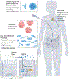
Fig. 2 ∣. The influence of the microbiome in T1D.
Several mechanisms have been proposed to explain associations of the microbiome with type 1 diabetes (T1D). An analysis of stool samples established a link between enterovirus infection and T1D, and persistent infection is associated with islet autoimmunity and progression to overt clinical disease. Invariant immune cells such as mucosal-associated invariant T cells, innate lymphoid cells and others have a role in the maintenance of the intestinal barrier. Their dysfunction and loss of microbiome diversity can lead to loss of tolerance to commensal bacteria and allow the outgrowth of inflammation-inducing species that may affect local tolerance mechanisms, enhance the function of autoreactive cells or even activate immune cells that have cross-reactivity between commensal bacteria and autoantigens. In addition, metabolic products of the microbiome, such as short-chain fatty acids (SCFA), may affect systemic immune regulation, including via direct effects on mucosal-associated invariant T (MAIT) cells,,,,. Antibodies to commensal microbiota have been identified in individuals at risk for T1D, and in non-obese diabetic mice, cross-reactive antigens that are recognized by CD8+ T cells were found (not shown),, Moreover, microbial exposure may also affect the efficacy of immunotherapies for T1D (ref. 39).
Fig. 3 ∣
Fig. 3 ∣. Insulitis and CD8+ T cells in type 1 diabetes.
a, Confocal microscopy of islets from a 12-week-old non-obese diabetic (NOD) mouse. Islets were stained for insulin (green), CD45 (red) and nuclei (blue). The two examples shown are of an islet without insulitis (left) and with immune cell infiltration (right) from the same mouse, illustrating the heterogeneity of β-cell destruction during type 1 diabetes pathogenesis. (Image taken in the Yale Center for Cellular & Molecular Imaging Confocal Facility). b, Stem-like autoreactive CD8+ memory T cells targeting β-cell antigens in type 1 diabetes have properties of both stem memory T (TSCM) cells and effector memory T (TEM) cells. Middle row: the stem-like population has a genome methylation profile that reflects this intermediate phenotype, which is reflected by the methylation patterns of key transcription factors related to differentiation (EOMES, TBX21, TCF7), effector functions (PRF1, GZMK, IFNγ) and exhaustion (TOX, BATF). Bottom row: in NOD mice, this stem-like population of T cells resides in the pancreatic lymph nodes, wherein the cells can differentiate into short-lived TEM cells that migrate to the pancreas and destroy β-cells.
Fig. 4 ∣
Fig. 4 ∣. T cell phenotypes associated with B cell help are linked to T1D.
Elevated levels of T cells with a follicular helper (TFH) phenotype and increased IL-21 production by T cells have been noted in mouse models and individuals with T1D (ref. 101). In mouse models of diabetes, insulin-specific T cells can exhibit a TFH phenotype and provide help to B cells that mount insulin-specific antibody responses. Insulin-specific T cells with a TFH phenotype have also been documented in individuals who have recently developed islet-targeted autoantibodies. T cells with a T peripheral helper (TPH) phenotype are also increased in individuals at onset of T1D and in at-risk individuals who go on to develop diabetes. Like TFH cells, TPH cells expresses inducible T cell costimulator (ICOS) and PD1 but lack expression of CXCR5 and instead can express chemokine receptors associated with migration to inflammatory sites (for example, CCR2 and CCR5). Whether TPH cells recognizes T1D-associated antigens such as insulin remains unclear. TCR, T cell receptor.
Fig. 5 ∣
Fig. 5 ∣. Disease-relevant hybrid insulin peptides (HIPs).
The figure shows HIPs that were confidently identified from pancreatic islets by mass spectrometry and for which disease-relevant HIP-specific T cells were also identified in peripheral blood mononuclear cells. For example, the CD4+ T cell clone E2 was isolated from peripheral blood mononuclear cells of individuals with recent-onset T1D (ref. 136) or from residual islets of organ donors with T1D (such as T cells expressing the TCR GSE.8E3). Moreover, experiments in NOD mice identified various diabetes-triggering CD4+ T cell clones that target HIPs bearing a distinct C-peptide (InsC) fragment (ending in the amino acid sequence LAL) linked to the N-termini of peptides derived from chromogranin A (ChgA), proinsulin (C-peptide) or islet amyloid polypeptide (IAPP). These HIPs are formed by the aspartic protease cathepsin D through a reversed proteolytic transpeptidation reaction. Peptide sequences highlighted in blue originate from C-peptide, whereas sequences highlighted in red originate from ChgA, IAPP or InsC.
Fig. 6 ∣
Fig. 6 ∣. Drugs and mechanisms that have shown efficacy in T1D.
The targets of these therapies have included Innate and inflammatory mediators and pathways, B cells, costimulatory molecules, T cells and β-cells,,,,,,,,,. The principle targets of drug action are highlighted in red. The T cells depicted are generic and may include several different subsets (for example, CD4+,CD8+, TFH cells and others). mAb, monoclonal antibody.
Fig. 7 ∣
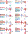
Fig. 7 ∣. Mechanism of action of the CD3-targeting monoclonal antibody teplizumab.
Early studies in animal models of the Fc receptor (FcR) non-binding monoclonal antibody (mAb) teplizumab have shown that by binding CD3, it induces a partial T cell receptor signal that induces clonal anergy and, thereby, inhibits β-cell killing. In vitro and in vivo experiments have demonstrated that it induces a relative expansion of CD8+ T cells and the secretion of IL-10; similar results were also found with a FcR non-binding version of the CD3-targeted mAb. Moreover, teplizumab induced a transient decline in circulating lymphocytes, which reflected the migration of T cells to the gut wall, wherein activation signals led to the induction of CD4+TGFβ+ or CD8+IL-10+ T cells,. Moreover, FcR non-binding CD3-targeted mAbs induced a selective preservation of Treg cells, and another study has reported that teplizumab induced TGF β+ Treg cells that have a role in the restoration of self-tolerance in the pancreatic draining lymph nodes,. a, Observations from the initial clinical trials showed activation of CD8+ T cells in vivo, and a transcriptome analysis of bulk RNA from peripheral blood mononuclear cells (PBMCs) from clinical responders in the AbATE trial, showed changes in CD8+ T cells that were later found to involve the induction of the transcription factor EOMES and increased expression of the receptors KLRG1 and TIGIT, suggesting that the cells were ‘partially exhausted’,,. b, A long-term follow-up study of responders from the AbATE trial has shown an increased expression of PD1 on CD8+ T cells and an increase in PD1+ central memory CD8+ T cells and anergic CD8+ T cells, which may explain the long-term effects of teplizumab. Autoantigen-reactive T cell receptors from CD8+ T cells have weak avidity for the antigen–MHC complex. The partial agonist signal delivered by the FcR non-binding CD3-targeted mAb may preferentially affect TCR signalling in response to such weak interactions, given that responses to conventional antigens such as viral antigens recover quickly after drug administration. APC, antigen-presenting cell.

References

    1. Gregory GA et al. Global incidence, prevalence, and mortality of type 1 diabetes in 2021 with projection to 2040: a modelling study. Lancet Diabetes Endocrinol. 10, 741–760 (2022). - PubMed
    1. Evans-Molina C et al. β Cell dysfunction exists more than 5 years before type 1 diabetes diagnosis. JCI Insight 3, e120877 (2018). - PMC - PubMed
    1. Sims EK et al. Teplizumab improves and stabilizes beta cell function in antibody-positive high-risk individuals. Sci. Transl. Med 13, eabc8980 (2021). - PMC - PubMed
    1. Turtinen M. et al. Characteristics of familial type 1 diabetes: effects of the relationship to the affected family member on phenotype and genotype at diagnosis. Diabetologia 62, 2025–2039 (2019). - PMC - PubMed
    1. Redondo MJ, Steck AK & Pugliese A. Genetics of type 1 diabetes. Pediatr. Diabetes 19, 346–353 (2018). - PMC - PubMed