. 2024 Jun;20(6):742-750.
doi: 10.1038/s41589-023-01535-8. Epub 2024 Feb 2.

Antibody discovery identifies regulatory mechanisms of protein arginine deiminase 4

Affiliations

Antibody discovery identifies regulatory mechanisms of protein arginine deiminase 4

Xin Zhou et al. Nat Chem Biol. 2024 Jun.

Abstract

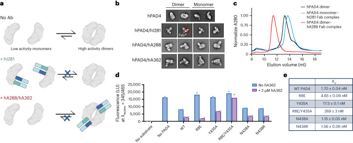
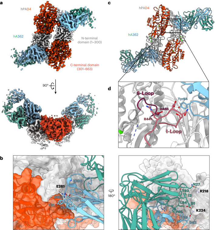
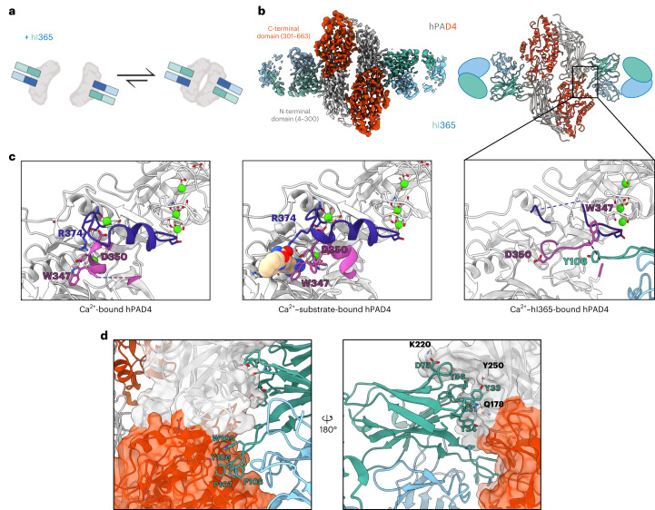
Unlocking the potential of protein arginine deiminase 4 (PAD4) as a drug target for rheumatoid arthritis requires a deeper understanding of its regulation. In this study, we use unbiased antibody selections to identify functional antibodies capable of either activating or inhibiting PAD4 activity. Through cryogenic-electron microscopy, we characterized the structures of these antibodies in complex with PAD4 and revealed insights into their mechanisms of action. Rather than steric occlusion of the substrate-binding catalytic pocket, the antibodies modulate PAD4 activity through interactions with allosteric binding sites adjacent to the catalytic pocket. These binding events lead to either alteration of the active site conformation or the enzyme oligomeric state, resulting in modulation of PAD4 activity. Our study uses antibody engineering to reveal new mechanisms for enzyme regulation and highlights the potential of using PAD4 agonist and antagonist antibodies for studying PAD4-dependency in disease models and future therapeutic development.


Conflict of interest statement

The authors declare the following competing interests: X.Z., S.K., and J.A.W., and the regents of the University of California have filed a provisional patent application related to this project.

Figures

Fig. 1
Fig. 1. PAD4–antibody selection and hits characterization.
a, PAD4 phage display schematic. The Fab-phage library (1) was depleted of nonspecific streptavidin binders (2); the remaining soluble Fab-phage were allowed to bind to PAD4 immobilized on streptavidin in the presence of 1 mM Ca2+ or 1 mM EDTA with TCEP and washed (3); the bound Fab-phage were eluted (4), amplified (5) and subjected to additional selection rounds before final characterization of individual hits (6). b, Schematic of fluorescent-substrate PAD4 activity assay (derived from Sabulski et al.). Higher fluorescent signal is indicative of lower PAD4 activity. c, Characterization of Fab binding effect on PAD4 activity from hPAD4 selection 1 and 2 and mPAD4 selection 4 (Extended Data Fig. 3a) via fluorescent-substrate activity assay. Highlighted clones hI281, hA288, hA362, hI364, hI365 and mA342 are described in the main text. d, Activity of hPAD4 in the presence of control antibody, an inhibitory Fab to human PAD4 (hI281) or activating Fabs (hA288, and hA362) measured by citrullination of protein substrate H3. hI281 reduces the activity of PAD4 while hA288–hA362 increases PAD4 activity. e, Activity of mPAD4 in the presence of control antibody, an activating IgG to murine PAD4 (mA342) and its variant mA342-c4 measured by citrullination of protein substrate H3. Representative blots were reproduced three times before inclusion in text. GFP, green fluorescent protein. Source data
Fig. 2
Fig. 2. Antibodies that modulate hPAD4 dimerization influence enzymatic activity.
a, Summary diagram of antibodies influencing PAD4 activity through modulating its dimerization. Monomeric PAD4 is less active than dimeric PAD4, and hI281 blocks dimerization while hA288 and hA362 promote dimerization. b, NSEM 2D micrographs showing monomeric state of PAD4 in complex with hI281 and dimeric state in complex with hA288 or hA362. c, SEC traces of PAD4 alone, in complex with Fab-hI281 and in complex with Fab hA362. PAD4 alone exists in both the monomer and dimer form, while addition of hI281 promotes the monomer form and hA362 promotes the dimer form as evidenced by elution time. d, Binding is correlated with antibody function using a trypsin substrate assay. Biological duplicates are shown. Fabs binding to mutants with less affinity are less able to inhibit enzymatic activity (right). e, Table displaying measured binding affinities of activating Fab hA362 to PAD4 mutants (left). Binding is drastically decreased on mutation of several residues at the PAD4 dimer interface. Source data
Fig. 3
Fig. 3. hA362 directly contributes to the PAD4 dimerization interface and helps order the substrate-binding site.
a, Two views of the cryo-EM map of PAD4 in complex with Fab hA362 (PDB 8SMK). PAD4 monomers are in shades of gray (N-terminal domain) and red (C-terminal domain), hA362 heavy and light chains are teal and blue. b, Model of PAD4–hA362 derived from the cryo-EM map shown as ribbon. Boxed region delineates Fab CDR interaction with the I- and S-loop on PAD4. c, Zoomed-in view of the boxed region in b shows hA362 reaching across the PAD4 monomer to interact with the I-loop on the other PAD4 monomer. This helps order the S-loop via the R441-D465 salt bridge. d, Detailed hA362–PAD4 interactions. Both chains of the Fab pack a large number of aromatics against both monomers of PAD4 dimer. Ion and hydrogen bonds marked with dashed lines.
Fig. 4
Fig. 4. Cryo-EM structure illustrates the mechanism of calcium dependency and inhibitory function of hI365.
a, Schematic showing that inhibitory Fab hI365 is able to bind both the monomeric and dimeric form of hPAD4. b, Cryo-EM map (left, PDB 8SML) and the resulting model (right) of hPAD4 (N-terminal domain, gray; C-terminal domain, red) in complex with hI365 (HC, teal; LC, blue). The Ab primarily binds an N-terminal Ca2+-coordinated region, but the H3 loop extends to the C-terminal pocket and induces conformational change. c, Comparison of the Ca2+-bound hPAD4 (PDB 1WD9), Ca2+–substrate bound hPAD4 (PDB 1WDA), and the Ca2+–hI365 bound hPAD4. The interaction of hI365 with residues 340–352 in hPAD4 (magenta) alters the structure and orientation of this fragment disrupting the organization of several residues (D350, R374, W347) involved in calcium and substrate binding. d, Detailed hA365–PAD4 interactions. The Fab interacts predominantly with one chain against one monomer of PAD4, burying a considerable number of aromatics with some hydrogen bonds.
Fig. 5
Fig. 5. Antibody engineering strategies used to improve affinity and inhibition activity of the calcium-dependent binder, hI365.
a, hPAD4–hI365 binding interface with CDRs L3, H1–H4 highlighted. CDR loops H1–H4 are forming contacts with PAD4, but L3 is too short to facilitate any contact with the enzyme. b, SEC traces of WT hI365 and affinity matured clones. Selected hits show improved SEC profiles as indicated by earlier elution times. A280, absorbance at 280 nm. c, CDR sequences of WT hI365 aligned with residues to be mutagenized via soft randomization; L3 is randomized to contain nine or ten amino acids. d, CDR sequences of the two top engineered binders with good affinity and solubility. e, PAD4 activity as measured by the fluorescent-substrate assay shows that several Ab clones identified from soft randomization inhibit PAD4 more potently than WT hI365. 2 mM Ca2+ was used in this assay to mimic physiological extracellular conditions. Error bars represent mean ± s.d. of three biological replicates. f, IC50 of lead candidates E3 and E6 as measured by PAD4 citrullination of H3. IC50 of E3 and E6 determined to be 95 and 13 nM, respectively, and represent mean ± s.e.m. of three biological replicates. Source data
Extended Data Fig. 1
Extended Data Fig. 1. Expression and biophysical characterization of hPAD4.
(a) Map of hPAD4 gene expressed in C43 or BL21 E. coli. N-terminal 6x His tag and Avi tag are separated by a protease cleavage site to hPAD4 to remove tags. (b) Gel shift assay of full length hPAD4 (left, 74 kDa) and biotinylated hPAD4 bound to NeutrAvidin (right, 134 kDa). (c) PAD4 enzyme contains 19 free cysteines (colored in yellow), requiring storage with reducing agent to prevent oxidation of cysteines. (d) SDS–PAGE showing reduction of the inter-heavy and light chain disulfide of a 4G104D5 antibody upon addition of reducing agents. The 4G104D5 antibody adopts the antibody scaffold of the Fab-phage library used for PAD4 antibody selection. 50 kDa band represents disulfide-linked Fab while 25 kDa bands represent separated heavy and light chains following TCEP reduction. (e) Biolayer Interferometry (BLI) showing binding of Fab 4G104D5 to its cognate antibody is not influenced by various reducing agents. (f) Immunoblotting detection of PAD4-mediated Histone 3 citrullination (Cit-H3) showing PAD4 activity is calcium dependent. (g) Differential scanning fluorimetry (DSF) plot showing melting temperatures of hPAD4 in the presence and absence of Ca2+. (h) Table listing PAD4 melting temperatures measured by DSF. Source data
Extended Data Fig. 2
Extended Data Fig. 2. Phage ELISA for characterizing hPAD4/mPAD4 cross reactivity and multi-point biolayer interferometry (BLI) binding data of lead candidate IgGs to hPAD4.
(a) Binding of Fab-phage to hPAD4/mPAD4 measured by ELISA A280 signal. Very few clones from hPAD4 or mPAD4 phage selections (left, middle) were cross reactive. By taking the round 3 phage pool and performing selections with the other antigen for two additional rounds, nearly all phage clones from round 5 are cross reactive with hPAD4 and mPAD4 (right). (b) Multi point BLI data show that hI364 and hI365 are dependent on calcium to bind hPAD4. hI281 and hA362 are calcium-independent binders although binding to the calcium-bound form of the enzyme is stronger. Binding of all clones to PAD4 at 2 mM Ca2+ is comparable to their binding at 10 mM Ca2+. KDs reported as <1 nM are due to bottoming out of off-rate measurements on BLI. Source data
Extended Data Fig. 3
Extended Data Fig. 3. Functional characterization of mPAD4 and h/mPAD4 cross reactive antibodies.
(a) hmI400 was identified as a functional inhibitor of both hPAD4 and mPAD4. (b) BLI results show mA342 interacts with the calcium bound form of mPAD4 but binds minimally to the calcium-free form. (c) SEC trace showing improved biophysical properties of mA342-c4 as compared to WT mA342. (d) BLI comparison of WT mA342 and its improved variant mA342-c4 in 10 mM Ca2+. Source data
Extended Data Fig. 4
Extended Data Fig. 4. Biophysical characterization of PAD4 monomerization mutants and effect of PAD4 monomerization on antibody binding.
(a) Computational modeling of the PAD4 dimer interface to identify key residues that contribute to dimerization (highlighted in red). (b) Key residues R8, Y435, and W548 highlighted at the dimer interface in red. The selected control residue N438 is highlighted in blue. (c) SEC trace showing earlier elution of dimeric, WT PAD4 compared to monomerization-promoting mutants R8E and Y435A. Control mutants, N438A and N438R (gray), elute with WT PAD4. (d). DSF plot of PAD4 mutants in the presence of 10 mM Ca2+ (green) or 1 mM EDTA (yellow). (e) Estimated Tms of PAD4 mutants from DSF plots. (f) BLI measurements of hA362 binding to WT hPAD4 and monomerization mutants. Source data
Extended Data Fig. 5
Extended Data Fig. 5. Detailed view of Ca2+ ions and Fab/hPAD4 interactions with cryo-EM map.
(a) Three Ca2+ ions in the N terminal domain of hPAD4-hA362 structure. Model in ribbon and atoms on top, overlayed with cryo-EM map on the bottom. (b) Ca2+ ion in the C terminal domain of the hPAD4-hA362 structure. One Ca2+ was removed from analysis as the electron density was very weak. (c) Zoomed in view of the hA362 interactions with the hPAD4 I-loop with corresponding cryo-EM map. Adapted from Fig. 3c. (d) Ca2+ ions in the N terminal domain of the hPAD4-hA365 structure. (e) Zoomed in view of the hI365 interactions with the hPAD4 active site with corresponding cryo-EM map. Adapted from Fig. 4c, right panel.
Extended Data Fig. 6
Extended Data Fig. 6. Rosetta antibody design (rAbD) guided optimization of hI365.
(a) Several G58 mutants are predicted to reduce interface energy of hI365 and hPAD4. (b) BLI measurements showing that point mutant G58D binds PAD4 with similar affinity as WT hI365. (c) SEC trace showing improved biophysical properties of mutant G58D as compared to WT hI365, as evidenced by the earlier elution time. (d) Position of CDR L3 at the PAD4 binding interface and rAbD workflow for designing L3 variants. Though the loop sits at the interface, it is not forming any favorable interactions with PAD4 due to its short length. High resolution structure of PAD4/hI365 interface served as the input model for rAbD. CDR L3 of the relaxed model was allowed to ‘Graft’ and ‘Sequence’ design or only ‘Sequence’ design with or without flexible backbone design. Neighboring CDRs L1 and H3 were repacked to avoid clashes and optimize interface interactions. (e) ‘Graft’ and ‘Sequence’ design generated models (blue) with low totalscore and interface energies shown by dG_separated (in REU). ‘Sequence’ design alone did not improve the interface with all models (orange) having poor dG_separated score. The dG_separated score of the cryo_EM structure of PAD4/hI365 interface is ∼−54.4 REU (f) Rosetta antibody design (rAbD) predicted CDR L3 at lengths of 9 and 10 aa to have the most optimal interface energies. Different CDR L3 lengths and corresponding dG separated scores are shown. (g) SEC chromatagram showing poor performance of L3 mutants compared to parent (hI365) and control antibodies (hI356). (h) Fluorescent substrate activity assay showing inhibition of PAD4 in the presence of WT hI365 and the predicted L3 variants by rAbD. Error bars represent mean ± standard deviation of three biological replicates. Source data
Extended Data Fig. 7
Extended Data Fig. 7. Soft randomization of hI365.
(a-b) Sequence logos of library 1 and 2 showing amino acid preference for each CDR. (c-d) Binding of top Fab-phage clones to PAD4 as measured by direct and competition ELISAs. Top clones exhibit similar or improved binding to WT hI365 at both 20 nM and 5 nM PAD4. Top clones are less likely to be competed off PAD4, indicating a lower relative koff.(e) Multi-point octet trace of leading clones, E3 and E6. Both appear to bind PAD4 at nanomolar affinity. Source data
Extended Data Fig. 8
Extended Data Fig. 8. Anti-PAD4 antibody specificity and effect on PAD4 on whole cell lysate detect via anti-modified citrulline western blot.
(a) Binding of hI365, E3, and E6 to PAD4, PAD2, PAD3, and Trastuzumab negative control measured by BLI. All PAD4 antibodies bind only to PAD4 and not PAD2 or PAD4. (b) Whole cell lysate treated with PAD4 +/- inhibitors. Functional antibodies, hI365, E3, and E6 inhibit PAD4-catalyzed citrullination on a variety of cytosolic, nuclear, and membrane-bound proteins. Trastuzumab IgG and pan-PAD inhibitor Cl-amidine included as negative and positive controls. An anti-actin loading control is included. Source data
Extended Data Fig. 9
Extended Data Fig. 9. Cryo-EM map statistics for hPAD4/hA362.
(a) Map colored by local resolution. (b) Global resolution FSC plots generated from half maps. Blue line indicates FSC = 0.143 cutoff. (c) Directional FSC plots. Left: Histogram showing the percentage of per angle FSC vs resolution, showing agreement with global FSC and sphericity of 0.966. Right: Directional FSC curves. (d) Left: Model colored by per residue Q score; Right: Fab CDR loop showing model/map fit. PDB: 8SMK. (e) Cryo-EM data processing workflow for PAD4-hA362.
Extended Data Fig. 10
Extended Data Fig. 10. Cryo-EM map statistics for hPAD4/hI365.
(a) Map colored by local resolution. (b) Global resolution FSC plots generated from half maps. Blue line indicates FSC = 0.143 cutoff. (c) Directional FSC plots. Left: Histogram showing the percentage of per angle FSC vs resolution, showing some degree of anisotropy, but overall agreement with the global FSC and sphericity of 0.928. Right: Directional FSC curves demonstrating better than 3 Å resolution in two directions and ∼4.5 Å in third direction. (d) Left: Model colored by per residue Q score; Right: Fab CDR loop showing model/map fit. PDB: 8SML. (e) Cryo-EM data processing workflow for PAD4-hI365.

References

    1. Witalison EE, Thompson PR, Hofseth LJ. Protein arginine deiminases and associated citrullination: physiological functions and diseases associated with dysregulation. Curr. Drug Targets. 2015;16:700–710. doi: 10.2174/1389450116666150202160954. - DOI - PMC - PubMed
    1. Fujisaki M, Sugawara K. Properties of peptidylarginine deiminase from the epidermis of newborn rats. J. Biochem. 1981;89:257–263. doi: 10.1093/oxfordjournals.jbchem.a133189. - DOI - PubMed
    1. Watanabe K, Shenshu T. Isolation and characterization of cDNA clones encoding rat skeletal muscle peptidylarginine deiminase. J. Biol. Chem. 1989;26:15255–15260. doi: 10.1016/S0021-9258(19)84818-4. - DOI - PubMed
    1. Leshner M, et al. PAD4 mediated histone hypercitrullination induces heterochromatin decondensation and chromatin unfolding to form neutrophil extracellular trap-like structures. Front. Immunol. 2012;3:307. doi: 10.3389/fimmu.2012.00307. - DOI - PMC - PubMed
    1. Wong SL, Wagner DD. Peptidylarginine deiminase 4: a nuclear button triggering neutrophil extracellular traps in inflammatory diseases and aging. FASEB J. 2018;32:6358–6370. doi: 10.1096/fj.201800691R. - DOI - PMC - PubMed

Publication types

MeSH terms