

Acoustic Cell Patterning for Structured Cell-Laden Hydrogel Fibers/Tubules
- PMID: 38308105
- PMCID: PMC11005686
- DOI: 10.1002/advs.202308396
Acoustic Cell Patterning for Structured Cell-Laden Hydrogel Fibers/Tubules
Abstract
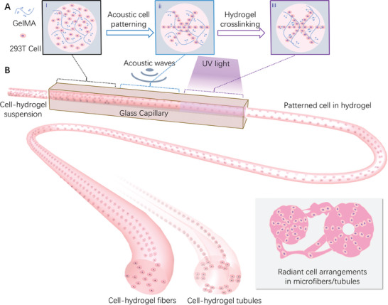
Cell-laden hydrogel fibers/tubules are one of the fundamentals of tissue engineering. They have been proven as a promising method for constructing biomimetic tissues, such as muscle fibers, nerve conduits, tendon and vessels, etc. However, current hydrogel fiber/tubule production methods have limitations in ordered cell arrangements, thus impeding the biomimetic configurations. Acoustic cell patterning is a cell manipulation method that has good biocompatibility, wide tunability, and is contact-free. However, there are few studies on acoustic cell patterning for fiber production, especially on the radial figure cell arrangements, which mimic many native tissue-like cell arrangements. Here, an acoustic cell patterning system that can be used to produce hydrogel fibers/tubules with tunable cell patterns is shown. Cells can be pre-patterned in the liquid hydrogel before being extruded as cross-linked hydrogel fibers/tubules. The radial patterns can be tuned with different complexities based on the acoustic resonances. Cell viability assays after 72 h confirm good cell viability and proliferation. Considering the biocompatibility and reliability, the present method can be further used for a variety of biomimetic fabrications.
Keywords: acoustofluidic; biofabrication; cell patterning; hydrogel fibers.
© 2024 The Authors. Advanced Science published by Wiley‐VCH GmbH.
Conflict of interest statement
The authors declare no conflict of interest.
Figures


Similar articles
-
Combining electrospun nanofibers with cell-encapsulating hydrogel fibers for neural tissue engineering.J Biomater Sci Polym Ed. 2018 Sep;29(13):1625-1642. doi: 10.1080/09205063.2018.1479084. Epub 2018 Jun 3. J Biomater Sci Polym Ed. 2018. PMID: 29862935 Free PMC article.
-
Tendon Tissue Engineering: Effects of Mechanical and Biochemical Stimulation on Stem Cell Alignment on Cell-Laden Hydrogel Yarns.Adv Healthc Mater. 2019 Apr;8(7):e1801218. doi: 10.1002/adhm.201801218. Epub 2019 Feb 6. Adv Healthc Mater. 2019. PMID: 30725521
-
Minimally-Invasive and In-Vivo Hydrogel Patterning Method for In Situ Fabrication of Implantable Hydrogel Devices.Small Methods. 2023 Sep;7(9):e2300032. doi: 10.1002/smtd.202300032. Epub 2023 May 18. Small Methods. 2023. PMID: 37199695
-
Manufacturing of hydrogel biomaterials with controlled mechanical properties for tissue engineering applications.Acta Biomater. 2017 Oct 15;62:42-63. doi: 10.1016/j.actbio.2017.07.028. Epub 2017 Jul 20. Acta Biomater. 2017. PMID: 28736220 Review.
-
Cell-laden hydrogels for osteochondral and cartilage tissue engineering.Acta Biomater. 2017 Jul 15;57:1-25. doi: 10.1016/j.actbio.2017.01.036. Epub 2017 Jan 11. Acta Biomater. 2017. PMID: 28088667 Free PMC article. Review.
Cited by
-
Control of myotube orientation using ultrasonication.Sci Rep. 2024 Oct 28;14(1):25737. doi: 10.1038/s41598-024-77277-x. Sci Rep. 2024. PMID: 39468262 Free PMC article.
-
Outermost Cationic Surface Charge of Layer-by-Layer Films Prevents Endothelial Cells Migration for Cell Compartmentalization in Three-Dimensional Tissues.Adv Sci (Weinh). 2025 May;12(19):e2417538. doi: 10.1002/advs.202417538. Epub 2025 Feb 22. Adv Sci (Weinh). 2025. PMID: 39985273 Free PMC article.
-
Acoustofluidics: Technology Advances and Applications from 2022 to 2024.Anal Chem. 2025 Apr 8;97(13):6847-6870. doi: 10.1021/acs.analchem.4c06803. Epub 2025 Mar 25. Anal Chem. 2025. PMID: 40133046 Free PMC article. Review. No abstract available.
-
Scalable acoustic virtual stirrer for enhanced interfacial enzymatic nucleic acid reactions.Sci Adv. 2025 Mar 7;11(10):eadt6955. doi: 10.1126/sciadv.adt6955. Epub 2025 Mar 5. Sci Adv. 2025. PMID: 40043123 Free PMC article.
-
Acoustic black hole effect enhanced micro-manipulator.Microsyst Nanoeng. 2024 Oct 12;10(1):144. doi: 10.1038/s41378-024-00789-z. Microsyst Nanoeng. 2024. PMID: 39394206 Free PMC article.
References
-
- Khademhosseini A., Langer R., Nat. Protoc. 2016, 11, 1775. - PubMed
MeSH terms
Substances
Grants and funding
- 12032015/National Natural Science Foundation of China
- 12121002/National Natural Science Foundation of China
- 2019-01-07-00-02-E00030/Innovation Program of Shanghai Municipal Education Commission
- 21TQ1400203/Shanghai Pilot Program for Basic Research - Shanghai Jiao Tong University
- JLU-cncr-202304/Opening Project of the Key Laboratory of CNC Equipment Reliability, Ministry of Education, Jilin University
LinkOut - more resources
Full Text Sources