. 2024 Feb 2;22(1):97.
doi: 10.1186/s12964-023-01440-6.

Ferritin-mediated neutrophil extracellular traps formation and cytokine storm via macrophage scavenger receptor in sepsis-associated lung injury

Affiliations

Ferritin-mediated neutrophil extracellular traps formation and cytokine storm via macrophage scavenger receptor in sepsis-associated lung injury

Hao Zhang et al. Cell Commun Signal. .

Abstract

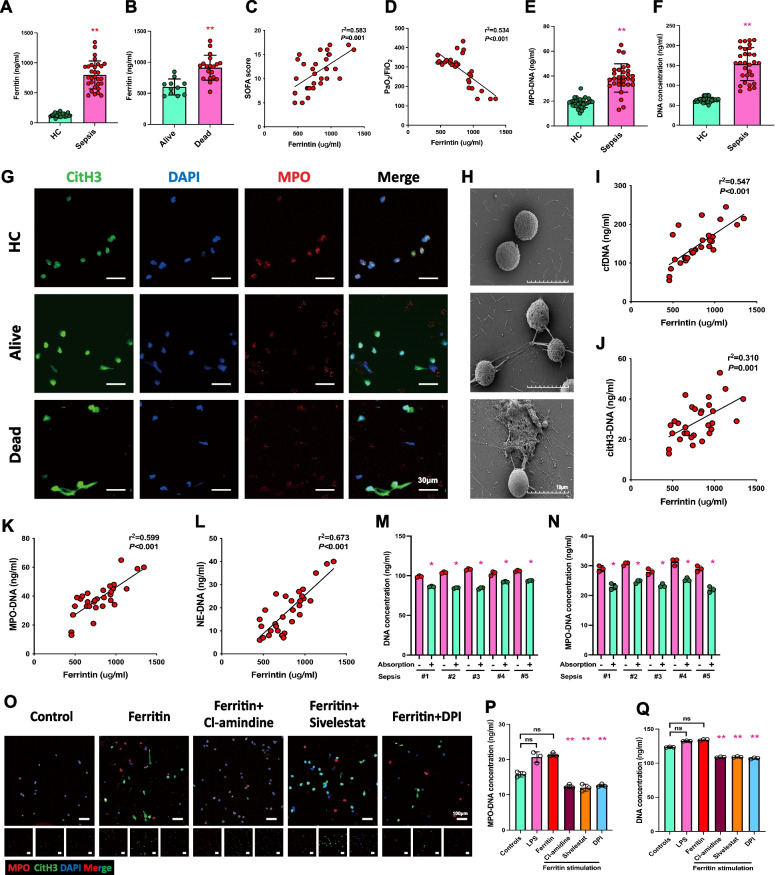
Background: Sepsis is a severe systemic inflammatory disorder manifested by a dysregulated immune response to infection and multi-organ failure. Numerous studies have shown that elevated ferritin levels exist as an essential feature during sepsis and are able to suggest patients' prognoses. At the same time, the specific mechanism of ferritin-induced inflammatory injury remains unclear.

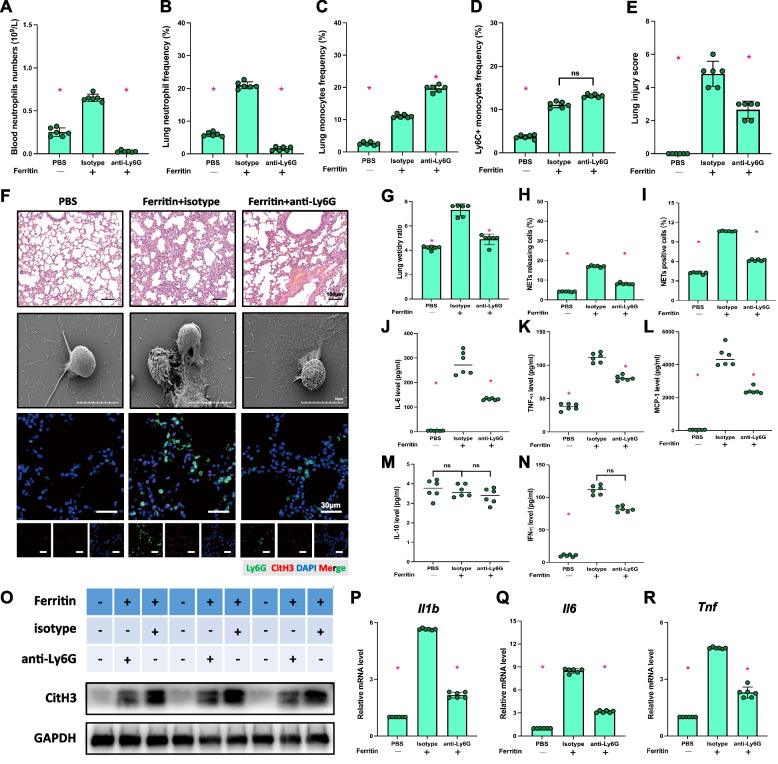
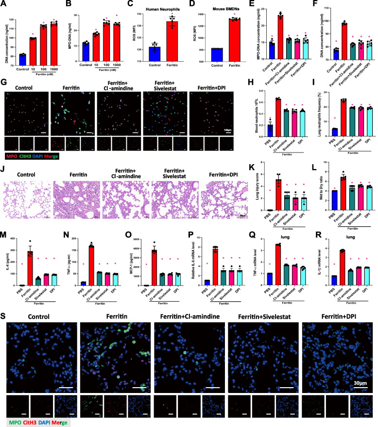
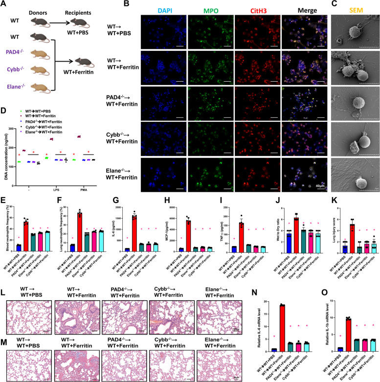
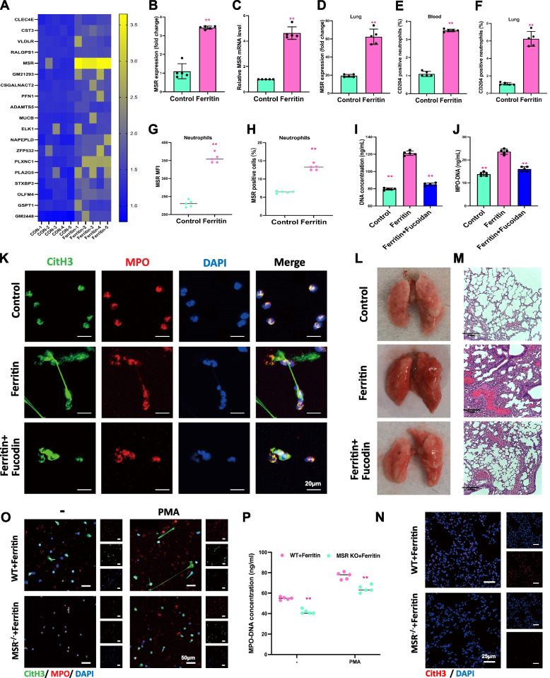
Methods: Hyper-ferritin state during inflammation was performed by injecting ferritin into a mouse model and demonstrated that injection of ferritin could induce a systemic inflammatory response and increase neutrophil extracellular trap (NET) formation.Padi4-/-, Elane-/- and Cybb-/- mice were used for the NETs formation experiment. Western blot, immunofluorescence, ELISA, and flow cytometry examined the changes in NETs, inflammation, and related signaling pathways.

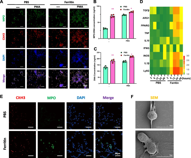
Results: Ferritin induces NET formation in a peptidylarginine deiminase 4 (PAD4), neutrophil elastase (NE), and reactive oxygen species (ROS)-dependent manner, thereby exacerbating the inflammatory response. Mechanistically, ferritin induces the expression of neutrophil macrophage scavenger receptor (MSR), which promotes the formation of NETs. Clinically, high levels of ferritin in patients with severe sepsis correlate with NETs-mediated cytokines storm and are proportional to the severity of sepsis-induced lung injury.

Conclusions: In conclusion, we demonstrated that hyper-ferritin can induce systemic inflammation and increase NET formation in an MSR-dependent manner. This process relies on PAD4, NE, and ROS, further aggravating acute lung injury. In the clinic, high serum ferritin levels are associated with elevated NETs and worse lung injury, which suggests a poor prognosis for patients with sepsis. Our study indicated that targeting NETs or MSR could be a potential treatment to alleviate lung damage and systemic inflammation during sepsis. Video Abstract.

Keywords: Ferritin; Macrophage scavenger receptor; Neutrophil extracellular traps; sepsis-associated acute lung injury.


Conflict of interest statement

The authors declare no competing interests.

Figures

Fig. 1
Fig. 1
Ferritin induces systemic inflammation and lung injury in vivo, resulting in lung neutrophil accumulation. Mice were treated with ferritin injections for the corresponding times (3 h, 6 h, 12 h, 24 h, 48 h). A Lung tissues’ HE staining of ferritin-injected mice. B Lung injury scores of ferritin-treated mice (n = 6). C and D Neutrophils frequency and cell number in peripheral blood of mice (n = 6). E Lung wet to dry rate (n = 6). F and G Frequency and number of peripheral blood monocytes. H and I Frequency and number of Ly6C+ monocytes in peripheral blood (n = 6). J Splenic appearance of mice 12 h after ferritin treatment. K, L, P, and Q Plasma inflammatory factor levels in mice (IL-6, TNF-α, IL-1β, and MCP-1, n = 4). M, N, O, R, S, and T Lung tissue neutrophil, monocyte, Ly6C+ monocyte, macrophage, T-cell, and B-cell frequencies (n = 6) *p < 0.05
Fig. 2
Fig. 2
Ferritin promotes the formation of NETs. Neutrophils isolated from ferritin-treated and control mice were treated with PMA. A Representative immunofluorescence staining of spontaneous, PMA-induced NET formation in ferritin-treated mice at 12 h BMDNs. Neutrophils were stained with MPO (green), CitH3 (red) antibodies and DAPI (blue). Scale bar, 30 μm. B Serum MPO-DNA complexes and (C) dsDNA concentration in cell culture supernatant. D Heatmap of RNA-seq differential analysis of BMDNs in PBS and ferritin-injected mice. E Immunofluorescence staining images of mice lung tissue after PBS or ferritin treatment. (MPO, green; CitH3, red; DAPI, blue. Bar = 30 μm). F Image of SEM (scanning electron microscopy) showed the morphology of extracellular meshes NETs formation after ferritin injection. (bar = 10 μm) *p < 0.05
Fig. 3
Fig. 3
Ferritin-induced systemic inflammatory response and lung injury were dependent on neutrophils. Co-administration of anti-Ly6G antibody to ferritin-treated mice for 12 h. A, B, C and D Neutrophil number, frequency, monocyte frequency and Ly6C+ neutrophil frequency in mouse lungs. E Lung injury score. F HE staining of 12 h mouse lung tissue (Scale bar = 100 μm), SEM images of BMDN neutrophil NETs (Scale bar = 10 μm), and IF images of lung tissue (Ly6G, green; CitH3, red; DAPI, blue. Scale bar = 30 μm). G lung wet to dry rate. H The proportion of cells releasing NETs in peripheral blood. I The proportion of NETs-positive cells in lung tissue. J, K, L, M and N Mouse plasma levels of inflammatory factors: IL-6, TNF-α, MCP-1, IL-10 and IFN-γ. O Immunoblot of mouse lung tissue for CitH3 protein expression. P, Q and R Il1b, Il6, Tnf mRNA levels in lung tissue. *p < 0.05
Fig. 4
Fig. 4
PAD4, NE and ROS are required for the formation of NETs induced by ferritin. Ferritin-injected mice were treated with PAD4, NE, and ROS inhibitors. A DNA concentration and (B) MPO-DNA complex were measured in cell culture supernatants after exposing healthy human neutrophils to indicated concentrations of ferritin. C ROS levels in healthy human neutrophils after ferritin stimulation for 12 h in vitro. D ROS levels in mouse BMDNs after 12 h of ferritin stimulation. E and F the concentration of MPO-DNA in human neutrophils’ culture supernatants was measured after being stimulated with ferritin alone or in combination with Cl-amidine, sivelestat, and DPI. G IF images of the effect of ferritin alone and combined with inhibitors on the formation of neutrophil NETs. (MPO, red; CitH3, green; DAPI, blue. Bar = 100 μm). H and I Neutrophil counts and frequencies in peripheral blood and lung tissue of mice after application of ferritin alone and in combination with various inhibitors. J HE staining of mouse lung tissue (Scale bar = 50 μm). K and L Lung injury score and wet-to-dry ratio. M, N and O Plasma concentrations of inflammatory factors IL-6, TNF-α and MCP-1 in mice. P, Q and R Lung tissue mRNA levels of inflammatory factors including Il-6, Tnf and Il-b. S IF-stained images of NETs markers in lung tissue. (MPO, green; CitH3, red; DAPI, blue. Bar = 30 μm)
Fig. 5
Fig. 5
Bone marrow of donors with PAD4, NE, ROS gene defects inhibited ferritin-induced NET production in WT recipient. A Overview of the experiment: WT, Padi4−/−, Elane−/−, and Cybb−/− mice had their bone marrow transplanted into WT recipients. The recipients were treated with ferritin after 4 weeks. B IF staining of BMDNs after ferritin injection in bone marrow transplanted mice (MPO, green; CitH3, red; DAPI, blue. Scale bar = 40 μm). C Scanning electron microscope images of BMDNs (Scale bar = 10 μm). D Peripheral blood DNA concentration. E and F Peripheral blood and lung neutrophil frequency. G-I Serum inflammatory factors (IL-6, MCP-1 and TNF-α) levels. J and K Lung wet-to-dry ratios and lung injury scores. L and M HE staining and Masson staining images of lung tissue. N and O Inflammatory factor-associated mRNA levels in lung tissue
Fig. 6
Fig. 6
Ferritin promotes the production of NETs by activating Msr. A Heatmap of differentially expressed mRNA from BMDNs extracted for RNA-seq after ferritin stimulation applied to mice. B and C WB and qPCR results of Msr protein and mRNA levels in BMDNs. D Msr protein expression levels in lung tissues. E and F Proportion of neutrophils in peripheral blood and lungs of mice after ferritin stimulation. G and H Mean fluorescence intensity of MSR and percentage of Msr-positive cells in ferritin-treated human neutrophils. I-J DNA concentration and MPO-DNA concentration after ferritin injection using the Msr inhibitor Fucodin combined with ferritin. K IF images of BMDNs in mice after ferritin injection using the Msr inhibitor Fucodin combined with ferritin (MPO, red; CitH3, green; DAPI, blue. Scale bar = 20 μm). L and M Morphological and HE-stained images of lungs in mice. N IF images of CitH3 in lung tissue of WT and Msr ablated mice treated with ferritin (CitH3, red; DAPI, blue. Scale bar = 25 μm). O IF staining and (P) MPO-DNA complex concentration of BMDNs from ferritin-treated WT and Msr−/− mice with PMA stimulation in vitro
Fig. 7
Fig. 7
MSR ablation prevents systemic and pulmonary inflammation caused by ferritin. A and B Neutrophil frequency in peripheral blood and lung tissue of WT and MSR KO mice after ferritin treatment. C and D After ferritin treatment, lung tissue monocytes and Ly6C+ neutrophils frequency in WT and MSR KO mice. E Lung wet-to-dry ratio of WT and MSR KO mice with 12 h ferritin stimulation. F and G HE and morphological images of ferritin-injected WT and MSR KO mice. H, I, J and K After ferritin injection, serum levels of inflammatory factors including IL-6, MCP-1, TNF-α and IFN-γ in WT and MSR KO mice
Fig. 8
Fig. 8
Up-regulation of ferritin in patients with sepsis leads to increased NETs production and exacerbates sepsis-associated acute lung injury. A Serum ferritin in healthy people and patients with sepsis (n = 30) and controls (n = 30). B Serum ferritin was detected in alive and dead sepsis patients. Alive (n = 11) and dead (n = 19). C and D Evaluate the correlation between serum ferritin concentration and the degree of lung damage (SOFA score and PaO2/FiO2) in sepsis patients. E and F MPO-DNA and DNA concentration in healthy people and patients with sepsis (n = 30) and controls (n = 30). G Representative NETosis images of alive-, dead-sepsis patients in comparison with healthy control. (MPO, red; CitH3, green; DAPI, blue. Scale bar = 30 μm). H SEM images depicting the release of NETs in septic or healthy neutrophils. (Scale bar = 10 μm). I, J, K and L The correlation between ferritin and NETs-related indicators (cfDNA, citH3-DNA, MPO-DNA and NE-DNA). M and N Stimulation of healthy donor neutrophils was performed using serum from five septic patients (serum ferritin absorbed and unabsorbed, respectively). Cell-free DNA and MPO-DNA complexes were quantified. O, P and Q Inhibition of ferritin-induced NET formation by PAD4, NE, ROS, and Msr1 inhibitors detected by IF of MPO (green), citH3 (red), and DAPI (blue) (Scale bar = 100 μm) in corresponding with MPO-DNA and cell-free DNA complexes

References

    1. Kernan KF, Carcillo JA. Hyperferritinemia and inflammation. Int Immunol. 2017;29:401–409. doi: 10.1093/intimm/dxx031. - DOI - PMC - PubMed
    1. Cullis JO, Fitzsimons EJ, Griffiths WJ, et al. Investigation and management of a raised serum ferritin. Br J Haematol. 2018;181:331–340. doi: 10.1111/bjh.15166. - DOI - PubMed
    1. Mahroum N, Alghory A, Kiyak Z, et al. Ferritin - from iron, through inflammation and autoimmunity, to COVID-19. J Autoimmun. 2022;126:102778. doi: 10.1016/j.jaut.2021.102778. - DOI - PMC - PubMed
    1. McCullough K, Bolisetty S. Iron homeostasis and ferritin in Sepsis-associated kidney injury. Nephron. 2020;144:616–620. doi: 10.1159/000508857. - DOI - PMC - PubMed
    1. Huang M, Cai S, Su J. The pathogenesis of Sepsis and potential therapeutic targets. Int J Mol Sci. 2019;20 10.3390/ijms20215376. - PMC - PubMed

Publication types

Substances