. 2024 Feb 2;22(1):126.
doi: 10.1186/s12967-024-04930-4.

Repurposing of Zika virus live-attenuated vaccine (ZIKV-LAV) strains as oncolytic viruses targeting human glioblastoma multiforme cells

Affiliations

Repurposing of Zika virus live-attenuated vaccine (ZIKV-LAV) strains as oncolytic viruses targeting human glioblastoma multiforme cells

Carla Bianca Luena Victorio et al. J Transl Med. .

Abstract

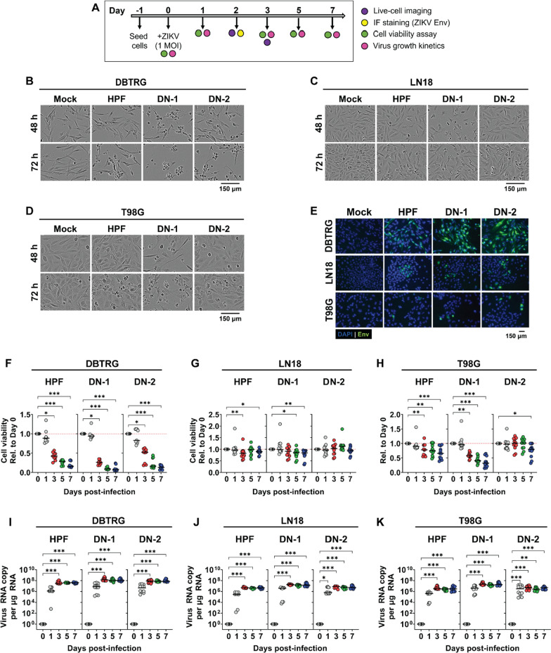
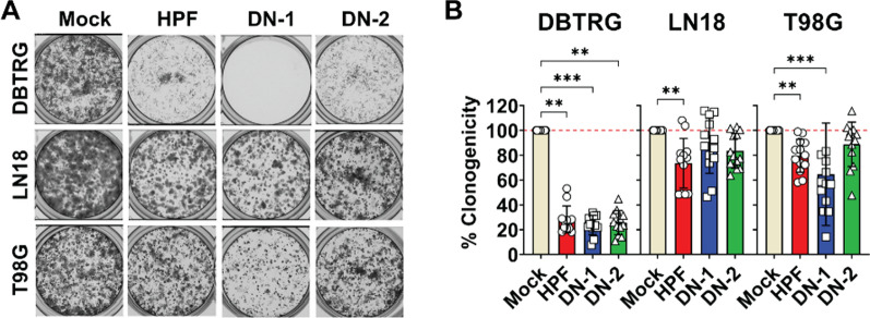
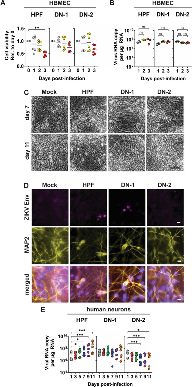
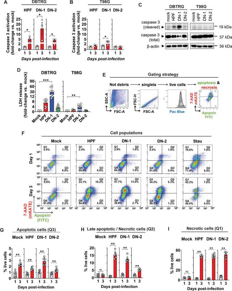
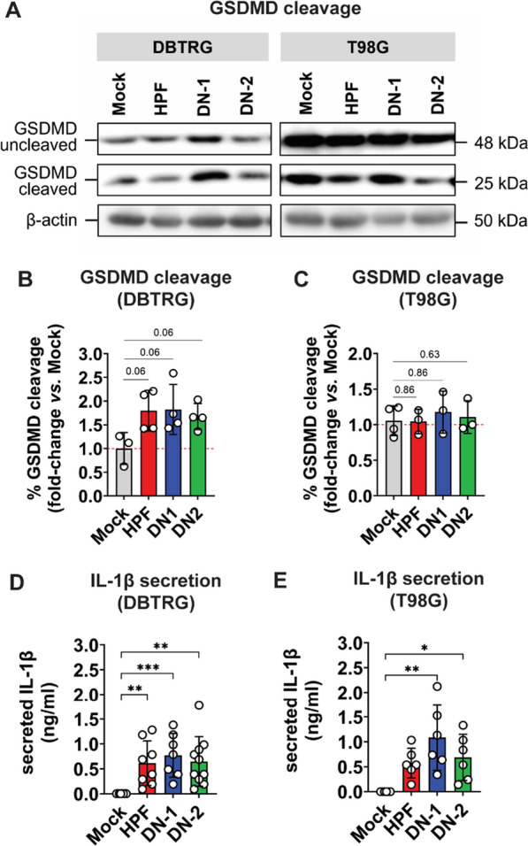
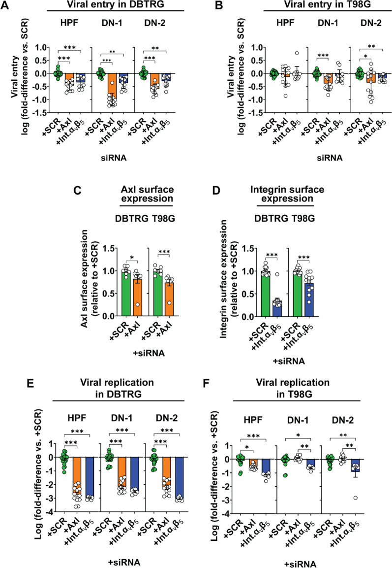
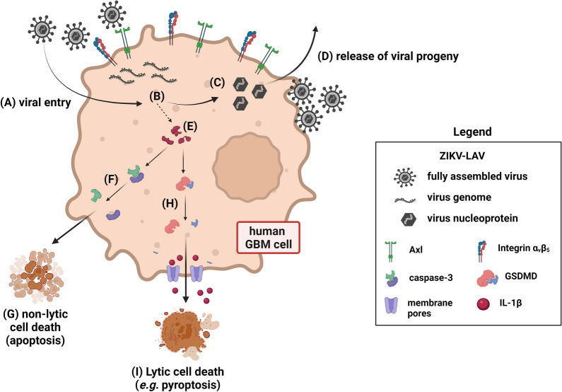
Glioblastoma multiforme (GBM) is the most common malignant primary brain cancer affecting the adult population. Median overall survival for GBM patients is poor (15 months), primarily due to high rates of tumour recurrence and the paucity of treatment options. Oncolytic virotherapy is a promising treatment alternative for GBM patients, where engineered viruses selectively infect and eradicate cancer cells by inducing cell lysis and eliciting robust anti-tumour immune response. In this study, we evaluated the oncolytic potency of live-attenuated vaccine strains of Zika virus (ZIKV-LAV) against human GBM cells in vitro. Our findings revealed that Axl and integrin αvβ5 function as cellular receptors mediating ZIKV-LAV infection in GBM cells. ZIKV-LAV strains productively infected and lysed human GBM cells but not primary endothelia and terminally differentiated neurons. Upon infection, ZIKV-LAV mediated GBM cell death via apoptosis and pyroptosis. This is the first in-depth molecular dissection of how oncolytic ZIKV infects and induces death in tumour cells.

Keywords: GBM; Glioblastoma; Immunogenic cell death; Live-attenuated vaccine; Oncolytic virus; ZIKV; Zika virus.


Conflict of interest statement

The authors declare no competing interests.

Figures

Fig. 1
Fig. 1
ZIKV-LAV strains productively infect human GBM multiforme (GBM) cell lines. A Timeline of in vitro infection experiments. Cells were inoculated with either ZIKV-LAV strains, DN-1 and DN-2, or parent strain HPF, at 1 plaque-forming unit (pfu) per cell. Infected cells were observed for 7 days. BD Representative live-cell images of human GBM cell lines B DBTRG, C LN18, and D T98G at 48 h and 72 h post-infection. E Representative fluorescence images of infected cells probed for the expression of ZIKV envelope (Env) protein. FH Kinetics of cell death in F DBTRG, G LN18, and H T98G cells over 7 days after virus infection. IK Virus growth kinetics following infection in I DBTRG, J LN18, and K T98G cells. Data are presented as individual points. Horizontal bars represent medians. Non-parametric Kruskal–Wallis test with Dunn’s post-hoc correction was used to compare groups. p-values are shown accordingly: *p < 0.05. **p < 0.005, ***p < 0.001
Fig. 2
Fig. 2
ZIKV-LAV infection inhibits clonogenic reproduction of human GBM cells. Human GBM cells (DBTRG, LN18, and T98G) were inoculated with 1 pfu per cell of either ZIKV-LAV strains (DN-1 and DN-2) or parent HPF strain. Cells were trypsinized and re-seeded at 5,000 cells per well on day 5 post-infection. A Representative clonogenicity plates of infected cells. B Quantification of clonogenicity data, which are presented as mean ± SEM. Non-parametric Kruskal–Wallis test with Dunn’s post-hoc correction was used to compare groups. p-values are shown accordingly: *p < 0.05. **p < 0.005, ***p < 0.001
Fig. 3
Fig. 3
ZIKV-LAV strains exhibit limited infection in normal brain cells. AB ZIKV-LAV infection of human brain microvascular endothelia (HBMEC) cells over 3 days, evaluated by measuring changes in cell viability A and viral copies B detected in infected cells. CE ZIKV-LAV infection of cultured human neurons over 11 days. Representative C live-cell images of cultured human neurons at day 7 and day 11 post-infection; and D immunofluorescence images of cultured human neurons at day 11 post-infection. Infected human neurons were probed for the expression of virus envelope protein (Env) and microtubule-associated protein 2 (MAP2). E Viral copies detected from the supernatant of human neuronal culture at 11 days post-infection with ZIKV-LAV. Data are presented as mean ± SEM. Non-parametric Kruskal–Wallis test with Dunn’s post-hoc correction was used to compare groups. p-values are shown accordingly: *p < 0.05. **p < 0.005, ***p < 0.001. Scale bar = 250 µm
Fig. 4
Fig. 4
ZIKV-LAV infection induces apoptotic and necrotic cell death in human GBM cells. AB Caspase-3 activation for apoptosis in virus-infected A DBTRG and B T98G cells detected by luminescence assay on days 1 and 3 post-infection. C Western blot detection of cleaved caspase-3 in infected cell lysates at day 2 post-infection. D Lactose Dehydrogenase (LDH) release by necrotic cells at day 3 post-infection. E Gating strategy for the simultaneous detection of apoptotic and necrotic DBTRG cells by flow cytometry. F Representative scatter dot-plots of DBTRG cells during early (day 1) and late (day 3) stages of infection with ZIKV-LAV. Cells were co-stained and detected with apopxin-FITC and 7-AAD-mKATE. GI Quantification of the fraction of G apoptotic, H late apoptotic/necrotic, and I necrotic DBTRG cells by flow cytometry during late stage of infection with ZIKV-LAV. Values are presented as % of live cells. Data are presented as mean ± SEM. Non-parametric Mann–Whitney test or Kruskal–Wallis test with Dunn’s post-hoc correction was used to compare groups. p-values are shown accordingly: *p < 0.05. **p < 0.005, ***p < 0.001
Fig. 5
Fig. 5
Pyroptotic cell death induced by ZIKV-LAV infection in human GBM cells. A Western blot detection of gasdermin D (GSDMD) cleavage in infected DBTRG and T98G cell lysates at day 5 post-infection. BC Quantification of GSDMD cleavage from western blot data normalized to β-actin expression. Data are shown as fold-change in percentage of (%) GSDMD cleavage relative to mock-infected cells. DE Luminescence-based detection of IL-1β secreted by infected cells. Data are shown as mean ± SD. Means were compared by Kruskal–Wallis test with Dun’s post-hoc correction. *p < 0.05; **p < 0.005; ***p < 0.001
Fig. 6
Fig. 6
ZIKV-LAV entry in human GBM cells is mediated by Axl and integrin αvβ5. Evaluation of the effect of siRNA-mediated knockdown of either Axl or integrin αvβ5 gene on AB viral entry in DBTRG (A) and T98G (B) cells; CD protein expression of Axl (C) and integrin αvβ5 (D) on the cell surface; and EF intracellular viral replication in DBTRG (E) and T98G (F) cells. SCR, scrambled siRNA. Int.αvβ5, integrin αvβ5. Data are presented as mean ± SEM. Non-parametric Mann–Whitney test or Kruskal–Wallis test with Dunn’s post-hoc correction was used to compare groups. p-values are shown accordingly: *p < 0.05. **p < 0.005, ***p < 0.001
Fig. 7
Fig. 7
Proposed model of human GBM cell death mediated by ZIKV-LAV infection. AD General virus infection life cycle. A ZIKV-LAV enters human GBM cells through Axl and integrin αvβ5 cellular receptors. B The ZIKV-LAV genome is translated to express viral proteins and the viral genome is replicated. C The viral genome and viral proteins assemble the nucleoprotein in preparation for (D) release of progeny into the surroundings with concomitant viral incorporation of host cell membrane. E Cellular and viral proteins expressed during ZIKV-LAV infection are also responsible for cell death in human GBM. FG cleavage of caspase-3 leads to non-lytic or non-inflammatory cell death by apoptosis; HI cleavage of gasdermin-D (GSDMD) leads to the formation of membrane pore complexes that shuttles inflammatory IL-1β outside of the cells. GSDMD cleavage also leads to inflammasome activation and lytic and inflammatory cell death by pyroptosis. Image created with BioRender.com

Similar articles

Cited by

References

    1. Alexander BM, Cloughesy TF. Adult glioblastoma. J Clin Oncol. 2017;35(21):2402–9. 10.1200/JCO.2017.73.0119. - PubMed
    1. Brain GBD, C. N. S. Cancer Collaborators Other. Global, regional, and national burden of brain and other Cns cancer, 1990–2016: a systematic analysis for the global burden of disease study 2016. Lancet Neurol. 2019;18(4):376–93. 10.1016/S1474-4422(18)30468-X. - PMC - PubMed
    1. Thakkar JP, Dolecek TA, Horbinski C, Ostrom QT, Lightner DD, Barnholtz-Sloan JS, Villano JL. Epidemiologic and molecular prognostic review of glioblastoma. Cancer Epidemiol Biomarkers Prev. 2014;23(10):1985–96. 10.1158/1055-9965.EPI-14-0275. - PMC - PubMed
    1. Stupp R, Hegi ME, Mason WP, van den Bent MJ, Taphoorn MJ, Janzer RC, Ludwin SK, Allgeier A, Fisher B, Belanger K, Hau P, Brandes AA, Gijtenbeek J, Marosi C, Vecht CJ, Mokhtari K, Wesseling P, Villa S, Eisenhauer E, Gorlia T, Weller M, Lacombe D, Cairncross JG, Mirimanoff RO, Research European Organisation for, Tumour Treatment of Cancer Brain, Groups Radiation Oncology, and Group National Cancer Institute of Canada Clinical Trials. Effects of radiotherapy with concomitant and adjuvant temozolomide versus radiotherapy alone on survival in glioblastoma in a randomised phase iii study: 5-year analysis of the Eortc-Ncic Trial. Lancet Oncol. 2009;10(5):459–66. 10.1016/S1470-2045(09)70025-7. - PubMed
    1. Feng E, Sui C, Wang T, Sun G. Temozolomide with or without radiotherapy in patients with newly diagnosed glioblastoma multiforme: a meta-analysis. Eur Neurol. 2017;77(3–4):201–10. 10.1159/000455842. - PubMed

Publication types

Substances